Search Header Logo
A&P Integ Intro 10/21/25

A&P Integ Intro 10/21/25

Assessment

Presentation

•

Science

•

9th - 12th Grade

•

Hard

Created by

Jessica Prado

Used 1+ times

FREE Resource

3 Slides • 7 Questions

1

​

By Jessica Prado

media

2

Match

Match the following

Anatomy

Histology

Physiology

cytology

the study of the structure of an organism

microscopic study of living tissues

study of the functions of living organisms and their parts

the study of cells

3

Multiple Choice

Question image

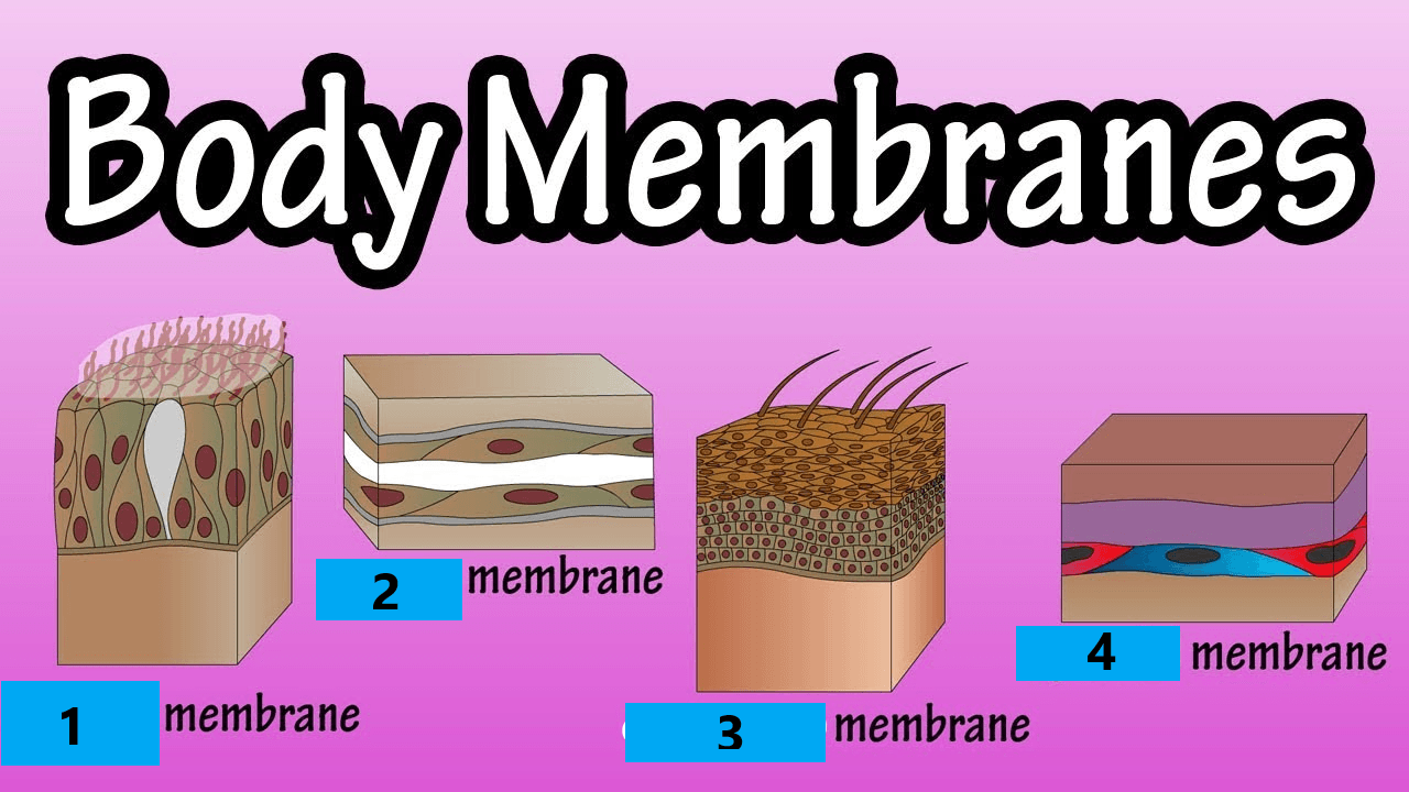
What type of body membrane is #1? They are "wet" or moist membranes. They line all body cavities that open to the exterior body surface like respiratory, digestive, and reproductive organs. They are adapted for absorption and secretion.

1

mucous membrane

2

serous membrane

3

cutaneous membrane

4

synovial membrane

4

Multiple Choice

Question image

What type of body membrane is #2? They are composed of surface simple squamous epithelium with underlying areolar CT. They line open body cavities that are open to the exterior of the body. They occur in pairs: parietal and visceral.

1

mucous membrane

2

serous membrane

3

cutaneous membrane

4

synovial membrane

5

Multiple Choice

Question image

What type of body membrane is #3? They are dry membranes because they are exposed to air. they are the outermost protective boundary. Have two parts: superficial epidermis and dermis.

1

mucous membrane

2

serous membrane

3

cutaneous membrane

4

synovial membrane

6

Multiple Choice

Question image

What type of body membrane is #4? Their function is the lubrication for friction; They are composed of connective tissue only. They line fibrous capsules surrounding joints.

1

mucous membrane

2

serous membrane

3

cutaneous membrane

4

synovial membrane

7

Open Ended

Question image

Describe how this student's view of the sample changed as they went from low to high power magnification on the microscope. You might want to click on the picture and zoom in to get a better view.

8

Labeling

This is a view of cheek cells you might see under our microscopes. It is the exact same placement on the slide, just at different magnifications. Match each view with its magnification as we would see with our microscopes.

Drag labels to their correct position on the image

1000x

40x

10x

400x

100x

9

Complete/ Redo Human Cells Lab
media
media

Remember to include magnifications and labels with your diagrams.
Answer the questions on the back of the paper
Glue into your journal and update your Table of Contents.

10

Complete the 2 Intro Videos
media
pattern-tertiary
​

By Jessica Prado

media

Show answer

Auto Play

Slide 1 / 10

SLIDE