
Font size
 Worksheets
6.2 Quiz: Microscopic Anatomy of Skeletal Muscles
Total questions: 10
Worksheet time: 10mins
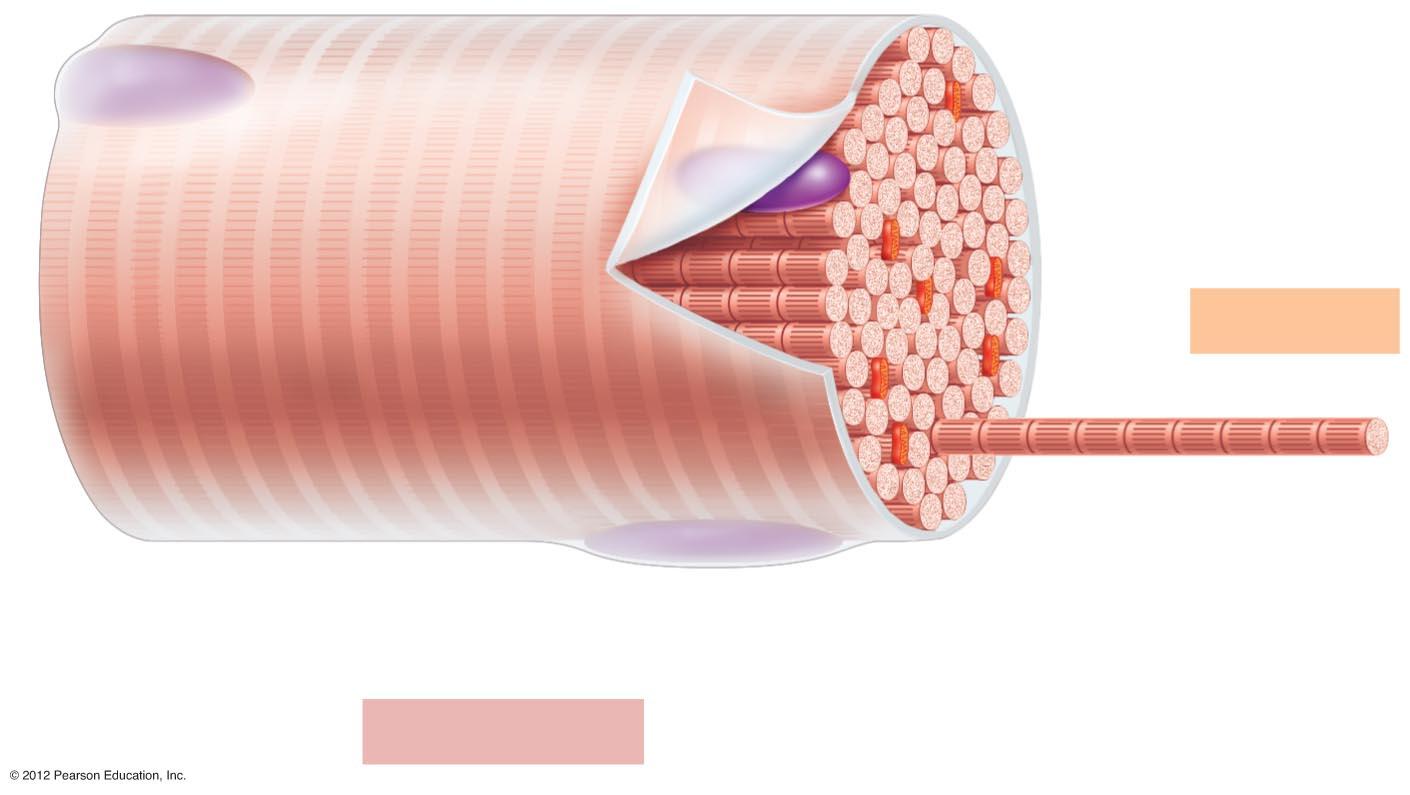
What is shown in the image?
neuron
myelin sheath
segment of a muscle fiber
blood vessel
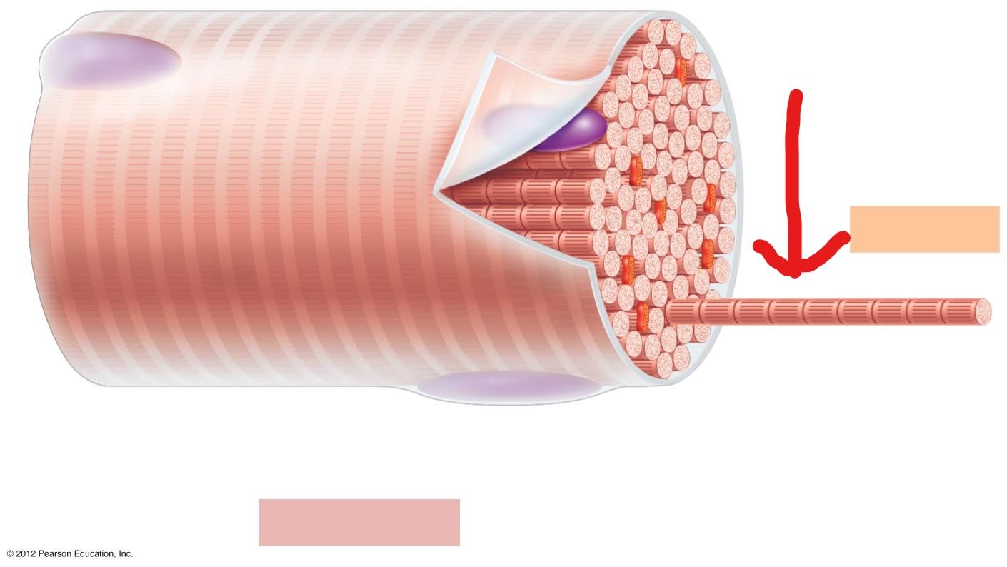
What is the arrow pointing at?
bare zone
thin filament
thick filament
myofibril
The plasma membrane of a skeletal muscle cell is called the:
sarcomere
centromere
sarcolemma
dilemma
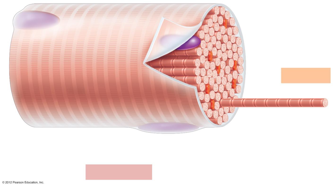
What feature(s) gives skeletal muscle cells their striped appearance?
Dark (A) band
thin (actin) filament
thick (myosin) filament
Light (I) band
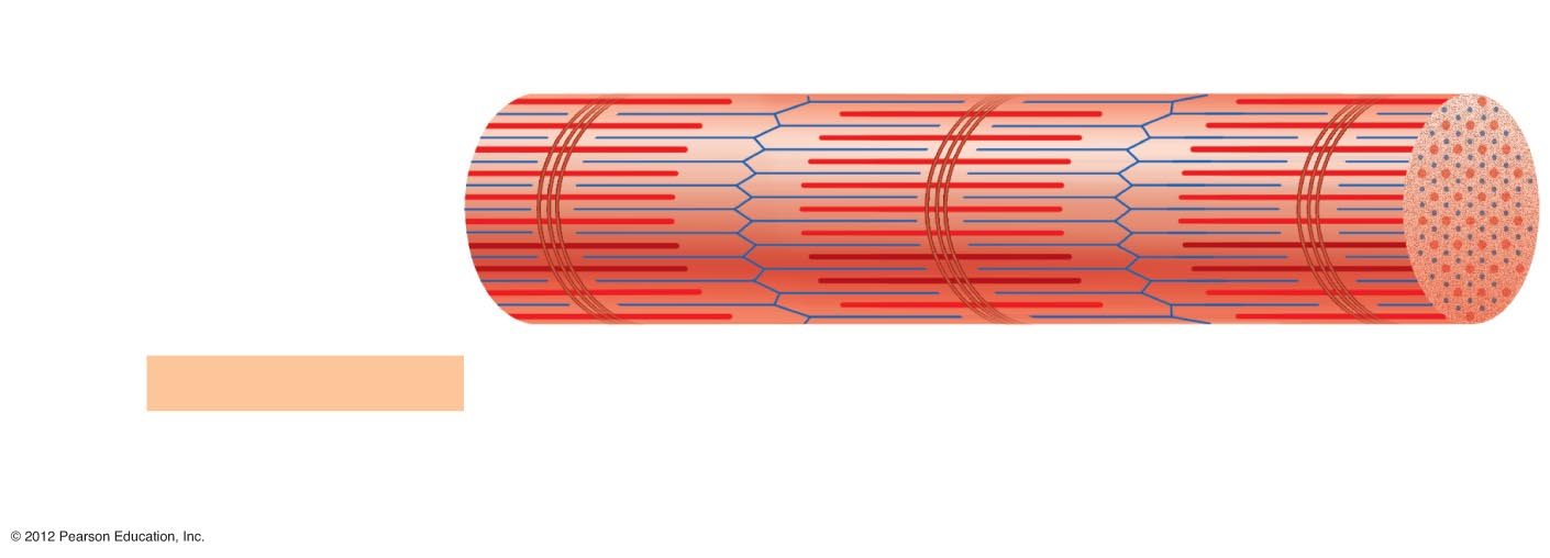
What is shown in the image?
myofibril
microfilaments
sarcoplasmic reticulum
cross bridges
Tiny contractile units that make up myofibrils are called:
sarcolemma
cross bridges
myosin
sarcomeres
There are two types of threadlike protein ______________________ within each of our "boxcar" sarcomeres.
cross bridges
myofilaments
microfilaments
sarcoplasmic reticulum
What feature of skeletal muscles "link the thick and thin filaments together during contraction"?
myosin
actin
cross bridges
sarcomeres
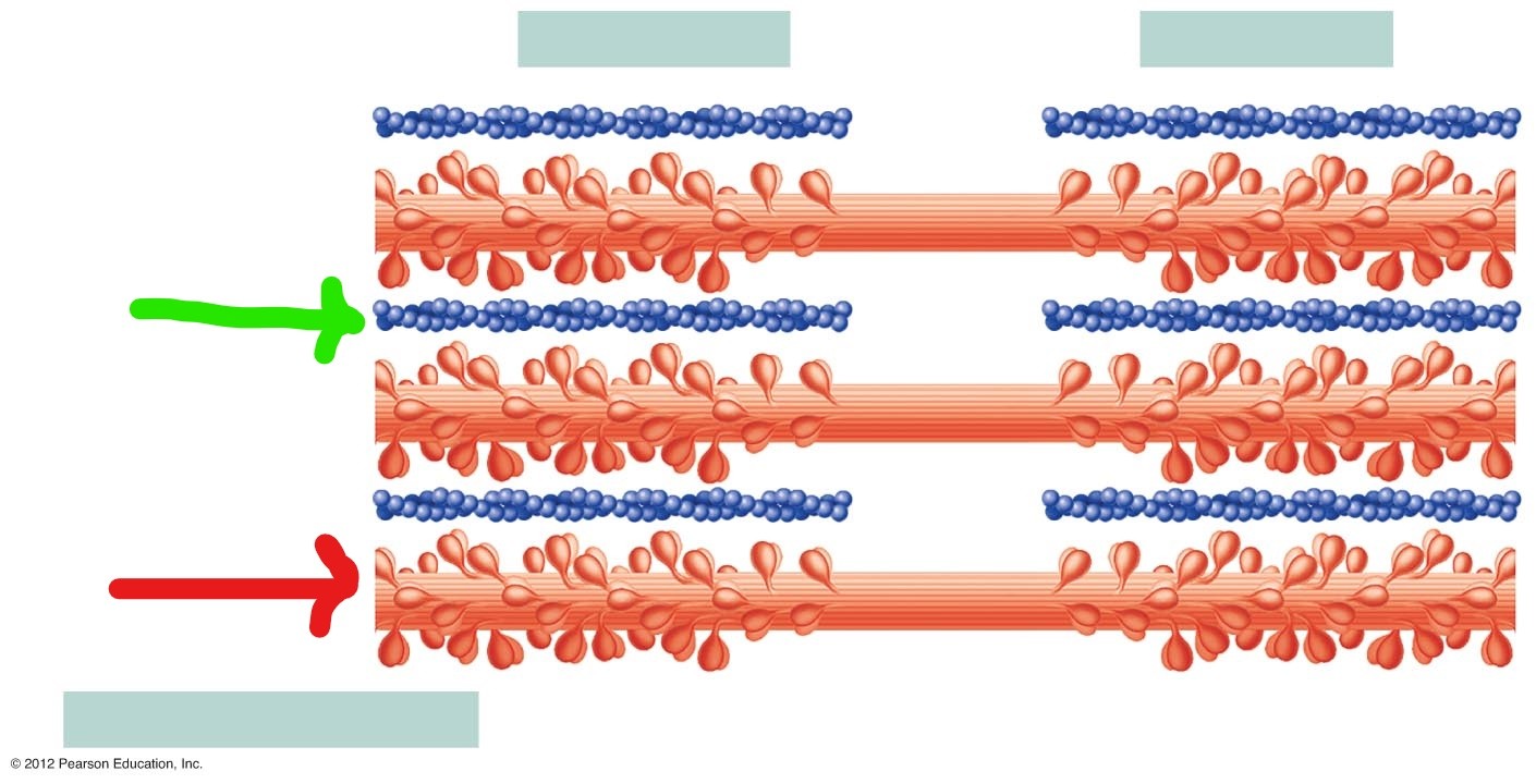
What feature of myofilaments is shown at the green arrow?
thick filaments, myosin
cross bridges
bare zone
thin filaments, actin
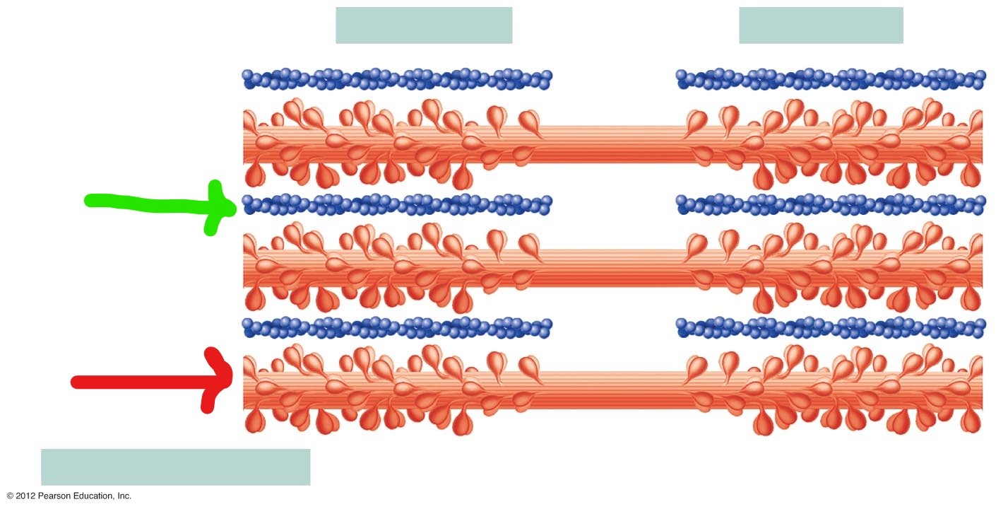
What feature of myofilaments is shown at the red arrow?
cross bridges
thick filaments, myosin
thin filaments, actin
bare zone

