
 Worksheets
EKG REVIEW 11/11/11
Total questions: 93
Worksheet time: 1hrs 13mins

Identify this rhythm
Sinus tachycardia
Normal Sinus Rhythm
Supraventricular Tachycardia
Atrial fibrillation

Interpret this Rhythm:
Normal sinus rhythm
Ventricular fibrillation
Sinus Brady with PAC's
Sinus Bradycardia

Identify this rhythm
Sinus bradycardia
1 degree heart block
Normal Sinus Rhythm
2nd Degree Type I heart block

Identify this rhythm
Supra-ventricular tachycardia
Atrial fibrillation
Sinus tachycardia
Atrial flutter

Identify the following rhythm
Electrical interference
Somatic tremor
Wandering baseline
Interrupted baseline

Which type of interference is represented by the photo?
Broken Recording
60second interference
Wandering Baseline
Somatic Tremor

Which type of interference is represented by the photo?
Broken Recording
60second interference
Wandering Baseline
Somatic Tremor

Name this rhythm
Multifocal Atrial Tachycardia
Wandering Atrial Pacemaker
Atrial Flutter
Atrial Fibrillation

EKG Strip?
Sinus Arrest
Sinus Arrhythmia
Uni-focal pvcs
Multi focal pvcs

Identify this rhythm
Supraventricular tachycardia
Sinus tachycardia
Atrial fibrillation
Normal Sinus Rhythm

Identify this rhythm
Sinus Tachycardia
Atrial fibrillation
Atrial flutter
Normal Sinus Rhythm
What typically happens to the rhythm after a pause in sinus arrest?
Cardiac Arrest
Usurpation
Escape
Ventricular Fibrillation
When the atrium takes over as the pacemaker of the heart, what is this action called?
Arrest
Usurpation
Escape
Fibrillation
Which characteristic of cardiac cells gives them the ability to send electrical impulses through the heart?
Contractility
Conductivity
Automaticity
Excitability
Which characteristic of cardiac cells gives them the ability to create impulses without any outside stimulation.?
Contractility
Conductivity
Automaticity
Excitability
Which characteristic of cardiac cells gives them the ability to contract the cardiac muscle?
Contractility
Conductivity
Automaticity
Excitability
Which characteristic of cardiac cells gives them the ability to respond to electrical impulses through depolarization?
Contractility
Conductivity
Automaticity
Excitability
What refers to the pressure inside the artery when the heart contracts and pumps blood through the body?
Systolic
Diastolic
Hypertension
Osmotic Pressure
What refers to the pressure inside the artery when the heart is at rest and filling with blood?
Systolic
Diastolic
Hypertension
Osmotic Pressure
What dominant pacemaker cells act as the conduction system in the heart?
Sinoatrial Node
Atrial Ventricular Node
Ventricles
Left Atrium
If the SA node fails, what will take its place?
Sinoatrial Node
Atrial Ventricular Node
Ventricles
Left Atrium
If the Atrial Ventricular node fails, what will take its place?
Sinoatrial Node
Atrial Ventricular Node
Ventricles
Left Atrium
A medical assistant is performing an EKG on a patient whose heart rate is 105 BPM. The assistant should identify this finding as an indication of which of the following conditions?
Tachycardia
Bradycardia
Sinus Rhythm
Atrial Fibrillation
A medical assistant is performing an EKG on a patient whose heart rate is 55 BPM. The assistant should identify this finding as an indication of which of the following conditions?
Tachycardia
Bradycardia
Sinus Rhythm
Atrial Fibrillation


A patient is experiencing electrocardiogram changes. Which wave should the nurse recognize as representing ventricular repolarization?
Q wave
R wave
S wave
T wave
A patient is admitted for diagnosis of an electrocardiogram abnormality. Which wave should the nurse plan care based upon changes in the patient’s ventricular depolarization?
P wave
QRS complex
U wave
T wave
The nurse is analyzing a client’s electrocardiogram tracing. Which of the following complexes is not normally seen on an electrocardiogram tracing?
P wave
QRS complex
T wave
U wave
In order to calculate the heart rate, what of the following is required?
Measure R - R interval
Calculate QTc
Measure ST segment
Measure P-R interval
Measure QRS complex
Leads I,II and III are
travel from a negative
bipolar
negative leads
positive leads
Leads AVL,AVR and AVF are
bipolar
unipolar
augmented
tracing
V1
LEFT side of the sternum, directly across from V1 at the FOURTH intercostal space
LEFT side of the chest, FIFTH intercostal space, midclavicular line
RIGHT side of the sternum at the FOURTH intercostal space
LEFT side of the chest, FIFTH intercostal space, midaxillary line
V2
LEFT side of the chest, FIFTH intercostal space, midclavicular line
LEFT side of the sternum, directly across from V1 at the FOURTH intercostal space
LEFT side of the chest, FIFTH intercostal space, midclavicular line
LEFT side of the chest, FIFTH intercostal space, anterior axillary line
V3
LEFT side of the chest, FIFTH intercostal space, midaxillary line
RIGHT side of the sternum at the FOURTH intercostal space
LEFT side of the chest, FIFTH intercostal space, anterior axillary line
LEFT side of the chest, midway between V2 and V4
V4
LEFT side of the chest, FIFTH intercostal space, midclavicular line
LEFT side of the chest, midway between V2 and V4
LEFT side of the chest, FIFTH intercostal space, midaxillary line
LEFT side of the sternum, directly across from V1 at the FOURTH intercostal space
V5
LEFT side of the chest, FIFTH intercostal space, anterior axillary line
LEFT side of the chest, FIFTH intercostal space, midaxillary line
LEFT side of the chest, FIFTH intercostal space, midclavicular line
LEFT side of the sternum, directly across from V1 at the FOURTH intercostal space
V6
LEFT side of the chest, FIFTH intercostal space, midclavicular line
LEFT side of the chest, FIFTH intercostal space, anterior axillary line
LEFT side of the chest, FIFTH intercostal space, midaxillary line
LEFT side of the sternum, directly across from V1 at the FOURTH intercostal space
A medical assistant is preparing a patient who has shortness of breath for at EKG. In which of the following positions should be assistant place the patient?
Prone
Semi- Fowler's
Supine
Sims'
A medical assistant is calibrating an EKG machine. Which of the following should the assistant identify as the standard speed of the recording?
25 mm/second
50 mm/second
10 mm/second
5 mm/second
A medical assistant is preparing a patient for an EKG. In which of the following locations should the assistant place the V4 lead?
5th intercostal space at the anterior axillary line
5th intercostal space at the midaxillary line
Midway between V3 and V5
5th intercostal space at the midclavicular line
A medical assistant notes a wandering baseline throughout an EKG tracing. Which of the following electrocardiograph issues could have resulted in this occurrence?
The electrode pads were defective or poorly attached
The machine was missing the grounding prong for the outlet
The machine did not have the proper standardization settings set
The cable tips were loose or had become separated
A provider requests a rhythm strip in addition to a regular EKG tracing for a patient. The medical assistant should identify that which of the following leads provides the clearest recording of the heart rhythm?
I
II
III
V2
which of the following is the normal PR interval
0.12-0.20 MM of voltage
0.12 -0.20 per second
0.16 -0.20 MM of voltage
0.02- 0.08 per second
Lead II is considered the
bipolar limb lead
augmented or unipolar limb lead
chest of precordial lead
none of the choices
Times where the conduction cells are “resistant” to stimuli.
Refractory Periods
Absolute Periods
Relative Periods
Supernormal Periods
Times where the cell cannot accept another impulse, still dealing with the last one.
Refractory Periods
Absolute Periods
Relative Periods
Supernormal Periods
Times where the cells strong stimulus will result in depolarization, but not as strong.
Refractory Periods
Absolute Periods
Relative Periods
Supernormal Periods
The nurse is determining the functioning of a patient’s AV node. How many times per minute should the nurse expect the node to normally fire?
20 to 40
25 to 55
40 to 60
60 to 100
The nurse is determining the functioning of a patient’s Purkinje Fibers/Ventricles. How many times per minute should the nurse expect the node to normally fire?
20 to 40
25 to 55
40 to 60
60 to 100
The Line from which waves and complexes take off.
Isoelectric Line
Usurpation
Escape
Fibrillation
The measurement of time between waves and complexes. (flat line)
Isoelectric Line
Segment
Interval
Fibrillation
The measurement of time that includes waves and complexes.
Isoelectric Line
Segment
Interval
Fibrillation
Every small square on EKG paper represents
0.04 sec
0.20 sec
5 mm
0,5 mv
Every large square on EKG paper represents
0.04 sec
0.20 sec
5 mm
0,5 mv
How many small square are in each large square of EKG paper?
5
10
20
25
A medical assistant is calibrating an EKG machine. Which of the following should the assistant identify as the standard speed of the recording for an infant, pediatric patient or patient with an accelerated heart rate?
25 mm/second
50 mm/second
10 mm/second
5 mm/second
What is the term that regulates the height of the complexes on the ekg paper
Gain
Lost
Positive
Negative
What should the default gain on an EKG machine be set to?
5 mm
10 mm
20 mm
25 mm
_________is unwanted interference.
Artifact
Somatic Tremors
Wandering Baseline
60 Second interference
This artifact is caused by a patient shivering and can be corrected by giving them a blanket
Somatic Tremor
Baseline Sway or wandering baseline
Broken Recording
60 second interference
This artifact can be removed by unplugging other machines close by, turning off phones,
Somatic Tremor
Baseline Sway or wandering baseline
Broken Recording
60 second interference
This artifact is caused by lotion or sweat on the skin.
Somatic Tremor
Baseline Sway or wandering baseline
Broken Recording
60 second interference
This artifact is due to bad wires, loose electrodes or cable.
Broken Recording
60second interference
Wandering Baseline
Somatic Tremor
This is a slight irregularity in the EKG rhythm associated with normal breathing patterns?
Sinus Bradycardia
Sinus Tachycardia
Sinus Dysrhythmia
Sinus Arrest
When should a medical professional be concerned with sinus arrest?
When it lasts less than 6 seconds
When it lasts more than 6 seconds
When it lasts more than 10 seconds
When it lasts less than 10 seconds
After performing an ECG on an adult client, the nurse reports that the PR interval reflects normal sinus rhythm. What is the PR interval for a normal sinus rhythm?
0.05 and 0.1 seconds.
0.12 and 0.2 seconds.
0.15 and 0.3 seconds.
0.25 and 0.4 seconds.
A nurse is caring for a client who's experiencing sinus bradycardia with a pulse rate of 40 beats/minute. The client's blood pressure is 80/50 mm Hg and the client reports dizziness. Which medication does the nurse anticipate administering to treat bradycardia?
Atropine
Dobutamine
Amiodarone
Lidocaine
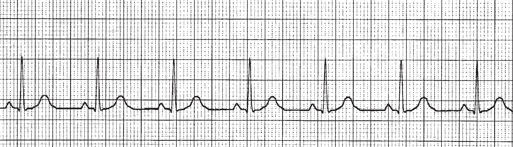
What is the hear rate of this ECG rhytm?
40
70
100
130
The QRS duration must be ___ seconds or less
0.11
0.12
0.13
0.20
0.25
What is the difference between sinus arrest and sinus block?
The rate
The width of the P waves
The number of pauses in the rhythm
The number of skipped P waves

Name this rhythm...
Atrial Fibrillation
Atrial Flutter
Sinus Bradycardia

What is this rhythm?
Sinus Bradycardia
Junctional Escape Rhythm
Bigeminy
Multiform PACs

what is this rhythm
Supraventricular Tachycardia
Atrial Flutter
Sinus Tachycardia
Sinus Rhythm

Identify the following rhythm
Normal sinus rhythm
Sinus bradycardia
Sinus tachycardia
Junctional

The following strip is a 6 second strip. What EKG rhythm is displayed here?
Atrial Fibrillation
Asystole
Normal Sinus Rhythm
Sinus Tachycardia

EKG Strip?
Sinus Arrest
Sinus Arrhythmia
Atrial Flutter
Atrial Fib.
Occurs when the natural pacemaker site shifts between SA node, the atria, and/or the AV node.
1st Degree AV Block
SA Block
Sinus Pause
Wandering Atrial Pacemaker

For the strip above, what is the HR?
60 bpm
40 bpm
65 bpm
70 bpm

For the above strip, what is the HR?
45 bpm
40 bpm
50 bpm
30 bpm

For the above strip, what is the HR?
120 bpm
125 bpm
130 bpm
135 bpm

For the above strip, what is the HR?
105 bpm
180 bpm
175 bpm
110 bpm

For the above strip, what is the EKG rhythm?
Sinus Tachycardia
Supraventricular Tachycardia
Junctional Tachycardia
Premature Atrial Contraction

For the above strip, what is the HR?
100 bpm
105 bpm
110 bpm
120 bpm

For the above strip, what is the EKG rhythm?
Asystole
Sinus Tachycardia
Paroxysmal Supraventricular Tachycardia
Junctional Tachycardia

For the above strip, what is the EKG rhythm?
Ventricular Fibrillation
Sinus Tachycardia
Atrial Flutter
Junctional Tachycardia

For the above strip, what is the EKG rhythm?
Normal Sinus Rhythm with PVC
Normal Sinus Rhythm with PAC
Normal Sinus Rhythm
Normal Sinus Rhythm with AVB
A QRS measurement of less than ______ seconds indicates a supraventricular pacemaker.
0.12
0.14
0.18
0.20

Name this Rhythm
Atrial Fibriallation
Sinus Tachycardia
Supraventricular Tachycardia
Multifocal Atrial Tachycardia

Name this Rhythm
Sinus Block
Sinus Arrest
Normal Sinus Rhythm w/ PAC's
Wandering Atrial Pacemaker

Name this Rhythm
Sinus Block
Sinus Arrest
Normal Sinus Rhythm w/ PAC's
Wandering Atrial Pacemaker

What is this rhythm?
Sinus Bradycardia
Trigeminy
Bigeminy
Multiform PACs