
 Worksheets
ECG/EKG Rhythm Strips
Total questions: 200
Worksheet time: 7hrs 40mins

Which of the following best describes this strip?
Tachycardia
Normal Sinus Rhythm
Bradycardia
Asystole

Which of the following best describes this strip?
Tachycardia
Normal Sinus Rhythm
Bradycardia
Asystole

Which of the following best describes this strip?
Tachycardia
Normal Sinus Rhythm
Bradycardia
Asystole

Which one is represent P wave
A
B
C
E
Which one is represent T wave
A
B
C
E
Which one is represent QRS complex
A
B
C
E
By relating events in ECG with phases in cardiac cycle, at which event the AV valve will be closed?
A
B
C
E
By relating events in ECG with phases in cardiac cycle, at which event both atria and ventricles will be relaxed?
A
B
C
E
By relating events in ECG with phases in cardiac cycle, at which event atria contract and ventricles relax?
A
B
C
E
By relating events in ECG with phases in cardiac cycle, at which event the second sound of heart "dup" will be produced?
A
B
C
E

How many cardiac cycle shown in the diagram
2
4
5
6
The reading of the ECG shown in the diagram is
from left to right (forward)
from right to left (backward)
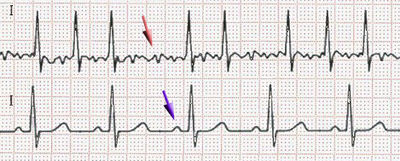
What is the ECG showing (Red Arrow)
Atrial Fibrillation
Ventricular Fibrillation
Asystole
Premature Ventricular Contraction
Heart Block

What is the ECG showing
Atrial Fibrillation
Ventricular Fibrillation
Asystole
Premature Ventricular Contraction
Heart Block

What is the ECG showing
Atrial Fibrillation
Ventricular Fibrillation
normal
Premature Ventricular Contraction
Heart Block
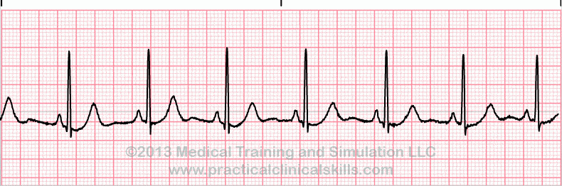
What is the ECG showing
Atrial Fibrillation
Ventricular Fibrillation
normal
Premature Ventricular Contraction
Heart Block

What is the ECG showing
Atrial Fibrillation
Ventricular Fibrillation
normal
Premature Ventricular Contraction
Heart Block

What is the ECG showing (arrow)
Atrial Fibrillation
Ventricular Fibrillation
normal
Premature Ventricular Contraction
Heart Block
On a typical ECG grid, 5 small squares, or 1 large square, represent 0.20 seconds of time.
True
False

Identify the ECG tracing.
Normal Sinus Rhythm
Atrial Fibrillation
Atrial Flutter
Ventricular tachycardia

Identify the ECG tracing.
Ventricular fibrillation
Ventricular tachycardia
Torsades de pointes
Asystole

What is the heart rate in this ECG tracing?
60 bpm
75 bpm
90 bpm
130 bpm

Identify the heart block, if any.
First-degree AV block
Second-degree AV block (type I)
Second-degree AV block (type II)
Third-degree AV block
Normal Sinus Rhythm
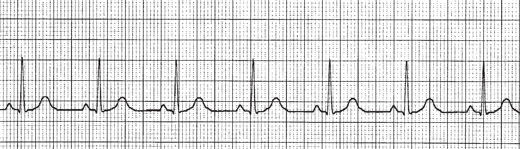
What is the hear rate of this ECG rhytm?
40
70
100
130
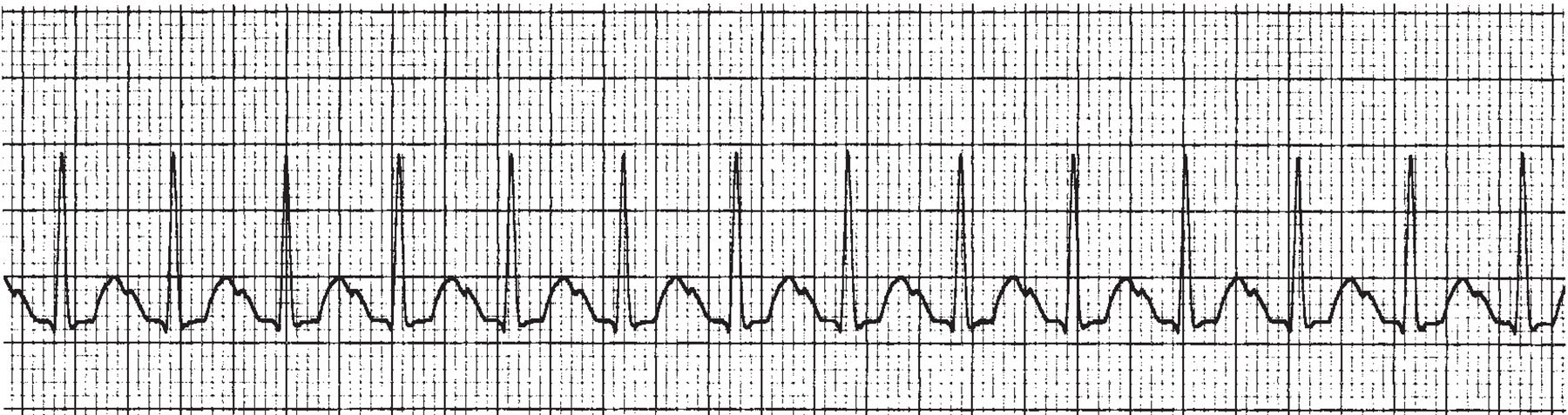
This ECG strip illustrates ____________.
Premature Atrial Contraction
Premature Ventricular Contraction
Atrial Fibrulation
Tachycardia

This ECG wave illustrates ____________.
An MI that occurred years ago
ST segment depression
An acute MI
Normal ECG wave
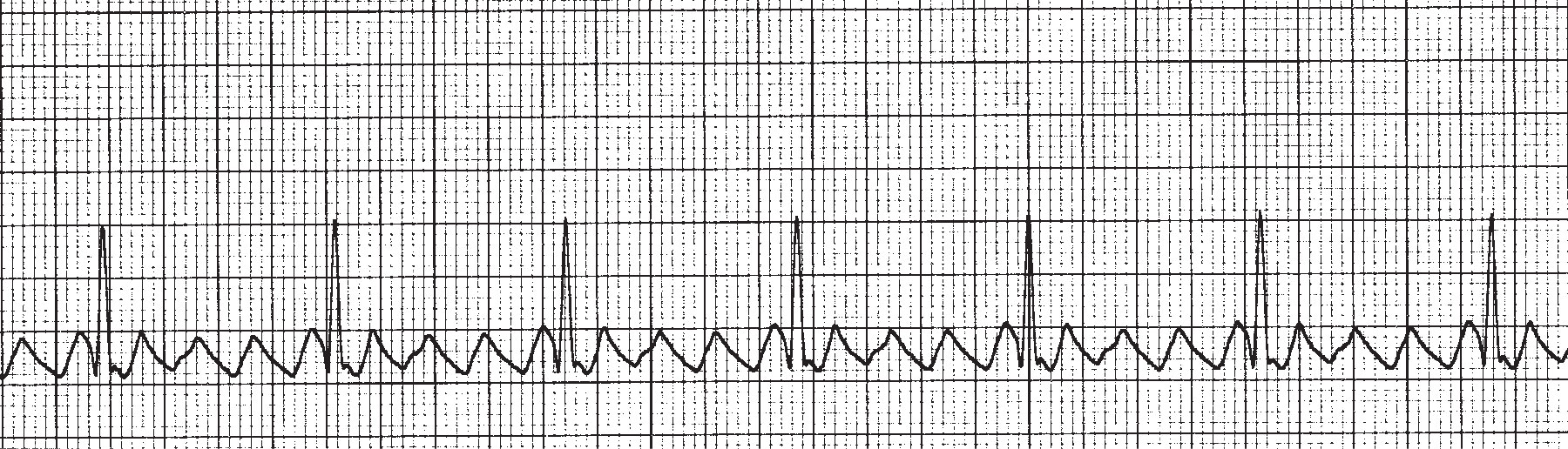
This ECG illustrates _______________.
Premature Atrial Contraction
Atrial Flutter
Atrial Fibrillation
Ventricular Fibrillation
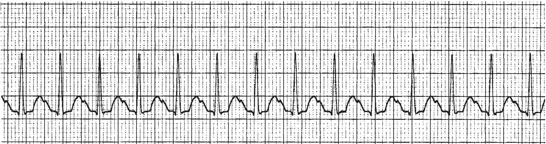
What is the HR of this ECG?
105pbm
135bpm
75bpm
165bpm
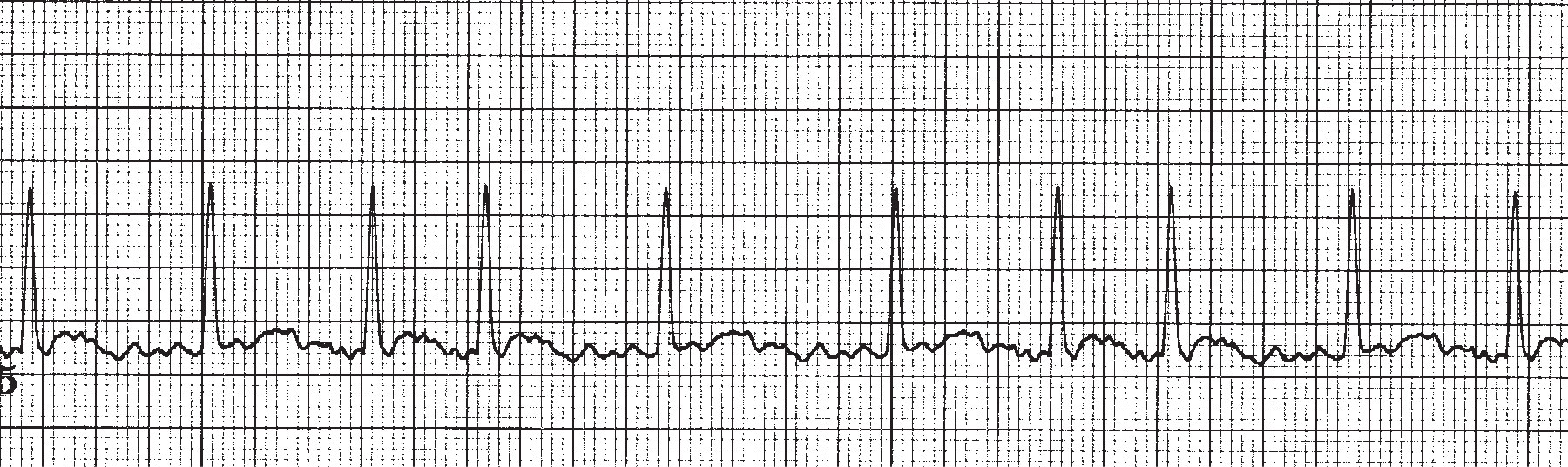
This ECG illustrates ____________________.
Atrial Flutter
Bradycardia
Atrial Fibrillation
Myocardial Infarct

This ECG illustrates ____________________.
1st Degree AV Block
2nd Degree AV Block
3rd Degree AV Block
4th Degree AV Block

This ECG illustrates ________________.
1st Degree AV Block
2nd Degree AV Block
3rd Degree AV Block
4th Degree AV Block

This ECG wave illustrates ______________.
Normal Wave Form
Ectopic Foci
Previous MI
Fibrillation

This ECG illustrates _________________.
Trigemany
Ventricular Fibrillation
Premature Ventricular Contractions
Ventricular Tachycardia

Identify The Tracing
Supraventricular Tachycardia
Premature Atrial Complex
First Degree Heart Block
Atrial Fibrillation

Identify The Tracing
Premature Vent. Complex: Trigeminy
Wandering Atrial Pacemaker
Atrial Flutter
2nd degree AV block, Mobitz I

Identify the tracing
2nd Degree Heart Block Mobitz I
Ventricular Tachycardia Polymorphic
ST Segment Depression
2nd Degree Heart Block Mobitz II

Identify the tracing
Premature Vent. Complex: Quadrigeminy
AV block: 3rd degree (complete heart block)
ST Segment Depression
Ventricular Tachycardia Polymorphic

Identify the tracing
AV block: 2nd degree
Atrial Fibrilation
Junctional Tachycardia
Sinus Bradycardia

Identify the tracing
Atrial Flutter
AV block: 1st degree
Atrial Fibrilattion
Sinus Tachycardia

Identify the tracing
Total AV Block
Atrial Flutter
Torsades de Pointes
Av block 1st Degree

Identify the tracing
Ventricular Fibrillation
PEA
Ventricular Tachycardia
Asystole
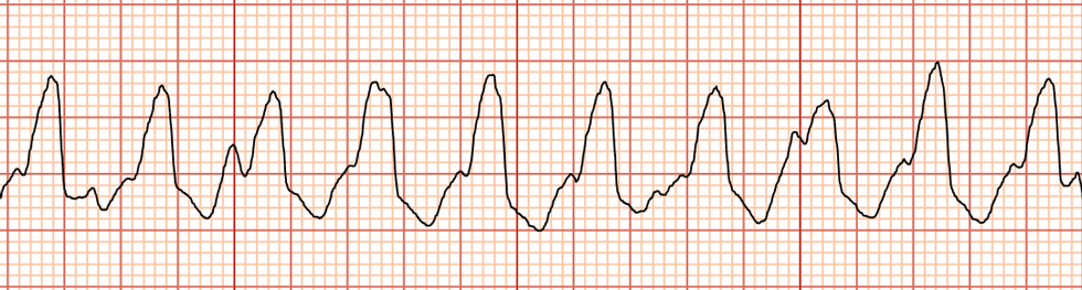
Identify the tracing
Atrial Fibrillation
Sinus Tachycardia
Ventricular tachycardia
Supraventricular tachycardia

What is your possible diagnosis
Sinus Tacchycardia
Sinus arrythmia
Atrial fibriilation
WPW syndrome

your correct diagnosis is
Atrila Fibrillation
Ventricular fibriilation
Sinus Tacchycardia
Abnormal ECG
What is the ECG showing ?
Atrial Fibrillation
Ventricular Fibrillation
normal
Premature Ventricular Contraction
Heart Block
What is the ECG showing?
Atrial Fibrillation
Ventricular Fibrillation
normal
Premature Ventricular Contraction
Heart Block
What is the ECG showing?
Atrial Fibrillation
Ventricular Fibrillation
normal
Premature Ventricular Contraction
Heart Block

Please choose the right electrical event according to the labels
A: Pacemaker generate wave of signals to contract
B: Signals delayed at AV node
C: Signals pass to heart apex
D: Signals spread throughout ventricles
A: Pacemaker generate wave of signals to contract
B: Signals spread throughout ventricles
C: Signals delayed at AV node
D: Signals pass to heart apex
A: Pacemaker generate wave of signals to contract
B: Signals delayed at AV node
C: Signals pass to heart apex
D: Signals spread throughout ventricles
A: Signals pass to heart apex
B: Signals delayed at AV node
C: Pacemaker generate wave of signals to contract
D: Signals spread throughout ventricles
Letter B is identified as the:
P wave
PR interval
PR segment
QRS complex
Letter D is identified as the:
PR interval
ST segment
PR segment
QRS complex

3rd Degree AV Block
2nd Degree AV Block
1st Degree AV Block

Sinus Tachycardia
Supraventricular Tachycardia
Ventricular Tachycardia

Atrial rate of >350 bpm, No definitive P wave, QRS complex usually normal
Ventricular Fibrillation
SVT
Atrial Fibrillation

Find the heart rate. Select the nearest number in your answer.
78 bpm
70 bpm
75 bpm
81 bpm

What part of the hearts conduction system is shown in the diagram?
SA node
AV Node
AV bundle
Purkinje Fibers

What part of the hearts conduction system is shown in the diagram?
SA node
AV Node
AV bundle
Purkinje Fibers

What part of the hearts conduction system is shown in the diagram?
SA node
AV Node
AV bundle
Purkinje Fibers

What part of the hearts conduction system is shown in the diagram?
SA node
AV Node
AV bundle
Purkinje Fibers

What is the name of the circled area of the ECG in the diagram?
P Wave
QRS Complex
T Wave
Cardiac Cycle

What is the name of the circled area of the ECG in the diagram?
P Wave
QRS Complex
T Wave
Cardiac Cycle

What is one complete contraction of the heart as represented in the ECG diagram called?
Coronary Cycle
Cardiac Output
ECG Cycle
Cardiac Cycle

What part of an ECG represents the depolarization of the ventricles?
P Wave
QRS Complex
T Wave

Identify the tracing
Atrial Fibrillation
Sinus Tachycardia
Ventricular tachycardia
Supraventricular tachycardia

Identify this Rhythm
2nd Degree AV Block type 1
2nd degree Block type 2
3rd degree AV Block
First degree AV Block

2nd Degree AV Block Type 1
2nd Degree AV Block Type 2
3rd Degree AV Block
First Degree AV Block

3rd Degree AV Block
2nd Degree AV Block Type 1
2nd Degree AV Block Type 2
First Degree AV Block

Atrial Fibrillation
2nd Degree AV Block Type 1
2nd degree AV Block Type 2
Atrial Flutter

First Degree AV Block
2nd Degree AV Block Type 1
2nd Degree AV Block Type 2
3rd Degree AV Block

3rd Degree AV Block
2nd Degree AV Block Type 1
2nd Degree AV Block Type 2
First Degree AV Block

What is this rhythm?

What is this rhythm?
Normal Sinus Rhythm
Accelerated Junctional Rhythm
Sinus Rhythm with 1AVB
2nd Degree AV Block
What is the approximate amount of time for the QRS complex to record on an ECG?
0.06-0.10 seconds
0.04-0.08 seconds
0.04-0.12 seconds
0.04-0.06 seconds
The P wave of an ECG indicates
atrial contraction.
the heart at rest.
ventricular contraction.
ventricular relaxation.
Which of the following rhythms identified on an ECG prompts the medical assistant to notify the provider immediately?
sinus bradycardia
premature ventricular contraction
ventricular fibrillation
sinus tachycardia
Which of the following indicates a normal sinus rhythm?
PR interval of 0.22 second, QRS complex of 0.10 second, QT interval of 0.40 second and HR of 78
PR interval of 0.22 second, QRS complex of 0.12 second, QT interval of 0.42 second and HR of 72
PR interval of 0.14 second, QRS complex of 0.10 second, QT interval of 0.40 second and HR of 78
PR interval of 0.14 second, QRS complex of 0.16 second, QT interval of 0.42 second and HR of 72

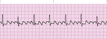
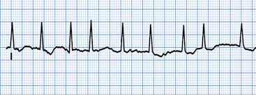
Which of the following rhythms is shown in the strip?
normal sinus rhythm
sinus bradycardia
sinus tachycardia
sinus arrhythmia
In how many body areas are leads placed in order to obtain a 12 lead ECG?
8
12
10
14
What is computer based monitoring also known as?
ambulatory monitoring
telemetry
out patient monitoring
inpatient monitoring
In single channel ECG machines, how many leads are recorded at one time?
12
6
3
1

What does this EKG show?
V-fib
Hypocalcemia
Hyperkalemia
Torsades de pointe
Which are the 2 shockable rhythms?
Vfib
Vtach
PEA
Asystole
A wide QRS complex is determined by a measurement of:
0.12 seconds or greater
0.04 seconds or greater
0.11 seconds or less
0.08 seconds or less
The PR interval is considered normal if it is between:
0.20 and 0.24 seconds
0.04 and 0.08 seconds
0.12 and 0.20 seconds
0.02 and 0.12 seconds
The point where the QRS complex meets the ST segment is commonly referred to as the:
J point
Q point
S point
P point
The normal heart rate is _________ beats per minute.
40 - 120
60 - 100
20 - 40
40 - 60
Step one of the 5-step approach for EKG interpretation is the:
Heart rate
Heart rhythm
PRI
QRS complex
R to R intervals that exhibit no similarity are termed:
Occasionally irregular
Regular
Irregularly irregular
Regularly irregular
The sequential beating of the heart as a result of the generation of electrical impulses is called:
Heart regularity
Heart system
Heart rhythm
Heart rate
A normal or narrow QRS complex indicates that the impulse was not formed in the ventricles and is termed:
Supraventricular
Aberrant conduction
Idioventricular
Midventricular
Heart rate below 60 beats per minute is termed:
Idioventricular
Tachycardia
Bradycardia
Supraventricular
The term __________ is utilized when an impulse may be "above the ventricles."
Idioventricular
Supraventricular
Enterventricular
Medventricular
Artifact is defined as EKG waveforms from sources outside the heart and is most often caused by all of the following EXCEPT:
Electrical impulses
Defective electrodes
Defective cables
Patient movement
A rhythm that originated in the SA node is determined by a:
P wave with a negative deflection
P wave that is not upright
P wave that is inverted
P wave with a positive deflection
When utilizing the 6-second method, you count the number of R waves in a six-second strip and multiply by:
20
15
5
10

The rate of sinus tachycardia is:
Less than 60 bpm
40 - 80 bpm
60 - 100 bpm
Greater than 100 bpm
An ECG rhythm strip shows a ventricular rate of 46, a regular rhythm, a PR interval of 0.14 second, a QRS duration of 0.06, and one upright P wave before each QRS. This rhythm is:
Sinus rhythm
Sinus bradycardia
Sinus arrest
Sinoatrial block
An ECG rhythm strip shows a ventricular rate of 128, a regular rhythm, a PR interval of 0.16 second, a QRS duration of 0.08, and one upright P wave before each QRS. This rhythm is:
Sinus arrhythmia
Sinus bradycardia
Sinus rhythm
Sinus tachycardia

Sinus Rhythm
Sinus Brady
Junctional
WPW
The patient's monitor shows a flat line but is awake and responding appropriately. You should
Call for help
Begin CPR
Check the leads
No action is needed
60 to 80 beats per minute (beats/min)
Sinus tachycardia
Sinus bradycardia
Normal sinus rhythm
none of the above

The following strip is a 6 second strip. What EKG rhythm is displayed here?
Atrial Fibrillation
Asystole
Normal Sinus Rhythm
Sinus Tachycardia

EKG Strip?
Sinus Arrest
Sinus Arrhythmia
Uni-focal pvcs
Multi focal pvcs

EKG Strip?
Sinus Arrest
Sinus Arrhythmia
Atrial Flutter
Atrial Fib.
Normal PR Interval
Normal QRS Interval
P wave is upright and uniform.
There is one p wave before each QRS complex.
Rhythm is regular.
Rate is 72.
sinus rhythm
sinus tachycardia
sinus bradycardia
sinus arrhythmia
Normal PR Interval
Normal QRS Interval
P wave is upright and uniform.
There is one p wave before each QRS complex.
Rhythm is regular.
Rate is 122.
sinus rhythm
sinus tachycardia
sinus bradycardia
sinus arrhythmia
Normal PR Interval
Normal QRS Interval
P wave is upright and uniform.
There is one p wave before each QRS complex.
Rhythm is irregular.
Rate is 65.
sinus rhythm
sinus tachycardia
sinus bradycardia
sinus arrhythmia
Normal PR Interval
Normal QRS Interval
P wave is upright and uniform.
There is one p wave before each QRS complex.
Rhythm is regular.
Rate is 48.
sinus rhythm
sinus tachycardia
sinus bradycardia
sinus arrythmia
I am a rhythm with a rate of 104. I am irregular and my p waves are missing. Who am I?
sinus rhythm
sinus tachycardia
sinus bradycardia
sinus arrhythmia
none of the above
I am a rhythm with a rate of 94. I am irregular. My PR interval is normal. There is a QRS after each P wave. Who am I?
sinus rhythm
sinus tachycardia
sinus bradycardia
sinus arrhythmia
none of the above
I am a rhythm with a rate of 113. I am regular. My PR interval is 0.12 seconds. There is a QRS is 0.08 and there is a QRS complex after each P wave. Who am I?
sinus rhythm
sinus tachycardia
sinus bradycardia
sinus arrhythmia
none of the above
I am a rhythm with a rate of 53. I am regular. My PR interval is 0.14 seconds. There is a QRS is 0.10 seconds and there is a QRS complex after each P wave. Who am I?
sinus rhythm
sinus tachycardia
sinus bradycardia
sinus arrhythmia
none of the above
I am a rhythm with a rate of 73. I am regular. My PR interval is 0.18 seconds. There is a QRS is 0.10 seconds and there is a QRS complex after each P wave. Who am I?
sinus rhythm
sinus tachycardia
sinus bradycardia
sinus arrhythmia
none of the above
What is the most common rhythm that could produce blood clots.
Atrial fibrillation
Wandering baseline
Second Degree Heart Block
None of these
On an ECG, the P wave represents __________________________.
Contraction of the Atria
Contration of the Ventricle
The pause at the AV node
The impulse traveling through the bundle branches
What is the term for rapid heartbeat?
Angina Pectoris
Fibrillations
Tachycardia
Ischemia
Which of these represents an entire cardiac cycle?
QRS complex
P-R complex
R-R interval
QRST complex
Which of these could cause sinus arrythmias?
An inexperienced technician
Changes in breathing
A heart block
Not winning Homecoming Queen
away from the midline of the body
lateral
anterior
medial
vascular
pertaining to the heart
medial
vascular
cardio
atria
toward the midline of the body
lateral
septum
atria
medial
front of the body
posterior
lateral
anterior
vascular
300 ÷ 2 =
(a)
the initials for electrocardiogram are
ekg
aed
ict
ecg
the two lower chambers of the heart
lateral
endo/cardium
peri/cardium
ventricles
1500 ÷ 10 =
1500
150
100
50
back of the body
posterior
lateral
anterior
medial
outermost layer of the heart ; thin layer of connective tissue and fat and contains the coronary arteries
epicardium
myocardium
endocardium
cardio
away from the midline of the body
lateral
posterior
anterior
medial
pertaining to blood vessels
cardio
anterior
lateral
vascular
divides the right and left chambers of the heart
myocardium
lateral
medial
septum
atria
lower chambers of the heart
upper chambers of the heart
valves such as the mitral and tricuspid
valves such as aortic and pulmonic
difference in electrical charge on either side of a cell membrane for myocardial cells
resting phase
repolarization
action potential
Movement of molecules from an area of higher concentration to an area of lower concentration.
division
diffusion
defacement
salinity
The nurse is assessing the client’s electrocardiography (ECG). What does the P wave on the ECG tracing represent?
Contraction of the atria
Contraction of the ventricles
Depolarization of the atria
Depolarization of the ventricles
The nurse is reviewing a patient’s electrocardiogram. Which wave should the nurse recognize as being the first positive wave in a normal cardiac cycle?
P wave
Q wave
R wave
S wave
A patient is admitted for diagnosis of an electrocardiogram abnormality. Which wave should the nurse plan care based upon changes in the patient’s ventricular depolarization?
P wave
QRS complex
U wave
T wave
After reviewing an electrocardiogram the nurse determines that an electrical impulse originated in a patient’s the sinoatrial node. What did the nurse see on this tracing?
An upright T wave
An inverted T wave
A positive P wave before a QRS complex
A negative P wave before a QRS complex
A patient has a normal heartbeat of 72 beats per minute. What area of the conduction system should the nurse consider as functioning normally in this patient?
Bundle of His
Sinoatrial node
Purkinje’s fibers
Atrioventricular node
The nurse determines that a patient’s electrocardiogram is demonstrating normal sinus rhythm. What did the nurse observe to come to this conclusion? (Select all that apply.)
Regular rhythm
P wave before each QRS complex
Wide QRS intervals
Heart rate 88 beats per minute
The nurse is analyzing a client’s electrocardiogram tracing. Which of the following complexes is not normally seen on an electrocardiogram tracing?
P wave
QRS complex
T wave
U wave
Lead II is the standard lead in viewing the EKG.
True
False
Medical condition where SA node ceases to generate the electrical impulses that normally stimulate the tissues for heart conduction.
Sinus Arrhythmia
1st Degree AV Block
Sinus Arrest
Sino-Atrial Block
A common cardiac dysrhythmia characterized by premature heartbeats originating in the atria.
A-Fib
A-Flutter
PAC's
SA Block
Rate: less than 60 bpm
Rhythm: regular
Sinus Rhythm
Sinus Arrhythmia
Sinus Bradycardia
Sinus Tachycardia
Rate: 150 – 250 bpm
Rhythm: regular
Accelerated Junctional Rhythm
ST
Accelerated IVR
SVT
ST Depression
NSTEMI
STEMI
ST Elevation
NSTEMI
STEMI

The width of each small box represents:
0.04 seconds
0.02 seconds
0.40 seconds
0.20 seconds
outermost layer of the heart ; thin layer of connective tissue and fat and contains the coronary arteries
epicardium
myocardium
endocardium
cardio
difference in electrical charge on either side of a cell membrane for myocardial cells
resting phase
repolarization
action potential
Which picture shows the correct placement for a 12 lead EKG?


What is the heart rate on this EKG?
(a)

This element of the EKG represents repolarization of the ventricles
T Wave
P Wave
PRS Complex
ST segment
Rate: less than 60 bpm
Rhythm: regular
P waves: present and regular
PR Interval: WNL
QRS: WNL
Sinus Rhythm
Sinus Arrhythmia
Sinus Bradycardia
Sinus Tachycardia
The pacemaker of the heart is the..
SA node
AV node
Purkinje Fibers
Bundle branches
What is the best position for the patient when performing an EKG?
Prone
Supine
Lateral
Standing
Which of the following leads is usually used as a rhythm strip?
Lead I
Lead II
Lead avR
V4
Which test is an EKG that the patient wears for an extended period of time and keeps a diary of their activities while wearing it?
Exercise Stress Test
Long Term EKG
Holter Monitor
Chemical Stress Test
Which electrode is found 5th Intercostal Space, midclavicular line?
V1
V2
V3
V4
What does depolarization result in?
relaxation
contraction
repulsion
attraction
Everything is normal except the PR interval is greater then .20 seconds
Second Degree Heart Block
Third Degree Heart Block
First Degree Heart Block
Complete Heart Block
PR interval gets progressively longer and longer until there is a P wave and no QRS
First Degree Heart Block
Second degree Block Type I (Wenckebach/Mobitz I)
Second degree Block Type II (Classical/Mobitz II)
Third degree Block
PR interval is constantly the same, but some P waves have no QRS
First Degree Block
Second Degree Block Type I (Wenckebach/Mobitz I)
Third Degree Block
Second Degree Block Type II (Classical/MobitzII)
R to R interval will change (be irregular) in a cycle, not just one beat
Sinus bradycardia
Sinus arrhythmia
Sinus tachycardia
Sinus pacemaker
Extra P waves visible, PR constantly changes, block between atria and ventricles
First Degree Block
Second Degree Block
Third Degree Block
Fourth Degree Block
Ventricles are irritable and no longer contract but quiver, there are no definite complexes
A fib
V fib
V tach
Atrial flutter
Ventricles are contracting but at a very fast rate, only see wide and bizarre complexes
Atrial tachycardia
Ventricular Fib
Atrial Fib
V tach

EKG Interpretation:
Course V-Fib
Fine V-Fib
The nurse/Monitor tech determines that a patient’s electrocardiogram is demonstrating normal sinus rhythm. What did they observe to come to this conclusion? (Select all that apply.)
Regular rhythm
P wave before each QRS complex
Wide QRS intervals
Heart rate 88 beats per minute
The period of time from the onset of the P wave to the beginning of the QRS complex.
Diastole
Contraction
Relaxation
PR Interval
Seen as a result of the repolarization of the ventricles.
P Wave
T Wave
QRS Complex
ST Segment

Using the 6 second strip method, what is the HR of this EKG?
50
52
55
58
What part of the ECG tracing represents the repolarization of the purkinje fibers
T wave
PR interval
U wave
P wave
The waves on the ecg waveform are positive when they go up from the baseline and negative when they go down from the baseline.
true
false
The nurse is assessing the client’s electrocardiography (ECG). What does the P wave on the ECG tracing represent?
Contraction of the atria
Contraction of the ventricles
Depolarization of the atria
Depolarization of the ventricles
1. An ECG rhythm strip shows a ventricular rate of 125 beats/min, a regular rhythm, a PR interval of 0.16 second, a QRS duration of 0.08 second, and one upright P wave before each QRS. This rhythm is sinus _____.
Sinoatrial Block
Sinus arrest
Normal Sinus Rhythm
Sinus Tachycardia
Sinus Arrythmia

