
 Worksheets
Right Heart Anomalies / CHD
Total questions: 23
Worksheet time: 25mins
Most common type of right ventricular outflow tract obstruction
Truncus Arteriosus
Tetrology of Fallot
Pulmonary Stenosis
Double Outlet Right Ventricle
Stenosis that occurs below the pulmonic valve
Before valve. Associated with other defects such as Tetrology of Fallot, infundibular muscle bundle hypertrophy (double chamber RV).
Branch Stenosis
Subvalvular PS
Supravalvular PS
Valvular stenosis
Stenosis that occurs above the pulmonic valve
May occur in association with the genetic disorders (Noonan syndrome (affects connective tissue, CHD), Alagille syndrome (affects the liver, jaundice), Williams syndrome, etc.(heart and blood vessel disorder)) or as an isolated defect.
Valvular stenosis
Subvalvular PS
Supravalvular PS
Branch Stenosis
Stenosis that occurs in the left or right pulmonary artery branch
Branch PAs are found to be slightly smaller than normal with mild flow turbulence through the right and left PAs (usually <2 m/s). The stenosis is usually bilateral and tends to regress without the need for any intervention by about 3 months of age. Other forms of branch PA stenosis can be unilateral or bilateral and are often found in conjunction with other CHD (e.g., TOF, pulmonary atresia with VSD, other cyanotic heart defects), genetic syndromes (Williams, Noonan, Alagille), or acquired after cardiac surgery.
Subvalvular PS
Supravalvular PS
Branch Stenosis
Valvular stenosis
most commonly fusion of valve leaflets
Dysplastic PV
Doming PV
Abnormal number of cusps
thickened leaflets with poor mobility
Abnormal number of cusps
Doming PV
Dysplastic PV
Clinical results of PS include:
Over time, right-to-left shunting across a PFO if one is present
Over time, left-to-right shunting
Right ventricular hypertrophy (RVH)
Left ventricular hypertrophy (LVH)
Increased right ventricular systolic and diastolic pressures
Based on systolic peak velocity or maximal instantaneous pressure gradients:
peak systolic velocity less than 3 m/s, PG = 36mmHg
Mild PS
Moderate PS
Severe PS
Based on systolic peak velocity or maximal instantaneous pressure gradients:
peak systolic velocity, 3 to 4 m/s, PG = 36 to 64mmHg
Mild PS
Moderate PS
Severe PS
Based on systolic peak velocity or maximal instantaneous pressure gradients :
peak systolic velocity greater than 4 m/s, PG = 64mmHg
Mild PS
Moderate PS
Severe PS
The 4 characteristic features of TOF:
RVH
Pulmonary Stenosis
LVH
Overriding aorta
VSD
a cardiac malformation in which a single semilunar valve and great vessel emerge from the heart. Conotruncal abnormality.
Ebstein’s Anomaly
Double Outlet Right Ventricle
Truncus Arteriosus
Pulmonary Stenosis
There are 2 classification systems for Truncus Arteriosus:
Collett-Edwards
DeBakey Classification
Van Praagh Classification
Stanford Duke Classification
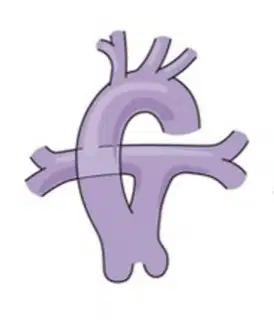
There are 2 classification systems for Truncus Arteriosus, Collett-Edwards:
A main PA segment is present
Type II
Type I
Type III

There are 2 classification systems for Truncus Arteriosus, Collett-Edwards:
The branch PAs arise separately but close to each other from the posterior aspect of the truncus, no MPA.
Type III
Type I
Types II

There are 2 classification systems for Truncus Arteriosus, Collett-Edwards:
The branch PAs arise separately from the right and left aspects of the truncus, like Type II but PAs are farther apart from each other.
Type II
Type III
Type I
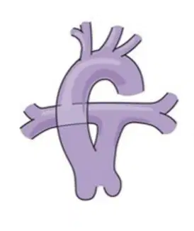
There are 2 classification systems for Truncus Arteriosus, Van Praagh Classification:
Pulmonary trunk arises from the truncus
A4
A1
A2
A3

There are 2 classification systems for Truncus Arteriosus, Van Praagh Classification:
2 PAs arise form the truncus
A2
A1
A3
A4
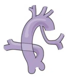
There are 2 classification systems for Truncus Arteriosus, Van Praagh Classification:
One PA arises from truncus, PDA or major aortopulmonary collateral artery (MAPCA).
A3
A1
A2
A4
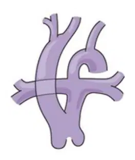
There are 2 classification systems for Truncus Arteriosus, Van Praagh Classification:
With interrupted aortic arch
A3
A2
A1
A4
Involves an assortment of lesions that share the common feature that both the aorta and PA arise from the RV. Embryological persistence of the subaortic conus. Most forms include a VSD
Ebstein’s Anomaly
Truncus Arteriosus
Double Outlet Right Ventricle (DORV)
Pulmonary Stenosis
Uncommon defect involving the abnormality of the tricuspid valve.
Tricuspid valve is abnormally formed and is more apically displaced than usual in the heart.
Apical displacement of the valves creates atrialization of a portion of the right ventricle.
Variable thinning of the wall of the “atrialized” RV (aRV) occurs, along with redundancy and tethering of the anterior leaflet of the TV.
Truncus Arteriosus
Ebstein’s Anomaly
Double Outlet Right Ventricle (DORV)
Tetrology of Fallot
True or False
The goal of imaging is to define the anatomy associated with Ebstein’s anomaly and to describe the severity of the lesion
False
True

