
 Worksheets
In the Zoom! A Dermoscopy Online Quiz Game
Total questions: 18
Worksheet time: 7mins
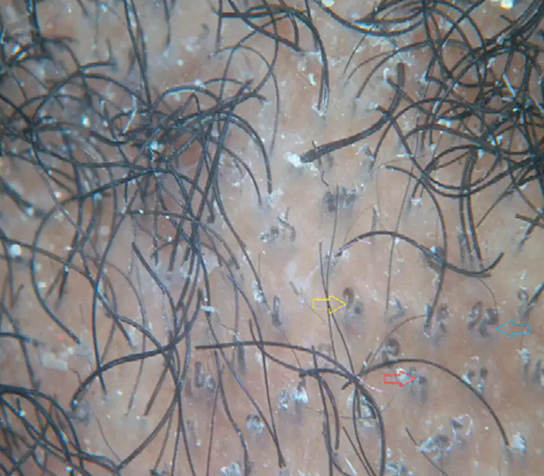
Dermatoscopy shows comma hairs (yellow arrow), broken hairs (red arrow), and corkscrew hairs (blue arrow)
Androgenetic Alopecia
Alopecia Areata
Trichotillomania
Tinea Capitis
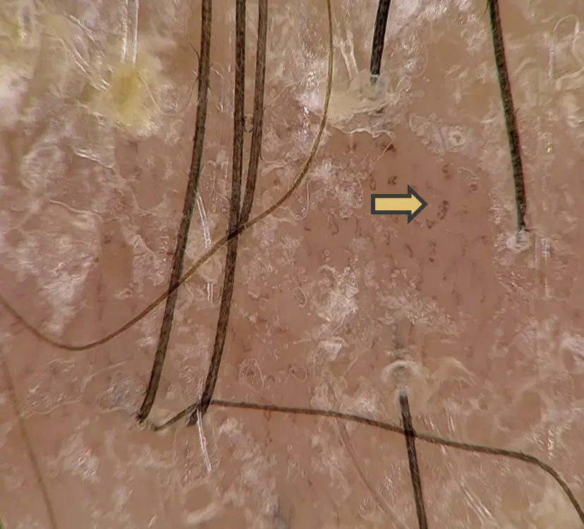
Identify the pointed structure.
arborizing vessels
globules
twisted capillaries
atypical red vessels
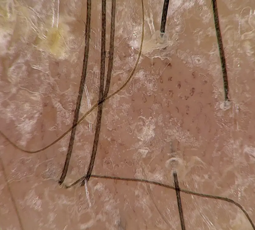
Name the disease entity.
Dermoscopy findings: White scales and twisted capillaries.
(a)
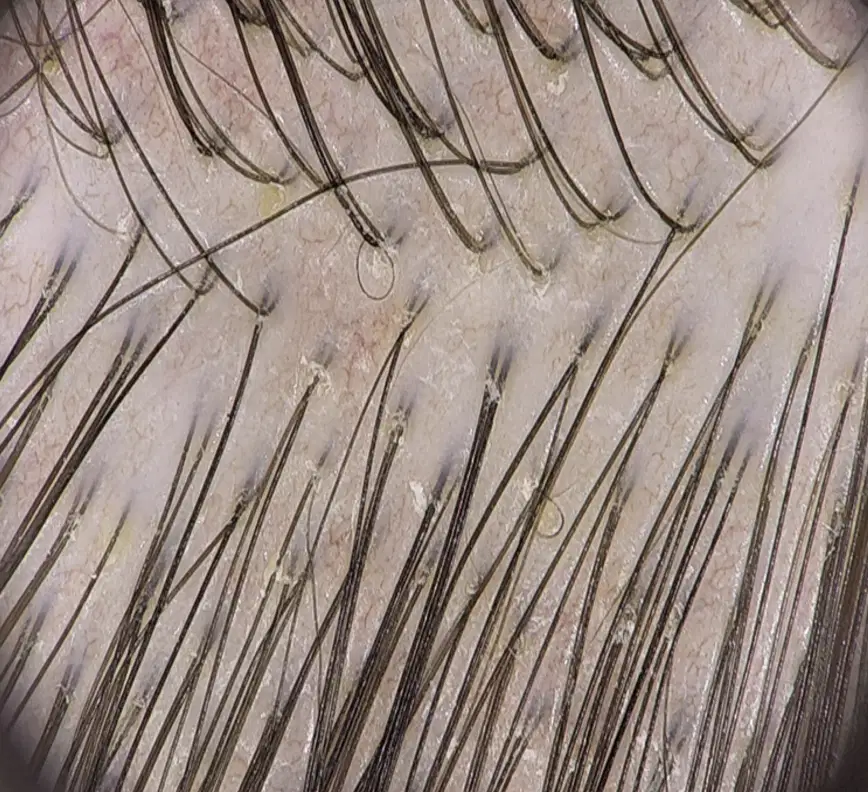
Which is the correct dermoscopy findings of Seborrheic Dermatitis?
Red dots and globules, twisted red loops and glomerular vessels
Diffuse scaling, loss of pigment network, and follicular keratotic plugs
Erythema, and peripilar casts around emerging hair shafts
Yellow and white scale and arborizing vessels; Absence of red dots and globules
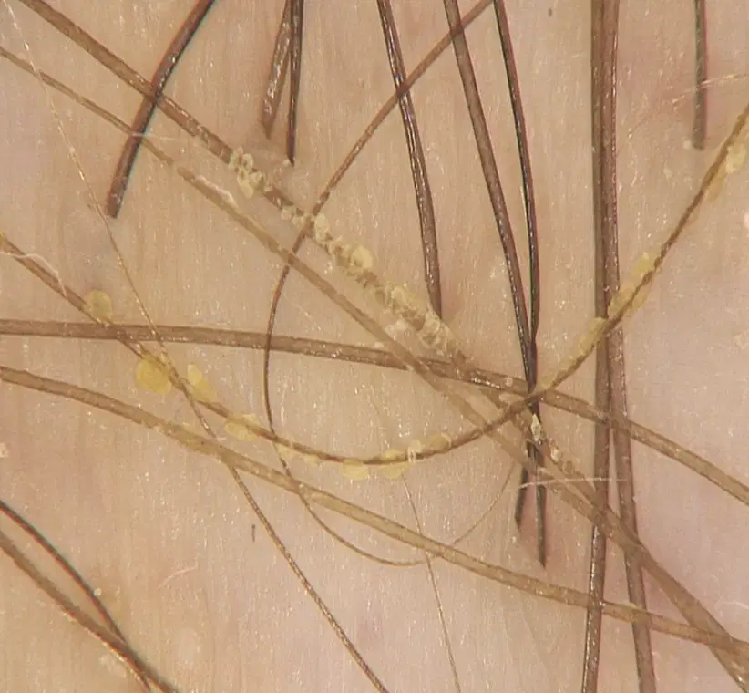
Name the disease entity:
Dermoscopy findings: yellow concretions on hair shafts.
(a)
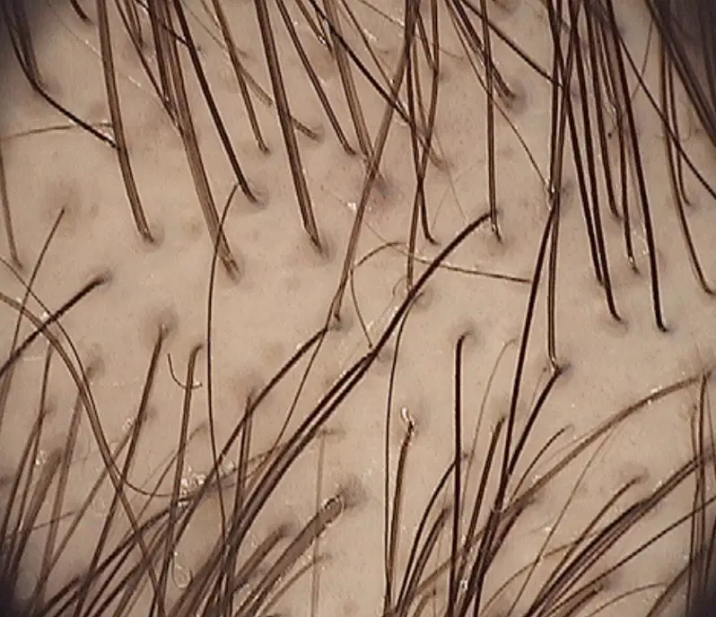
Name the disease entity:
Dermoscopic findings: Hair diameter variability and “peripilar signs” presenting as brown halos at follicular ostia. Hair shafts present with different degree of thinning.
(a)
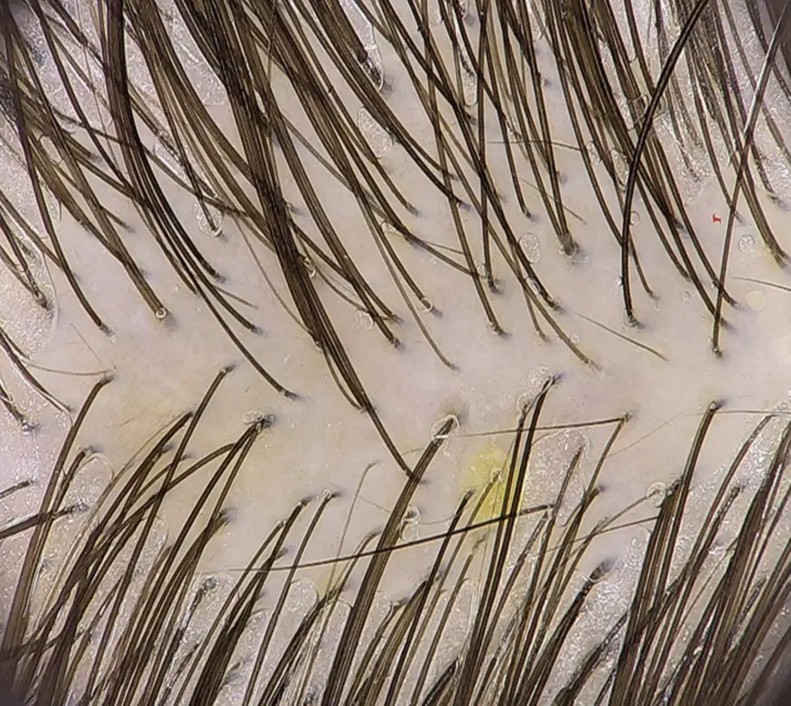
Name the disease entity:
Dermoscopy findings: Terminal hair shafts with uniform diameter and multiple short, regrowing hairs.
(a)
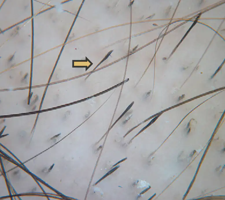
Identify the pointed structure.
(a)
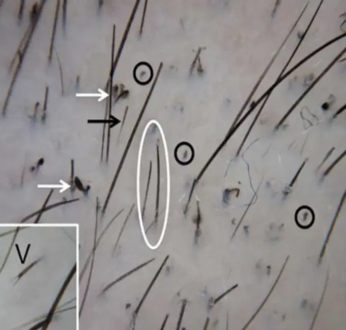
Name the disease entity.
Dermoscopy findings: Black dots (black circle), fraying ends (white circle), exclamation mark hair – similar to (black arrow), coiled hair (white arrow).
(a)
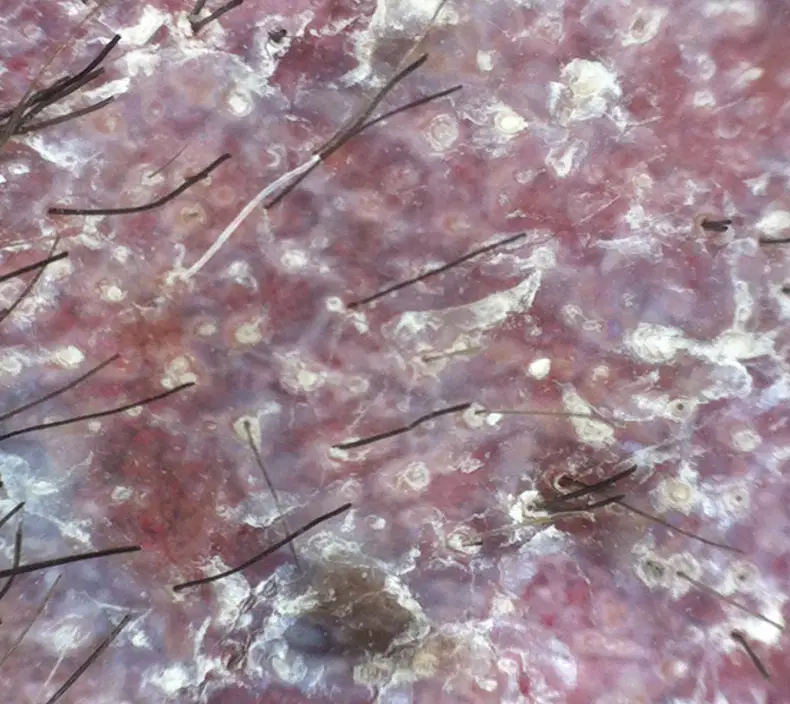
Dermoscopic findings:
Diffuse scaling, loss of pigment network, and follicular keratotic plugs
Centrifugal cicatricial alopecia
Discoid Lupus Erythematosus
Dissecting Cellulitis
Tinea Capitis
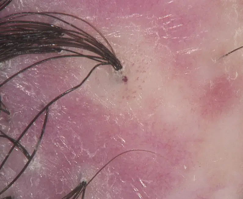
Name the disease entity.
Dermoscopic findings: Tufts of hair surrounded by thick peripilar casts.
(a)
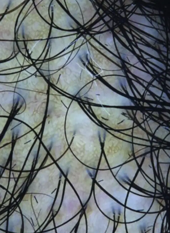
Dermoscopy shows peripilar white gray halo and interfollicular erythema
Frontal Fibrosing Alopecia
Central Centrifugal Cicatricial Alopecia
Lichen Planopilaris
Syphilitic Alopecia

Dermoscopic findings:
Loss of follicular openings, erythema, and "peripilar casts" around emerging hair shafts.
Syphilitic Alopecia
Discoid Lupus Erythematosus
Lichen Planopilaris

Name the disease entity.
Dermoscopy findings: hyperkeratotic and rough lesions with micropapules and small black dots.
(a)

Dermoscopy shows dorsal pterygium with formation from skin continuing with the skin of proximal nail fold (yellow asterisks) forming “V” shaped (blue arrows)
Nail lichen planus
Onychomadesis
Onychoschizia
Trachonychia
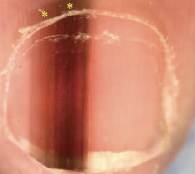
Dermoscopic findings: Longitudinal brown-black discoloration of nail plate with micro-Hutchinson sign (yellow asterisks)
What is this?
Subungal hematoma
Melanonychia
Nail matrix nevus
Beau's lines
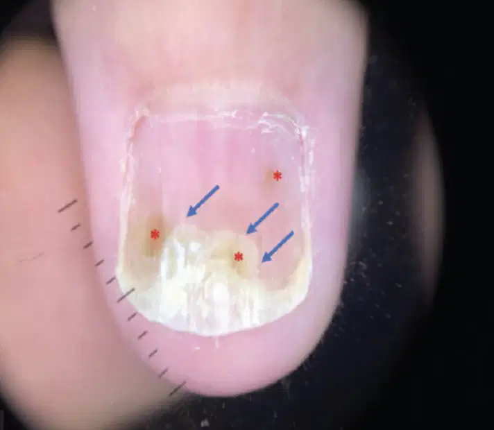
Dermoscopy shows oil spot or salmon patch on the left fourth fingernail with circular translucent yellow-red area (red asterisks) of discoloration surrounded by onycholysis.
Nail psoriasis
Onychorrhexis
Onychomycosis
Nail Trauma
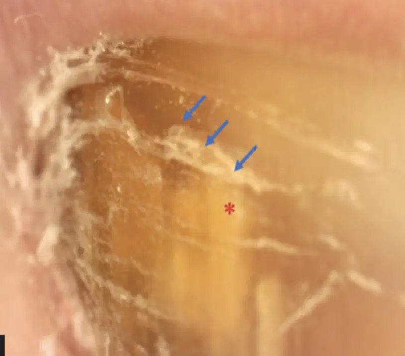
Dermoscopy findings: Nail plate showing a jagged proximal border of the onycholytic area (red asterisk), with yellow-spikes (blue arrows) projecting into the proximal nail plate
Nail Trauma
Onychorrhexis
Onychomadesis
Onychomycosis

