
Font size
 Worksheets
ankle and foot + knee joint
Total questions: 173
Worksheet time: 2hrs 19mins
What does "hallicus" mean?
Toes
Foot
Big toe
Heel

O: fibula I: lateral foot & ankle A: eversion of foot
Peroneal muscle
Fibularis Brevis
Fibularis Tertius
Extensor Hallucis Longus

O: Posterior tibia and fibula I: Posterior calcaneus A: Ankle plantar flexion
Soleus
Plantaris
Gastrocnemius
Tibialis Posterior

O: posterior condyle of femur I: Posterior calcaneus A: Knee flexion and ankle plantar flexion
Soleus
Plantaris
Gastrocnemius
Tibialis Posterior

O: upper lateral tibia I: medial cuneiform & 1st metatarsal A: dorsiflexion & inversion
Flexor Hallucis Longus
Flexor Digitorum Longus
Tibialis Anterior
Extensor Hallucis Longus


What arch is represented between A-B?
Transverse arch
Medial Longitudinal Arch
Lateral Longitudinal Arch
Lateral Transverse Arch

What bone is this?
Metatarsals
Intermediate Cuneiform
Lateral Cuneiform
Medial Cuneiform
Navicular

What bone is this?
Calcaneus
Intermediate Cuneiform
Lateral Cuneiform
Medial Cuneiform
Talus

What bone is this?
Calcaneus
Intermediate Cuneiform
Lateral Cuneiform
Medial Cuneiform
Talus

What bone is this?
Calcaneus
Intermediate Cuneiform
Lateral Cuneiform
Medial Cuneiform
Talus
The talocrural joint is also known as the
Joint in the ankle
Joints in the foot
Joint of the arch
Joint between the phalanges
Which bones is the medial malleolus located on
tibia
fibula
Inversion is turning outward laterally
True
False
What the 3 bones that make up the ankle joint?
Tibia, calcaneus, talus
Tibia, fibula, calcaneus
tibia, fibula, talus
tibia, fibula, navicular
How many bones make up the foot?
14
5
26
27
The most common type of ankle sprain is a...
Eversion sprain
Inversion sprain
Syndesmosis sprain
None of these answers
What bone has the landmark "Styloid Process"?
Talus
Calcaneus
Cuboid
5th metatarsal
The arch that runs from 1st toe to the 5th toe across the foot is called what?
Medial Longitudinal Arch
Lateral Longitudinal Arch
Transverse arch
None of these
The distal end of the fibula is called what (landmark)
Medial malleolus
Lateral Malleollus
Deltoid turbercle
calcaneal tuberosity
What is the anatomical name for the Ankle
Sub Talor joint
Talocrural joint
Syndesmosis
Tib-Fib joint
Name the Medial side ligament(s)
ATFL
Calcaneo-Fibular Lig
PTFL
Deltoid
A Jone's fracture is going to involve what structure?
Calcaneus
Fibula
5th Metatarsal
5th metacarpal

O: anterior inferior iliac spine I: superior patella & tibial tuberosity A: hip flexion & knee extension
Peroneus Muscle
Rectus Femoris Muscle
Vastus lateralis muscle
Biceps femoris

O: greater trochanter & linea aspera I: lateral patella & tibial tuberosity A: knee extension
Semimembranosus
Poplitus
Vastus intermedius
Vastus lateralis

O: anterior femur I: upper patella & tibial tuberosity A: knee extension
Tibialis anterior
Vastus intermedius
Gastrocnemius
Tibialis Posterior

O: linea aspera I: medial patella & tibial tuberosity A: knee extension
Vastus medialis
Rectus Femoris
Semitendinosus
Biceps femoris

O: ischial tuberosity I: pes asnerine A: knee flexion & hip extension
Vastus Lateralis
Semimebranosus
Semitendinosus
Tibialis Anterior

O: Ischial tuberosity I: posterior medial tibial condyle A: Knee flexion & hip extension
Semimembranosus
Gastrocnemius
Soleus
Popliteus

O: Ischial tuberosity I: head of fibula A: knee flexion & hip extension
Tibialis Anterior
Tibialis Posterior
Biceps Femoris
Rectus Femoris

O: posterior condyle of femur I: posterior tibia A: knee flexion & internal rotation
Popliteus
Hallucis Muscle
Peroneus
Extensor digitorum longus
Which is not a ligament of the deltoid
Calcaneofibular
tibionavicular
tibiotalar
tibocalcaneal

Which ligament is labeled 1
Calcaneofibular Ligament
Posterior Talofibular Ligament
Anterior Talofibular Ligament
Posterior Tibiofibular Ligament

Which ligament is labeled 2
Anterior tibiofibular ligament
Anterior talofibular ligmaent
Deltoid Ligament
Posterior Talofibular ligament

Number 5 represents the
talus
calcaneus
metatrsal
tibia
Number 2 represents the
lateral malleolus
styloid process
medial malleolus
fibula


6
Lateral Cuneiform
Medial Cuneiform
Intermediate Cuneiform
Trouble Cuneiform

Mark the spot of the subtalar joint

Match the color of the tarsal bones to their correct name.
Orange
Cuneiforms
Green
Cuboid
Blue
Navicular
Yellow
Talus
Purple
Calcaneus
Label the following lateral ankle ligaments
Calcaneofibular
Deltoid ligament
Posterior talofibular
Anterior talofibular
Match the following movements with their correct names.

Dorsiflexion

Plantarflexion

Inversion

Eversion

Circumduction

What is the blue bone called?
Cuboid
Calcaneus
Navicular
Talus
What color is the calcaneus bone?
Green
Purple
Yellow
Blue
Label the foot bones
Phalanges
Tarsals
Metatarsals

What bones are colored yellow?
Tarsals
Metatarsals
Phalanges
Metacarpals


Anterior Talofibular Ligament

Calcaneofibular Ligament

Posterior Talofibular Ligament
Which muscle(s) evert the foot?
peroneus longus and brevis
tibialis posterior
tibialis anterior
gastrocnemius
What muscle dorsiflexes the foot?
Gastrocnemius
Peroneus Longus
Anterior Tibial Shaft
Tibialis Anterior
body weight is transferred from?
fibula to calcaneus and talus
tibia to talus and calcaneus
talus to tibia and calcaneus
calcaneus to talus and tibia
where are the 2 sesamoid bones in the foot?
beneath then 2nd metatarsophangeal joint
3rd tarsal
beneath the 1st metatarsophangeal joint
1st metatarsal
which are the three ligaments of tibiofibular joint?
interosseus membrane, anterior tibiofibular, posterior tibiofibular
anterior tibiotalar, posterior tibiotalar, tibiocalcaneal
interosseus membrane, anterior tibiofibular, posterior tibiotalar
interosseus membrane, anterior talofibular, posterior talofibular
which are the 4 ligaments of the Lateral Ankle?
anterior talofibular, posterior talofibular, anterior tibiofibular, posterior tibiofibular
calcaneofibular, tibiocalcaneal, interosseus membreane, anterior tibifibular
anterior talofibular, posterior talofibular, calcaneofibular, tendon of peroneus brevis
anterior tibiotalar, posterior tibiotalar, tibiocalcaneal, tibionavicular
Which are the 4 deltoid ligaments of the medial ankle?
tibiocalcaneal, tibionavicular, anterior tibiofibular, posterior talofibular
anterior tibiotalar, posterior tibiotalar, tibiocalcaneal, calcaneofibular
anterior talofibular, posterior tibiofibular, tibionavicular, calcaneofibular
anterior tibiotalar, posterior tibiotalar, tibiocalcaneal, tibionavicular
the ankle has how many degrees of plantar flexion?
50
20
30
15
the ankle has how many degrees of dorsiflexion?
5
30
20
15
the subtalar joint has how many degrees of inversion?
20-30
10-15
5-10
15-20
the subtalar joint has how many degress of eversion?
30-50
5-10
20-25
5-15
what are the three arches of the foot?
medial longitudinal arch
longitudinal longitudinal arch
lateral longitudinal arch
transverse arch
pronation is the combination of?
ankle dorsiflexion
subtalar eversion
forefoot adduction
forefoot abduction
supination is a combination of?
ankle dorsiflexion
subtalar inversion
forefoot adduction
ankle plantar flexion
What bones make up the Sub Talor joint
Talus - Calcaneus
Talus - Tibia
Talus - Tibia - Fibula
Tibia - Fibula
The medical term for flat footed or low arch is...
Pes Cavus
Pes Planus


What is the name of ligament B?
tibiocalcaneal ligament
posterior talofibular ligament
anterior talofibular ligament
calcaneofibular ligament

The cushion between the femur and tibia is the ________.
Ligament
Meniscus
LCL
Patellar Tendon
The ligament on the inside of the knee is the MCL.
True
False
What does ACL stand for?
Atlanta Cruciate Ligament
Anterior Cruciate Ligament
Anterior Coolio Ligament
Select the muscle(s) that extend the hip & flex the knee.
Semimembranosus
Gastrocnemius
Biceps Femoris
Semitendinosus
What does MCL mean?
Fibula collateral ligament (lateral)
medial collateral ligament (tibial)
posterior cruciate ligament
Anterior cruciate ligament
what 3 bones make up the 2 joints of the knee complex?
Tibia, fibula, talus
Tibia, patella, femur
Tibia, Femur, fibula
Femur, Fibula, patella
What are the 2 joints that make up the knee complex?
Calcaneofibular, tibiotalar
tibiotalar, tibiocalcaneal
tibiofemoral, patellofemoral
Tibiofibular, talofibular
What's on top of the tibial plateau?
medial meniscus
lateral meniscus
Patella
Tibia
What two locations are the meniscus attached to?
Anterior horn
Patella
Posterior horn
Fibula
Anterior cruciate ligament does what?
(a)
Posterior cruciate ligament does what?
(a)
The knee joint has how many degrees of freedom?
(a)
The talocalcaneal joint is also called the
Talonavicular Joint
Midtarsal Joint
Subtalar Joint
Talocrural Joint
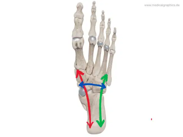
What arch does the blue arrow represent?
(a)
What arch does the green arrow represent?
(a)
What arch does the red arrow represent?
(a)
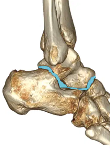
What joint is this?
(a)
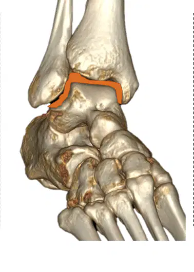
What joint is this?
(a)
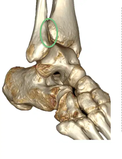
What joint is this?
(a)
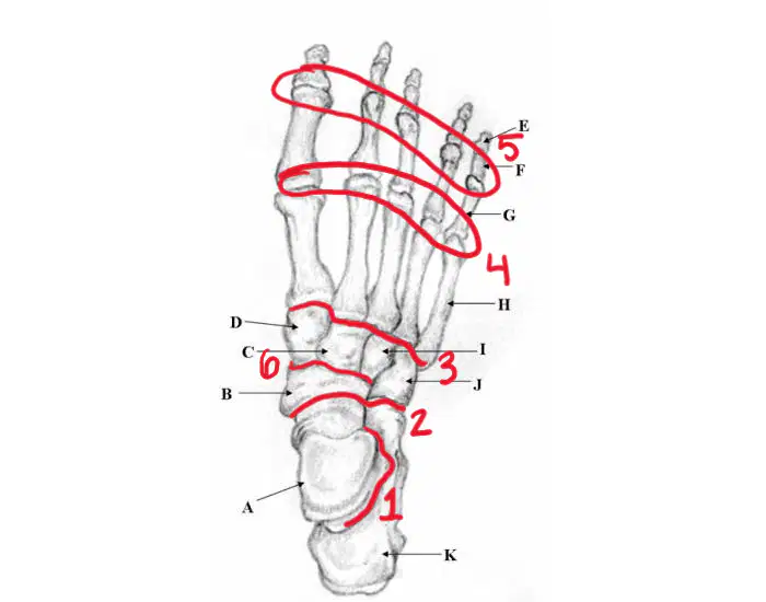
What joint is #6?
(a)
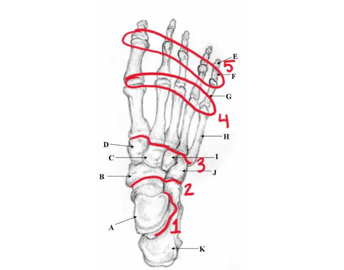
What joint is #5?
(a)
What joint is #3?
(a)
What joint is #4?
(a)
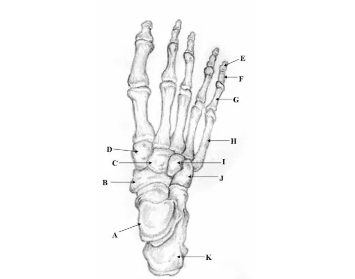
C.
(a)

Which ligament is highlighted in red
posterior talofibular ligament
anterior talofibular ligament
calcaneofibular ligament
deltoid

K
ACL
MCL
PCL
LCL
L
LCL
medial meniscus
MCL
lateral meniscus
M
LCL
ACL
MCL
PCL
N
LCL
ACL
MCL
patellar tendon
J
LCL
ACL
MCL
PCL
D
LCL
ACL
MCL
IT band
E
LCL
medial meniscus
MCL
lateral meniscus

What is M?
Fibular tuberosity
Lateral Condyle of femur
Lateral Condyle of tibia
Tibial tuberosity
What is K?
Medial condyle of tibia
Fibular fossa
Tibiaous fossa
Medial condyle of fibula
What is J?
Medial collateral ligament
Transverse ligament
Posterior Cruciate Ligament
Medial Meniscus
What is i?
Anterior Cruciate Ligament
Transverse ligament
Posterior Cruciate Ligament
Medial Collateral ligament
What is H?
Medial condyle of femur
Femurous fossa
Medial epicondyle of femur
Patella
What is G?
Anterior Cruciate Ligament
Transverse ligament
Posterior Cruciate Ligament
Medial Collateral ligament
What is F?
Head of fibula
Lateral Condyle of femur
Lateral Condyle of tibia
Head of tibia
What is E?
Head of fibula
Lateral Condyle of femur
Lateral Condyle of tibia
Head of tibia
What is D?
Lateral Meniscus
Medial Meniscus
Transverse Ligament
Lateral Collateral ligament
What is C?
Anterior Cruciate Ligament
Medial Meniscus
Posterior Cruciate Ligament
Lateral Collateral ligament
What is B?
Anterior Cruciate Ligament
Medial Meniscus
Posterior Cruciate Ligament
Lateral Collateral ligament
What is A?
Medial Condyle
Of femur
Medial Meniscus
Head of fibula
Lateral Condyle
of Femur
What ligament, inside the joint prevents forward motion of the tibia.
LCL
MCL
ACL
PCL
What are the 3 bursa sacks?
subcutaneous prepatella bursa
Suprapatella bursa
Inferior bursa
infrapatella bursa
What does the suprapatella bursa do?
provides cushion
reduces friction at quadriceps tendon
Decreases friction at patella tendon
Protect the patella
What does the subcutaneous prepatella bursa do?
Reduce friction
Protect the patella
Decreases friction
What does the infrapatella bursa do?
protect the patella
Decrease friction at patella tendon
reduces friction at quadriceps tendon
How many bands of tissue make up the MCL?
3
4
5
2
Knee flexion has how many degrees?
10 degrees
140 degrees
0 degrees
50
Knee extension has how many degrees?
0 degrees
140 degrees
30 degrees
10 degrees
Knee hyperextension is how many degrees?
0 degrees
30 degrees
140 degrees
10 degrees
What muscle helps the knee fully extend?
Vastus Lateralis
Vastus medialis
Vastus intermedius
Rectus femoris
what are the 3 specific locations of the Q angle?
Anterior superior iliac spine
behind patella
Center of patella
Tibial tuberosity
If the Q angle is greater than (a) degrees it indicates tight lateral structures
Label the diagram with the choices provided.
femur
patella
medial condyle; tibia
lateral condyle; tibia
neck of fibula
right
left
tibial tuberosity
head of fibula

Identify this landmark.
(a)

Identify this landmark.
(a)
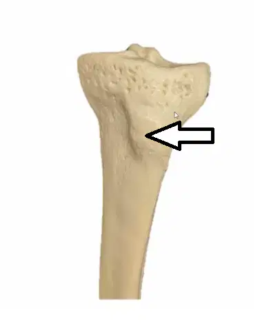
Identify this landmark.
(a)
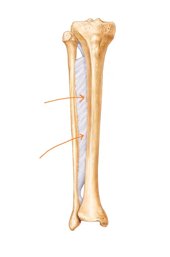
Identify this landmark.
(a)

What is N? (a)

What is M? (a)

What is K? (a)

Which bone is I? (a)

Which bone is H? (a)

What is G? (a)

What bone is B? (a)
Select the third metatarsal.
Select the calcaneus.
Select the talus.
Select the distal phalanx.
Select the first metatarsal.
Label this view of the foot.
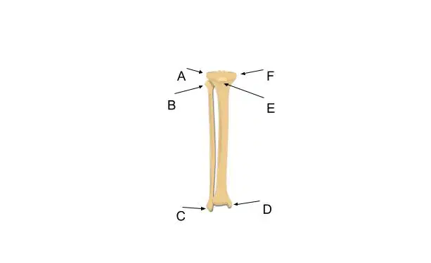
Use the letters in the picture to match the following
B
head of the fibula
C
lateral malleolus
D
medial malleolus
E
tibial tuberosity
A & F
condyles of the tibia

where is the lateral maleolus?
(a)
where is the medial malleolus?
(a)
where is the tibial tuberosity?
(a)
where is the lateral condyle?
(a)