
 Worksheets
OCT Seminari 2
Total questions: 16
Worksheet time: 26mins
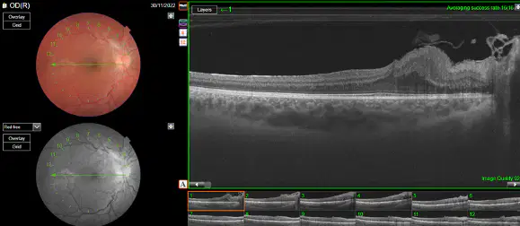
Is there a justification of the vision of the right eye (VA: 0.7) ?
Yes
No
Do you think that if he had correctly performed the amblyopia treatment (occlusion), vision would have improved?
Yes
No

¿Qué tipo de report podéis observar?
Line Report 6.0 mm
Radial Report 12.0 mm
5Line Cros Report 12.0 mm
Radial Report 6.0 mm
Line Report 12.0 mm
¿Qué podéis observar en este report?
Central hypopigmentation not observed on the red-free
Central hypopigmentation observed on the red-free
Central hyperpigmentation not observed on the red-free
Central hyperpigmentation observed on the red-free
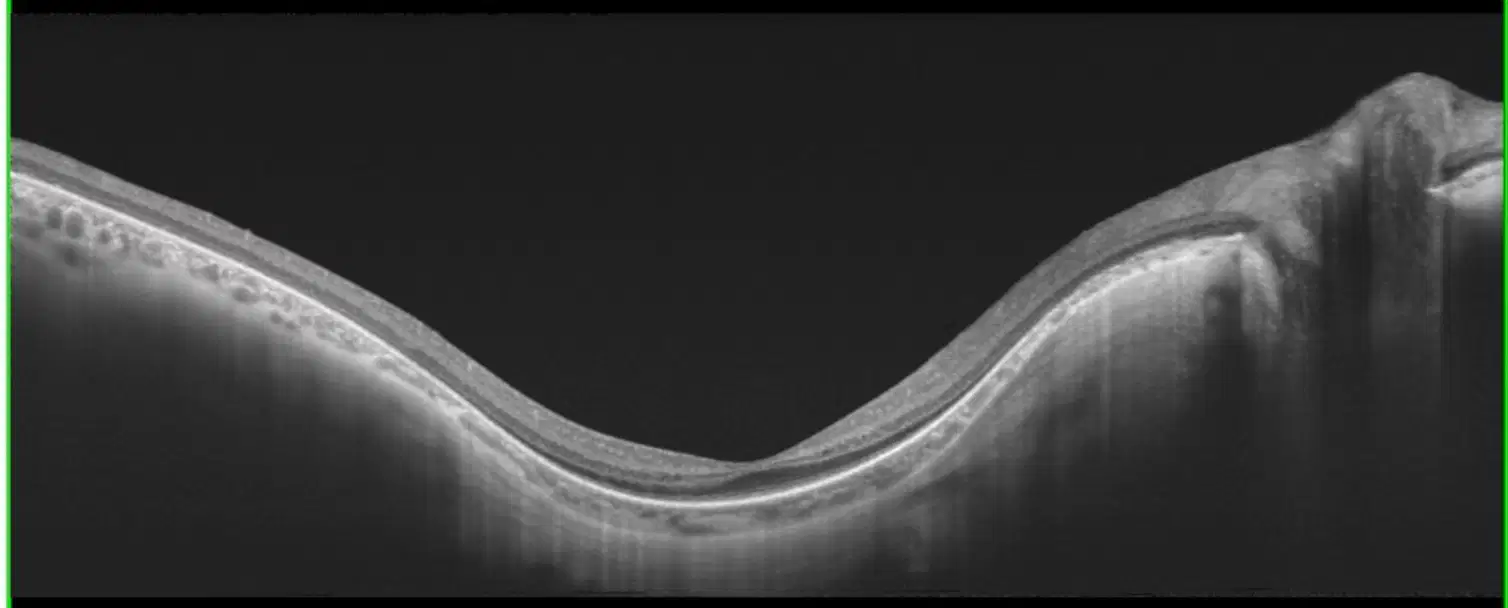
¿Qué podéis observar en la OCT?
Possible retinal staphyloma
Thinness of the veins with respect to the arteries
Epiretinal membrane
Preservation of the central photoreceptor line with peripheral loss

Describe any pertinent findings from this report
There is greater thinning of the RNFL layer of the RE than of the LE
There is a strong symmetry of the RNFL layers between both eyes
The signal strength is 4/10 and 6/10 in OD and OS respectively
This report suggests there is a thin RNFL superiorly and inferiorly in OU.

What do you see is different in RNFL Desviation map?
The OCT from 6 months shows that the RNFL thickness are within normal ranges.
The OCT from today suggested a thin RNFL, whereas the OCT from 6 months ago did not and instead shows that the RNFL thickness are within normal ranges.
The OCT from today suggested a thick RNFL, thickness are within normal ranges.
During the last 6 months there has been a loss of symmetry between both eyes
What do you see is different in RNFL Thickness Map?
The distribution and shape of the thickness (the yellow and red colours) in both eyes show progression from 6 months ago compared to today.
Very similar to today’s OCT compared to the OCT from 6 months ago
The distribution and shape of the thickness in both eyes are consistent from 6 months ago compared to today.
Worsening RNFL Thickness map OCT from 6 months ago compared to the OCT from today.
What could explain this discrepancy?
These findings are due to a large mobile posterior vitreous detachment floater, which was picked up within the neuroretinal rim.
The incorrect date of birth has been entered in the OCT ONH/RNFL from today’s visit.
The OCT ONH/RNFL from 6 months ago was performed on an unreliable machine; the new OCT from today is more accurate.
The findings today are due to poor signal strength and artefact and are nothing concerning.

What do you see in the images?
Peripalilar atrophy characteristic of myopic retina
Transparent crystalline
Preserved vascular arcades
Loss of photoreceptors in peripheral retina more marked in LE
Vitreous veil observed in RE
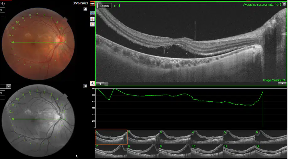
What do you observe in the RE OCT?
Neurosensory Retinal detachment
Edema macular
Central serous chorioretinopathy
Serous Posterior Epithelial Detachment (PED)
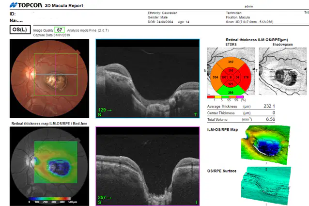
What do you observe in the RE OCT?
Pigmented lesion in the most central part of the retina. Coriorretinal scar
Foveolar loss and a huge excavation showing the atrophic lesion.
Increase central thickness of the retina, this time specified with average thickness values.
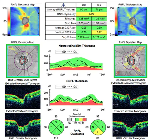
Which part of the OCT is not right?
RNFL Desviation Map RE
RNFL Desviation Map LE
RNFL Thickness Map RE
RNFL Thickness Map LE

What features on OCT suggest glaucoma in the left eye?
Cup-to-disc ratio of 0.72
Thin inferior and superior vulnerability zones
Reduced inferior neuro-retinal rim thickness
All of them

Which quadrant of the optic nerve is thinned by the progression of glaucoma?
Nasal
Superior
Temporal
Inferior
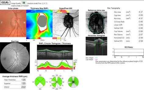
Is this OCT normal?
Yes
No

