
 Worksheets
Parasitology 1 Lab Test
Total questions: 25
Worksheet time: 8mins
What is the primary purpose of a centrifuge in parasitology concentration techniques?
To stain parasites
To concentrate parasites in a sample
To fix parasites on a slide
To visualize parasites under a microscope
Which concentration technique is commonly used to detect the presence of helminth eggs and larvae in fecal samples?
Zinc Sulfate technique
Acid-fast stain
Direct wet mount
Gram stain
In parasitology, what is the purpose of using a fecal flotation technique?
To stain parasites
To concentrate parasites in a sample
To identify protozoa
To perform blood smears
Which microscopic examination technique involves placing a drop of liquid sample on a slide and covering it with a coverslip for direct observation?
Kato-Katz method
Formalin-ether concentration
Direct wet mount
Fecal flotation
In parasitology, what is the purpose of the Giemsa stain?
To stain parasites
To concentrate parasites in a sample
To detect the presence of helminth eggs
To perform blood smears
What is the primary purpose of the Brine Flotation Technique in parasitology?
To stain parasites
To concentrate parasites in a sample
To fix parasites on a slide
To perform blood smears
What type of parasites is the Brine Flotation Technique particularly useful for detecting?
Helminths
Protozoa
Nematodes
Trematodes
Which of the following statements is true regarding the Brine Flotation Technique?
It relies on direct observation without staining.
It involves the use of a Baermann apparatus.
It is primarily used for detecting protozoa.
It uses a high-speed centrifuge for concentration.
In parasitology, why is the Trichome Staining Method commonly used for studying parasites?
To highlight parasite cysts
To stain the outer cuticle of parasites
To visualize parasite nuclei and structures
To detect parasite motility
Which type of parasites is particularly well-stained and visualized using the Iron Hematoxylin Staining Method?
Protozoa
Helminths
Arthropods
Nematodes
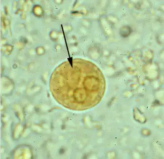
The picture is a cyst from a protozoan species. This species is well recognized as a pathogenic ameba, associated with intestinal and extraintestinal infections. What is the species of protozoan in the picture?
Brugia malayi
Balantidium coli
Entamoeba histolytica
Enterobius vermicularis
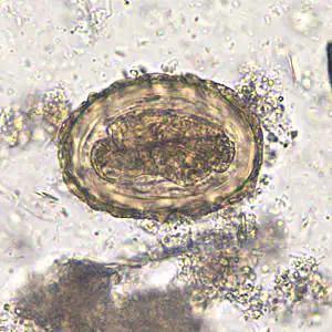
This egg belongs to a roundworm. Identify this egg.
Strongyloides stercoralis
Ascaris lumbricoides
Enterobius vermicularis
Fasciola hepatica
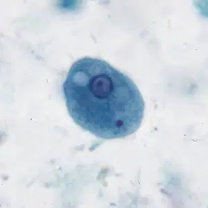
This parasite is species of amebae are capable of colonizing the human gastrointestinal tract, but not pathogenic. Identify this parasite.
Entamoeba coli
Entamoeba histolytica
Enterobius vermicularis
Gardia lambia
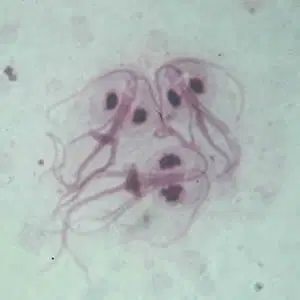
Identify the parasite in the picture.
Gardia sp.
trophozoites
Plasmodium sp.
trophozoites
Enterobius sp.
Entamoeba sp
trophoziotes
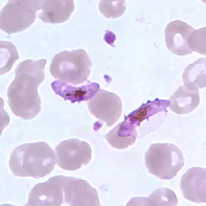
This parasite causes Malaria. What stage is this parasite?
Gametocytes
Trophozoites
Ring-form
Schizont
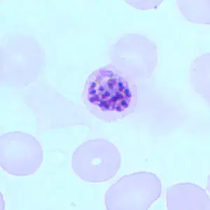
This parasite causes Malaria. What stage is this parasite?
Gametocytes
Trophozoites
Ring-form
Schizont
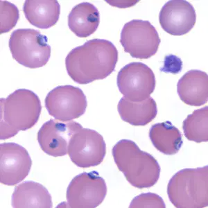
This parasite causes Malaria. What stage is this parasite?
Gametocytes
Trophozoites
Ring-form
Schizont
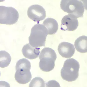
This parasite causes Malaria. What stage is this parasite?
Gametocytes
Trophozoites
Ring-form
Schizont
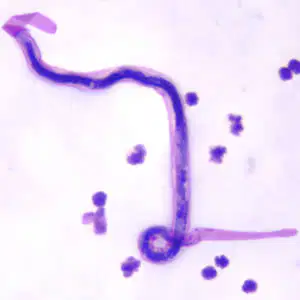
This parasite is a the causative agents of lymphatic filariasis, Identify this parasite.
Enterobius vermicularis
Hookworm
Ascaris lumbricoides
Brugia malayi,
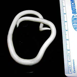
Adult females can grow up to 20 to 35 cm and adult males worm can grow up to 15 to 30 cm. This worm parasitize the human intestine. Identify this worm.
Ancylostomiasis (Hookworm)
Ascaris lumbricoides
Wuchereria bancrofti
Whipworm (Trichuriasis)
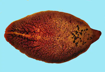
This parasite is commonly known as ?
Intestine fluke
lungs fluke
liver fluke
kidney fluke
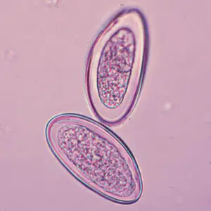
This eggs below to parasite X. Parasite X occurs worldwide, with infections occurring most frequently in school- or preschool-children and in crowded conditions. What is Parasite X.
B.malayi
A. lumbricoides
E. vermicularis
Hookworm
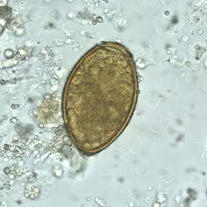
This egg is normally known as lung fluke. What is the name of the parasite?
Fasciola hepatica
Paragonimus westermani
Fasciolopsis buski
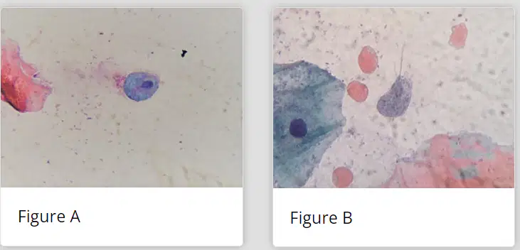
A 29-year-old female patient presented to her primary care physician reporting a 15-day history of whitish, vaginal discharge associated with itching. She also complained of lower abdominal pain. A vaginal, speculum examination revealed an inflamed cervix with red, punctate lesions. A Pap smear collected at the time of examination was sent to the pathology lab for a Papanicolaou stain. Figures A and B show what the pathologist observed using an oil immersion objective. The objects of interest measured 10-15 µm long. Identify this parasite
Brugia malayi
Trichomonas vaginalis
Gardia lambia
Plasmodium vivex
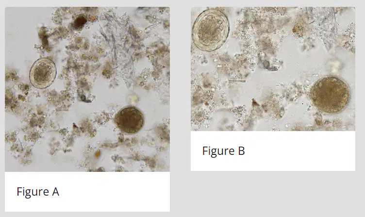
A 42-year-old pig farmer presented to his local medical facility with intermittent diarrhea and abdominal cramping. A stool specimen was collected in 10% formalin for laboratory testing. Figures A were captured at 200x magnification. Figures B a higher magnification (400x) of the same objects. What is your diagnosis?
Brugia malayi & Balantidium coli
Enterobius vermicularis & Balantidium coli
Ascaris lumbricoides & Balantidium coli
Entamoeba histolytica & Enterobius vermicularis

