
 Worksheets
Anatomy Muscles & Nervous System
Total questions: 248
Worksheet time: 62hrs 0mins
Types of Muscle Tissue
(a)
Striated & Voluntary
Skeletal Muscle
Smooth Muscle
Cardiac Muscle
Striated & Involuntary
Skeletal Muscle
Smooth Muscle
Cardiac Muscle
Untriated & Involuntary
Skeletal Muscle
Smooth Muscle
Cardiac Muscle
Muscles use _____ to contract.
Chemical Energy
Microscopic Energy
Physical Energy
Movement Energy
Muscles are _______; composed of skeletal
muscle tissue, nervous tissue, blood, and other
connective tissues
Organs
The Center of Strength
Reactive
Complex
__________ are large and are
covered by connective tissue called
endomysium. They contain myofibrils.
Muscle Fibers
Fascicles
Fascia
Tendons
________ are grouped (10-100) into fascicles
which are covered by perimysium.
Fibers
Fascicles
Fascia
Tendons
Fibers are grouped (10 - 100) into _________
which are covered by perimysium. Groups of _______ make up the muscle
which is surrounded by epimysium.
Muscle Fibers
Fascicles
Fascia
Tendons
Surround and Separate Muscles
Muscle Fibers
Fascicles
Fascia
Tendons
Connective tissue that extends beyond the
ends of muscles and becomes what are connect to the periosteum of bones.
Muscle Fibers
Fascicles
Fascia
Tendons
Types of Connective Tissue
Endomysium
Perimysium
Epimysium
Fascia
Tendon
Connective tissue that covers muscle fibers.
Endomysium
Perimysium
Epimysium
Fascia
Tendon
Covers Fascicles
Endomysium
Perimysium
Epimysium
Fascia
Tendon
Surrounds the Fascicles that make up Muscle
Endomysium
Perimysium
Epimysium
Fascia
Tendon
Sometimes muscles are connected to each
other by broad sheets of connective tissue
called _______.
Aponeuroses
Fibers
Epimysium
Endomysium
Each _________ is a single, long,
cylindrical cell.
Muscle Fiber
Sarcoplasm
Actin
Myosin
Sarcolemma
"Cell membrane" of muscle fibers with many
mitochondria and nuclei
Muscle Fiber
Sarcoplasm
Actin
Myosin
Sarcolemma
Inside the sarcolemma is ________ "cytoplasm"
that contains myofibrils.
Muscle Fiber
Sarcoplasm
Actin
Myosin
Sarcolemma
Thin Filaments within Myofibrils. Is a globular protein with myosin binding sites.
Muscle Fiber
Sarcoplasm
Actin
Myosin
Sarcolemma
Thick Filaments within Myofibrils. Consists of twisted strands with globular cross-bridges projecting outward along strands.
Muscle Fiber
Sarcoplasm
Actin
Myosin
Sarcolemma
The organization of actin and myosin produces what?
Muscle Fiber
Sarcoplasm
Actin
Striations
Sarcolemma
Extends from one Z-Line to the next.
Muscle Fiber
Sarcomere
Actin
Striations
Sarcolemma
Extends between the myofibrils as transverse tubules
Muscle Fiber
Sarcomere
Actin
Striations
Sarcolemma
Stored in Sarcoplasmic Reticulum
Muscle Fiber
Sarcomere
Actin
Calcium
Sarcolemma
(Light Bands) made up
of actin filaments are anchored
to Z lines.
I Bands
Z Lines
A Bands
H Zone
M Line
I Bands are anchored to these. They are dark lines in the I Bands.
I Bands
Z Lines
A Bands
H Zone
M Line
(Dark Bands) are made
up of overlapping thick and thin
filaments.
I Bands
Z Lines
A Bands
H Zone
M Line
In the center of A bands are these (One is a Dark Line & One are the two white lines on the sides of the dark line) consisting of myosin
filaments only.
I Bands
Z Lines
A Bands
H Zone
M Line
Actin & Myosin
Filaments
Sarcomere
A-Band, I-Band, & Z-Line
Filaments
Sarcomere
The _________ and
________ are involved in triggering
a muscle contraction when the fiber is
stimulated.
14
Sarcoplasmic Reticulum and
Transverse Tubules
Sarcolemma and Sarcoplasmic Reticulum
Transverse Tubules and Sarcolemma
Neurotransmitter for nerve & muscle.
Acetylcholine
Calcium
Creatine Phosphate
Glycogen
This (theory)
describes muscle contraction.
sliding filament model
filament sliding model
creatine model
contraction model
________ proteins
cover the binding sites when calcium ions
(Ca2+) are absent.
Tropomysosin and Troponin
Glycogen
Creatin
Chlorine
Tropomysosin and troponin proteins
cover the binding sites when ______ ions
(Ca2+) are absent.
Calcium
Glycogen
Creatin
Chlorine
________ bind and
pull on the actin filaments, causing
the sarcomeres to shorten.
Myosin cross-bridges
Creatine Phosphate
Glycogen
Calcium
Myosin crossbridges attach to ________ filaments and bend, pulling on the _____ filaments; then release and attach to
the next _________ and pull
again. Exposed binding sites on ______ allow the muscle
contraction cycle to occur
Binding Sites on Actin
Binding Sites on Myosin
Types of Muscle Attachment
Origin
Insertion
Proximal
Distal
ATP
(Energy for Contraction & Must be Regenerated in Mitochondrion)
ADP+P+Energy
(Energy Regeneration)
ADP + P ----> ATP
ADP + P ----> ATP
Mitochondrion
36 ATP/Glucose
(Longer)
2 ATP/Glucose
(Fast)
Creatine Phosphate
(Energy for Contraction & Must be Regenerated in Mitochondrion)
ADP+P+Energy
(Energy Regeneration)
ADP + P ----> ATP
ADP + P ----> ATP
Mitochondrion
36 ATP/Glucose
(Longer)
2 ATP/Glucose
(Fast)
Sugar
(Energy for Contraction & Must be Regenerated in Mitochondrion)
ADP+P+Energy
(Energy Regeneration)
ADP + P ----> ATP
ADP + P ----> ATP
Mitochondrion
36 ATP/Glucose
(Longer)
2 ATP/Glucose
(Fast)
Aerobic Respiration
(Energy for Contraction & Must be Regenerated in Mitochondrion)
ADP+P+Energy
(Energy Regeneration)
ADP + P ----> ATP
ADP + P ----> ATP
Mitochondrion
36 ATP/Glucose
(Longer)
2 ATP/Glucose
(Fast)
Anaerobic Respiration
(Energy for Contraction & Must be Regenerated in Mitochondrion)
ADP+P+Energy
(Energy Regeneration)
ADP + P ----> ATP
ADP + P ----> ATP
Mitochondrion
36 ATP/Glucose
(Longer)
2 ATP/Glucose
(Fast)
Energy Sources for Muscle Contraction
Protein, Fat, Glycogen (Sugar)
Anaerobic & Aerobic Respiration using Glucose
Sugar
Creatine Phosphate
ATP
Energy Sources for Muscle Contraction from Best to Worst
1. ATP
2. Creatine Phosphate
3. Sugar (Aerobic & Anaerobic Respiration)
4. Glycogen (Sugar)
5. Fat
6. Protein
1. ATP
2. Creatine Phosphate
3. Glycogen (Sugar)
4. Sugar (Aerobic & Anaerobic Respiration)
5. Fat
6. Protein
1. ATP
2. Creatine Phosphate
3. Glycogen (Sugar)
4. Sugar (Aerobic & Anaerobic Respiration)
5. Protein
6. Fat
1. Creatine Phosphate
2. ATP
3. Glycogen (Sugar)
4. Sugar (Aerobic & Anaerobic Respiration)
5. Protein
6. Fat
The amount of oxygen the liver cells need/require to convert accumulated lactic acid into glucose, plus the amount that muscle cells need to resynthesize ATP and Creatine Phosphate to their original concentration. This could take several hours.
Oxygen Debt
Slow-Oxidative Fibers
Fast-Glycolytic Fibers
Muscle Fatigue
Red Fibers, Need Oxygen, Aerobic Respiration, Slow-Twitch
Oxygen Debt
Slow-Oxidative Fibers
Fast-Glycolytic Fibers
Muscle Fatigue
White Fibers, Bulk Up, Anaerobic Respiration, Fast-Twitch
Oxygen Debt
Slow-Oxidative Fibers
Fast-Glycolytic Fibers
Muscle Fatigue
When muscles lose the ability to contract during strenuous exercise. This usually arises from accumulation of lactic acid in the muscle.
Oxygen Debt
Slow-Oxidative Fibers
Fast-Glycolytic Fibers
Muscle Fatigue
When you feel fatigued vs. When you actually are. (Your body only knows so much of what it can do because your brain makes you think you're done.) "Can't do it" - Long before you're done.
Oxygen Debt
Slow-Oxidative Fibers
Central Fatigue
Muscle Fatigue
Mediated by factors or signals from the central nervous system. Commonly Associated with Calcium or Electrolyte Imbalance.
Cramping
Heat Production
Threshold Stimulus
Twitch
Latent Period
40% - 50% of muscle contraction energy is lost within this. Muscles produce a good chunk of this.
Cramping
Heat Production
Threshold Stimulus
Twitch
Latent Period
The amount of stimulus needed to contract the muscle.
Cramping
Heat Production
Threshold Stimulus
Twitch
Latent Period
Single, Short Contraction involving a few motor units.
Cramping
Heat Production
Threshold Stimulus
Twitch
Latent Period
Brief delay between stimulation & contraction. _____III_____
Cramping
Heat Production
Threshold Stimulus
Twitch
Latent Period
Membrane Potential
Action Potential
Summation
Tetanus Summation
All-Or-None
When a muscle fiber contracts, it contracts to its full extent.
Action Potential
Summation
Tetanus Summation
All-Or-None
A twitch on top of another twitch.
Action Potential
Summation
Tetanus Summation
All-Or-None
Constant Muscle Contraction
Action Potential
Summation
Tetanus Summation
All-Or-None
A motor neuron and the muscle fibers it controls make up a _______. When stimulated, the muscle fibers of the _______ contract all at once.
Motor Unit
Recruitment
Muscle Tone
Origin
Insertion
Increase in the number of activated motor units within a muscle at higher intensities of stimulation.
Motor Unit
Recruitment
Muscle Tone
Origin
Insertion
Achieved by a continuous state of sustained contraction of motor units within a muscle. (Flabby vs. Hard)
Motor Unit
Recruitment
Muscle Tone
Origin
Insertion
Immovable end of a muscle.
Motor Unit
Recruitment
Muscle Tone
Origin
Insertion
Moveable End of Muscle & Contraction pulls this toward the origin.
Motor Unit
Recruitment
Muscle Tone
Origin
Insertion
The muscle doing the majority of the work.
Prime Mover/Agonist
Synergist
Antagonist
Smooth Muscle Fibers
Cardiac Muscle
"Helper Muscles"
Prime Mover/Agonist
Synergist
Antagonist
Smooth Muscle Fibers
Cardiac Muscle
Opposing Muscles to the Prime Movers
Prime Mover/Agonist
Synergist
Antagonist
Smooth Muscle Fibers
Cardiac Muscle
Lack Striations, elongated cells, tapered ends, walls of hollow organs, responsible for peristalsis.
Prime Mover/Agonist
Synergist
Antagonist
Smooth Muscle Fibers
Cardiac Muscle
Mechanism of contraction in the heart. Essentially the same thing as skeletal & Smooth Muscle, but some differences. Transverse Tubules & can contract for longer periods of time. Self-Exciting/Contracts as a Unit.
Prime Mover/Agonist
Synergist
Antagonist
Smooth Muscle Fibers
Cardiac Muscle
Named according to size, shape, location, action, number of attachments, or direction of fibers.
Prime Mover/Agonist
Synergist
Muscles
Smooth Muscle Fibers
Cardiac Muscle
The site where the motor neuron and
muscle fiber meet is the _______.
Neuromuscular Junction
Synaptic Vesicles
Motor End Plate
The muscle fiber membrane
forms a _______ in which
the sarcolemma is tightly folded
and where nuclei and mitochondria
are abundant.
Neuromuscular Junction
Synaptic Vesicles
Motor End Plate
The motor neuron contain numerous
________ storing
neurotransmitters.
Neuromuscular Junction
Synaptic Vesicles
Motor End Plate
Some muscles have more than one insertion & origin.
True
False
Label These Correctly
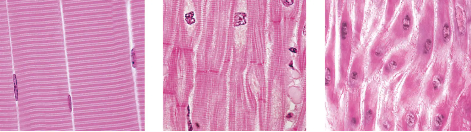
1. Skeletal Muscle
2. Cardiac Muscle
3. Smooth Muscle
S = Striated
U = Unstriated
Label These Correctly
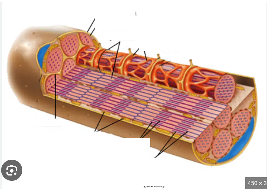
1. Bone
2. Tendon
3. Fascia
4. Epimysium
5. Perimysium
6. Endomysium
7. Fascicle
8. Nerve
9. Blood Vessel
10. Muscle Fiber
11. Sarcolemma
12. Nucleus
13. Sarcoplasmic Reticulum
14. Myofibril
15. Filaments

Label These Correctly
1. Sarcolemma
2. Sarcomere
3. Myofibrils
4. Sarcoplasmic Reticulum
5. T-Tubule
6. A-Band
7. Nucleus
8. Thin Filaments (Actin)
9. Thick Filaments (Myosin)
10. Mitochondria
11. H-Zone
12. I-Band
13. Openings into T Tubules
Label These Correctly
1. Z-Line
2. Sarcomere
3. M-Line
4. H-Zone
5. I-Band
6. A-Band

Label These Correctly
1. Cross-Bridges
2. Actin Filament (Thin)
3. Actin Molecule
4. Myosin Filament (Thick)
5. Myosin Molecule
6. Tropomyosin
7. Troponin

Label These Correctly
1. Origin of Biceps Brachii
2. Insertion of Biceps Brachii
3. Tendons

Label These Correctly
1. Trapezius
2. Triceps Brachii
3. Latissimus Dorsi
4. Hand & Finger Extensors
5. Calcaneal Tendon

Label These Correctly
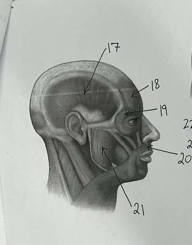
1. Orbicularis Oculi
2. Orbicularis Oris
3. Masseter
4. Sternoceidomastoid
5. Temporalis
6. Frontalis
Label These Correctly
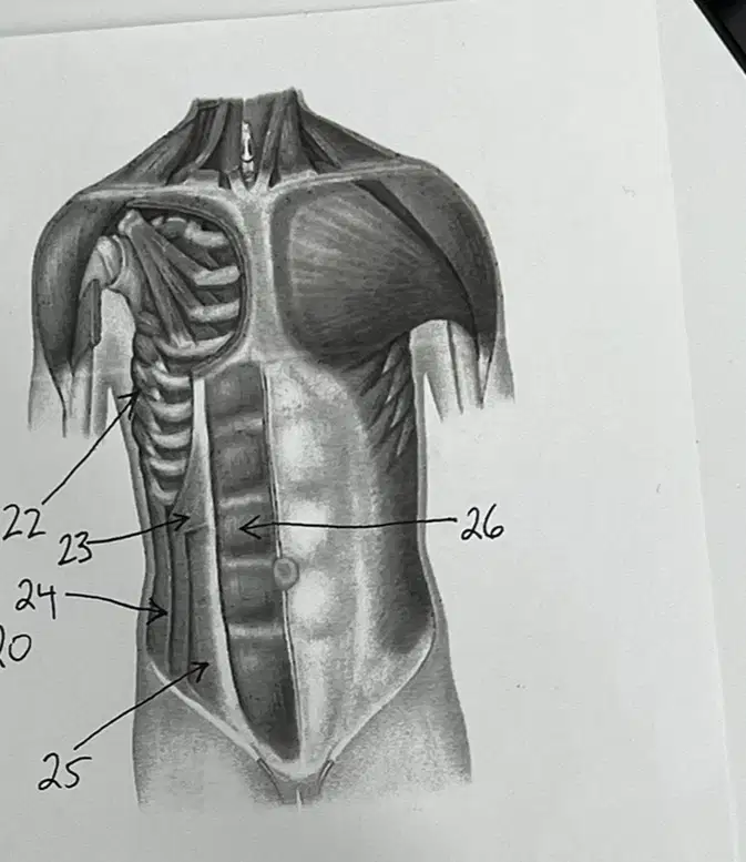
1. Rectus Abdominus
2. Serratus Anterior
3. Internal Oblique
4. Transverse Abdominus
5. External Oblique
Label These Correctly
1. Biceps Femoris
2. Semimembranosus
3. Gracilis
4. Semitendonosus
5. Gluteus Maximus
6. Gluteus Medius
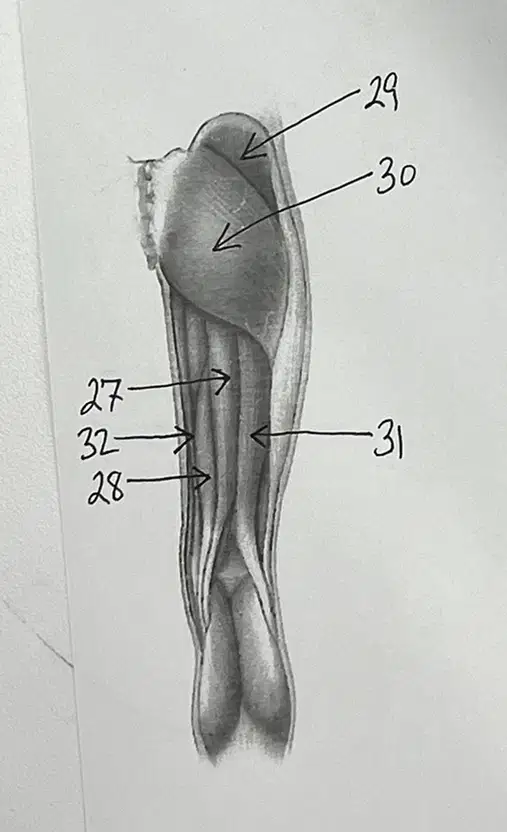
Label These Correctly
1. Sternocleomastoid
2. Deltiod
3. Biceps Brachii
4. Hand Flexors
5. Rectus Femoris
6. Vastus Medialis
7. Tibialis Anterior
8. Vastus Lateralis
9. Sartorius
10. Brachialis
11. Pectoralis Major

Label These Correctly
1. Gastronemius
2. Soleus

Label The Picture Correctly
1. Fibularis Longus
2. Tibialis Anterior
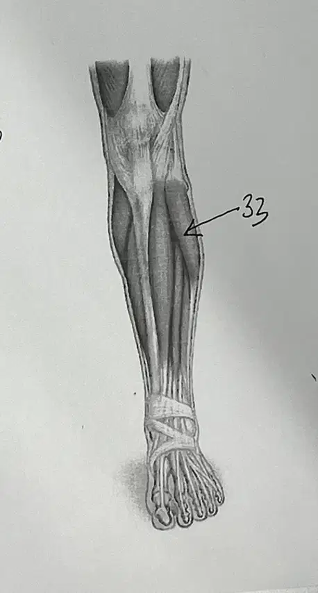
Answer the questions

Composed of neurons & neuroglia.
Nervous System/Tissue
Neurons
Cell Body
Axon
Dendrite
Transmit Nerve Impulses along nerve fibers to other neurons.
Nervous System/Tissue
Neurons
Cell Body
Axon
Dendrite
Nucleus of Neuron that makes the decision on whether or not to fire.
Nodes of Ranvier
Myelin Sheaths
Cell Body
Axon
Dendrite
______ carry impulses from other neurons (or from receptors) toward the cell body/Information comes into the cell from this.
Nodes of Ranvier
Myelin Sheaths
Cell Body
Axon
Dendrite
______ transmits the impulse
away from the cell body & splits into terminals. (1 mm-1 m+ long)
Nodes of Ranvier
Myelin Sheaths
Cell Body
Axon
Dendrite
Larger axons are enclosed by ___________ provided by Schwann cells and are myelinated fibers.
Nodes of Ranvier
Myelin Sheaths
Cell Body
Axon
Dendrite
Narrow gaps in the myelin sheath
between Schwann cells are called __________. These can make impulses travel faster, almost teleporting.
Nodes of Ranvier
Myelin Sheaths
Cell Body
Axon
Dendrite
Parts of Neurons
Nervous System/Tissue
Neurons
Cell Body
Axon
Dendrite
Made up of bundles of nerve fibers.
Nerves
Neurglia
Dendrite
Axon
Cell Body
Carries out a variety of functions to aid and protect components of the Nervous System.
Nerves
Neuroglia
Dendrite
Axon
Cell Body
Brain & Spinal Chord
Somatic and Autonomic Nervous System
Motor Division of Nervous System
Central Nervous System
Peripheral Nervous System
Sensory Division of Nervous System
Info into the Nervous System. Nerves in the Body that connect the CNS to the rest of the body. Separate Sensory and Motor Divisions.
Somatic and Autonomic Nervous System
Motor Division of Nervous System
Central Nervous System
Peripheral Nervous System
Sensory Division of Nervous System
Parts of the Peripheral Nervous System.
Somatic and Autonomic Nervous System
Motor Division of Nervous System
Central Nervous System
Peripheral Nervous System
Sensory Division of Nervous System
Carries Info Away from the CNS
Somatic and Autonomic Nervous System
Motor Division of Nervous System
Central Nervous System
Peripheral Nervous System
Sensory Division of Nervous System
Parts of the Motor Division of the PNS
Somatic
Nervous System
Autonomic Nervous System
Central Nervous System
Peripheral Nervous System
Sensory Division of Nervous System
Conscious Control (Skeletal Muscle)
Somatic
Nervous System
Autonomic Nervous System
Sensory Receptors
Schwann Cells
Microglial Cells
Unconscious Control (Smooth & Cardiac Muscle Glands)
Somatic
Nervous System
Autonomic Nervous System
Sensory Receptors
Schwann Cells
Microglial Cells
At the ends of Peripheral Nerves, Gather Information, and Convert into Impulses
Somatic
Nervous System
Autonomic Nervous System
Sensory Receptors
Schwann Cells
Microglial Cells
Produce Myelin on Axons
Somatic
Nervous System
Autonomic Nervous System
Sensory Receptors
Schwann Cells
Microglial Cells
Small Cells that phagocytize (Devour) bacterial cells and cellular debris.
Somatic
Nervous System
Autonomic Nervous System
Sensory Receptors
Schwann Cells
Microglial Cells
A type of tissue in your brain and spinal cord (central nervous system) that plays a crucial role in allowing you to function normally from day to day. It DOES NOT consists of high amounts of Myelin Sheath
Grey Brain Matter
White Brain Matter
Found in the deeper tissues of the brain (subcortical or PNS). It contains nerve fibers (axons). Many of these nerve fibers are surrounded by a type of sheath or covering called myelin. Myelin gives this its color.
Grey Brain Matter
White Brain Matter
Axon Terminals Go to These
Motor End Plate
Axon Terminals
Synapse
Synaptic Cleft
Part of the Axon. These can go to motor end plates on muscles, or other Dendrites.
Motor End Plate
Axon Terminals
Synapse
Synaptic Cleft
Junction b/t two communicating neurons at the axon terminals. This can by Nerve to Nerve, Nerve to Muscle, or Nerve to Gland.
Motor End Plate
Axon Terminals
Synapse
Synaptic Cleft
Site in which impulse must be conveyed.
Motor End Plate
Axon Terminals
Synapse
Synaptic Cleft
Chemicals that carry signals across the Synaptic Cleft. Excitatory or Inhibitory.
Neurotransmitters
Myelin Sheath
Myelinated Axons
Sensory Neurons
Motor Neurons
Insulation or "Hot Dog Bun" on the Axon that can increase the speed of a neural response. The space between these are important.
Neurotransmitters
Myelin Sheath
Myelinated Axons
Sensory Neurons
Motor Neurons
Humans have larger.....
Neurotransmitters
Myelin Sheath
Myelinated Axons
Sensory Neurons
Motor Neurons
Also known as Afferent & Carry Signals TO the Brain (ARRIVE)
Neurotransmitters
Myelin Sheath
Myelinated Axons
Sensory Neurons
Motor Neurons
Also known as Efferent & Carry Signals AWAY from the Brain (EXIT)
Neurotransmitters
Myelin Sheath
Myelinated Axons
Sensory Neurons
Motor Neurons
Two Types of Neuron Classification
Structural Neurons
Functional Neurons
Sensory Neurons
Motor Neurons
Polar Neurons
Bipolar, Unipolar, & Multipolar Neurons are classified as these. Most are Multipolar.
Structural Neurons
Functional Neurons
Sensory, Interneurons, & Motor Neurons are classified as these.
Structural Neurons
Functional Neurons
There are many types of Neurotransmitters
True
False
Label these with Arrows and the Correct Numbers
1. Cell Body/Nucleus
2. Dendrites
3. Axon
4. Axon Terminals
5. Draw Motor End Plate Where Muscle WOULD BE

Label these with Arrows and the Correct Numbers
1. Cell Body/Nucleus
2. Dendrites
3. Axon
5. Myelin Sheath

Label these with Arrows and the Correct Numbers
1. Axon Terminal
2. Synapse
3. Synaptic Cleft
5. Neurotransmitters
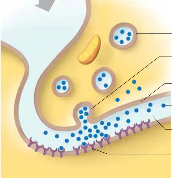
Draw the Correct Shape on the Correct type of Neuron
1. Circle the Multipolar
2. Rectangle on the Unipolar
3. Triangle on the Bipolar

Is usually polarized with a difference in electrical charge on each side. + on outside and - on inside.
Cell Membrane
Negative Net Charge
Positive Net Charge
Sodium (Na+)
Potassium (K+)
Calcium (Ca2+)
Hydrogen (H+)
Chlorine (Cl-)
Hydroxide (OH-)
Inside of membrane
Cell Membrane
Negative Net Charge
Positive Net Charge
Sodium (Na+)
Potassium (K+)
Calcium (Ca2+)
Hydrogen (H+)
Chlorine (Cl-)
Hydroxide (OH-)
Outside of membrane
Cell Membrane
Negative Net Charge
Positive Net Charge
Sodium (Na+)
Potassium (K+)
Calcium (Ca2+)
Hydrogen (H+)
Chlorine (Cl-)
Hydroxide (OH-)
Most Important + Charged Ions
Cell Membrane
Negative Net Charge
Positive Net Charge
Sodium (Na+)
Potassium (K+)
Calcium (Ca2+)
Hydrogen (H+)
Chlorine (Cl-)
Hydroxide (OH-)
Most Important - Charged Ions
Cell Membrane
Negative Net Charge
Positive Net Charge
Sodium (Na+)
Potassium (K+)
Calcium (Ca2+)
Hydrogen (H+)
Chlorine (Cl-)
Hydroxide (OH-)
Gives Charge to the Membrane, Cleans up Messes, and Controls the regulation of another answer.
Na+/K+ Pump
3 Na+ Out
2 K+ In
Resting Potential
Threshold Potential
Graded Potential
The separation of charge on a cell membrane. (When Equal)
Na+/K+ Pump
Action Potential
Resting Potential
Threshold Potential
Graded Potential
If sufficiently strong, depolarization occurs and this is achieved. Na+ Is let in until it is too much and hits this point before a ton is let in at once.
Na+/K+ Pump
Action Potential
Resting Potential
Threshold Potential
Graded Potential
Inward movement of Na+ depolarizes membrane, causing this. The trigger for action potential.
Na+/K+ Pump
Action Potential
Resting Potential
Threshold Potential
Graded Potential
May be reached when a series of subthreshold stimuli summate and reach and surpassed threshold. (Ex. Domino Tipping Point & A ton of Na+ Come In)
Na+/K+ Pump
Action Potential
Resting Potential
Threshold Potential
Graded Potential
Conduct impulses along entire membrane surface.
Unmyelinated Fibers
Myelinated Fibers
Salatory Conduction
All or None Response
Impulse Processing
Conduct impulses from one Node of Ranvier to the next.
Unmyelinated Fibers
Myelinated Fibers
Salatory Conduction
All or None Response
Impulse Processing
Impulse almost skips through myelinated fibers to make it almost faster than the conduction of unmyelinated because of the Nodes on Ranvier.
Unmyelinated Fibers
Myelinated Fibers
Salatory Conduction
All or None Response
Impulse Processing
The Nervous System is an....
Unmyelinated Fibers
Myelinated Fibers
Salatory Conduction
All or None Response
Impulse Processing
Dependent upon how neurons signal each other and are organized in the brain and spinal chord.
Unmyelinated Fibers
Myelinated Fibers
Salatory Conduction
All or None Response
Impulse Processing
Different Types of Impulse Processing
Antagonist Signals
Summation
Patterns of Connectivity
Excitation vs. Inhibition
Antagonist Signals
Summation
Patterns of Connectivity
Over Time or Space
Antagonist Signals
Summation
Patterns of Connectivity
Covergent or Divergent
Antagonist Signals
Summation
Patterns of Connectivity
Answer the Questions
Bone, Meninges, Blood, Cerebrospinal Fluid
Protective Layers of The Brain
Menings
Blood
CO2 Levels
Brain
Dura Mater, Arachnoid, & Piamater
Cell Membrane
Menings
Blood
CO2 Levels
Brain
Acts as a Brain Barrier
Cell Membrane
Menings
Blood
CO2 Levels
Brain
Affects blood because when dissolved into blood, it chemically reacts with water. This produces carbonic acid, Ph and biocarbonate ions.
Cell Membrane
Menings
Blood
CO2 Levels
Brain
H2O + CO2 ------> H2CO5 ------> H+ + HCO3-
Cell Membrane
Menings
Blood
CO2 in Blood
Brain
Has special capillaries to prevent leaking and has to go through the Cell Membrane.
Cell Membrane
Menings
Blood
CO2 in Blood
Brain
The Brain has special capillaries to prevent leaking. This has to go through the _______.
Cell Membrane
Menings
Blood
CO2 in Blood
Brain
The Brain has special capillaries to prevent leaking because the brain is a _______. So by adding other blood, it would mess up your brain processes.
Chemical Machine
Other Blood
Brian Processes
CO2 in Blood
Brain
The Brain has special capillaries to prevent leaking because the brain is a chemical machine. So by adding ________, it would mess up your brain processes.
Chemical Machine
Other Blood
Brian Processes
CO2 in Blood
Brain
Lebel the Cut-In-Half Brain
1. Cerebrum
2. Cerebellum
3. Diencephalon (Next 2 Make It Up)
4. Thalamus
5. Hypothalamus
6. Brain Stem (Next 3 Make It Up)
7. Midbrain
8. Pons
9. Medulla

Links the spinal cord and higher brain levels. Controls cardiovascular, respiratory, digestive, visual, and auditory reflexes. Consists of medulla, pons & midbrain.
Brain Stem
Thalamus
Hypothalamus
Pineal Gland
Cerebellum
Parts of the Diacephalon
Brain Stem
Thalamus
Hypothalamus
Pineal Gland
Cerebellum
Reinforces voluntary motor behavior initiated by motor cortex. “Relay station” and synaptic integrating center for sensory input. Helps direct attention to stimuli of interest. Capable of crude awareness of sensations but cannot distinguish their location or intensity.
Brain Stem
Thalamus
Hypothalamus
Pineal Gland
Cerebellum
Link between cerebrum and lower autonomic centers. Link between autonomic nervous and endocrine systems. Integrating center for homeostatic function. Brain area most involved in directly regulating internal environment. (Water Balance)
Brain Stem
Thalamus
Hypothalamus
Pineal Gland
Cerebellum
Biological Clock - Secretes Melatonin
Brain Stem
Thalamus
Hypothalamus
Pineal Gland
Cerebellum
Assist Cerebrum in motor and sensory integration, planning/coordination of skeletal muscle activity, controls posture, & maintains balance.
Brain Stem
Thalamus
Hypothalamus
Pineal Gland
Cerebellum
Higher Thought, 80% of total brain weight, 2 hemispheres connected by corpus callosum, divided into lobes, & outer surface is the convoluted cerebral cortex which contains 75% of neurons in the NS.
Cerebrum
Motor
Sensory
Association
Sensory & Motor Fibers
Both _________ CROSS OVER in the spinal chord or brain stem, so the centers in the right hemisphere are interpreting or controlling the left side of the body, and vice versa.
Cerebrum
Motor
Sensory
Association
Sensory & Motor Fibers
Functional area of the brain overlap, but the cortex can generally be divided into 3 areas called......
Cerebrum
Motor
Sensory
Association
Sensory & Motor Fibers
The left side of the brain controls the right and vice versa.
True
False
Label these with the correct number
1. Frontal Lobe
2. Occipital Lobe
3. Parietal Lobe
4. Temporal Lobe
5. Central Sulcus
6. Brain Stem
7. Cerebrum

Contains Visual Cortex
Occipital Lobe
Temporal Lobe
Parietal Lobe
Frontal Lobe
Central Sulcus
Houses the Auditory Cortex
Occipital Lobe
Temporal Lobe
Parietal Lobe
Frontal Lobe
Central Sulcus
Reception/Perception of Somatosensory Input
Occipital Lobe
Temporal Lobe
Parietal Lobe
Frontal Lobe
Central Sulcus
Voluntary Motor Movement
Occipital Lobe
Temporal Lobe
Parietal Lobe
Frontal Lobe
Central Sulcus
Part of the Parietal Lobe that delimits the boundary between motor and the sensory cortices, as well as the boundary between the frontal and parietal lobes.
Occipital Lobe
Temporal Lobe
Parietal Lobe
Frontal Lobe
Central Sulcus
Awareness of external world & self states. (maximal alertness, wakefulness, sleep, & coma)
Conciousness
Peripheral Nervous System
Somatic Nervous System
Autonomin Nervous System
Consist of the cranial and spinal nerves that arise from the CNS and travel to the remainder of the body. Made of the Somatic and Autonomic Nervous Systems.
Conciousness
Peripheral Nervous System
Somatic Nervous System
Autonomin Nervous System
Oversees Voluntary Activities
Conciousness
Peripheral Nervous System
Somatic Nervous System
Autonomin Nervous System
Oversees involuntary activity & is divided into the sympathetic and parasympathetic NS
Conciousness
Peripheral Nervous System
Somatic Nervous System
Autonomic Nervous System
Part of the Autonomic Nervous System that mainly stimulates "rest, repair, & digestive" activities.
Central Nervous System
Peripheral Nervous System
Parasympathetic Nervous System
Sympathetic Nervous System
Part of the Autonomic Nervous System that mainly stimulates "fright, fight, flight" responses.
Central Nervous System
Peripheral Nervous System
Parasympathetic Nervous System
Sympathetic Nervous System
Sensory receptors detect changes in the environment and send impulses through the _________ of the PNS to the Brain.
Afferent Division
Sensation
Somatic Senses
Special Senses
Specific
(Type, Location, Intensity) Is formed in the brain based on sensory input
Afferent Division
Sensation
Somatic Senses
Special Senses
Specific
Widely distributed throughout body
Afferent Division
Sensation
Somatic Senses
Special Senses
Specific
_____ of sensory organs IN HEAD
Afferent Division
Sensation
Somatic Senses
Special Senses
Specific
Each receptor is most sensitive to a ______ change, although it may react to an extreme level of other factors.
Afferent Division
Sensation
Somatic Senses
Special Senses
Specific
Are sensitive to change in chemical concentration (Nose and Mouth)
Chemoreceptors
Pain Receptors
Thermoreceptors
Mechanoreceptors
Photoreceptors
Are sensitive to change in chemical concentration (Nose and Mouth)
Chemoreceptors
Pain Receptors
Thermoreceptors
Mechanoreceptors
Photoreceptors
Detect intense mechanical, thermal, or chemical changes that can damage tissue.
Chemoreceptors
Pain Receptors
Thermoreceptors
Mechanoreceptors
Photoreceptors
Respond to temperature differences.
Chemoreceptors
Pain Receptors
Thermoreceptors
Mechanoreceptors
Photoreceptors
Respond to changes in pressure or movement (In ear and skin)
Chemoreceptors
Pain Receptors
Thermoreceptors
Mechanoreceptors
Photoreceptors
In the eyes respond to light energy
Chemoreceptors
Pain Receptors
Thermoreceptors
Mechanoreceptors
Photoreceptors
Feelings that occur when the brain interprets sensory impulses
Sensation
Projection
Sensory Adaptation
Tonic Receptors
Phasic Receptors
Used by the brain to create the impression that the sensation is coming from the area of stimulation. At the same time as the sensation is being formed.
Sensation
Projection
Sensory Adaptation
Tonic Receptors
Phasic Receptors
Sensory impulses are sent at decreasing rates until receptors fail to send impulses unless there is a change in strength of the stimulus.
Sensation
Projection
Sensory Adaptation
Tonic Receptors
Phasic Receptors
DO NOT adapt, or adapt SLOWLY
Sensation
Projection
Sensory Adaptation
Tonic Receptors
Phasic Receptors
DO Adapt; May have "Off" Signal
Sensation
Projection
Sensory Adaptation
Tonic Receptors
Phasic Receptors
Frequency of action potentials, which are triggered.
Intensity
Acuity
Tonic
Phasic
_________ of location detection depends on receptor field size and density. Can be reduced when nearby receptors are responding to weak stimuli. Can be enhanced through lateral inhibition of nearby receptors by the receptor that reacts to the strongest stimulation.
Intensity
Acuity
Tonic
Phasic
Receptors associated with skin, muscles, joints, and viscera
Somatic Senses
Free Nerve Endings
Tactile (Meissner's) Corpuscles
Lamellated (Pacinian) Corpuscles
3 Types of Receptors to Detect Touch & Pressure
Somatic Senses
Free Nerve Endings
Tactile (Meissner's) Corpuscles
Lamellated (Pacinian) Corpuscles
Associated with touch and pressure
Somatic Senses
Free Nerve Endings
Tactile (Meissner's) Corpuscles
Lamellated (Pacinian) Corpuscles
Abundant in hairless areas that are VERY sensitive to touch
Somatic Senses
Free Nerve Endings
Tactile (Meissner's) Corpuscles
Lamellated (Pacinian) Corpuscles
Resemble an onion and function to detect DEEP Pressure
Somatic Senses
Free Nerve Endings
Tactile (Meissner's) Corpuscles
Lamellated (Pacinian) Corpuscles
Temperature Senses, react within a range of temperatures, adapt quickly, and also stimulate pain receptors at extreme temps.
Warm Receptors
Cold Receptors
Touch Receptors
Smell Receptors
Consists of free nerve endings that are stimulated when tissues are damaged, and adapt little, if at all. Many stimuli can affect these.
Pain Receptors
Visceral Pain Receptors
Referred Pain
Acute Pain
Chronic Pain
The only receptors in the visceral that produce sensations
Pain Receptors
Visceral Pain Receptors
Referred Pain
Acute Pain
Chronic Pain
Occurs because the common nerve pathways leading from skin and internal organs
Pain Receptors
Visceral Pain Receptors
Referred Pain
Acute Pain
Chronic Pain
Type A Pain. This, Myelinated Fibers that carry impulses rapidly and cease when stimulus stops.
Pain Receptors
Visceral Pain Receptors
Referred Pain
Acute Pain
Chronic Pain
Type B or C pain. Thin, Unmyelinated fibers that conduct impulses slowly and continue sending impulses when stimulus stops.
Pain Receptors
Visceral Pain Receptors
Referred Pain
Acute Pain
Chronic Pain
Released in the dorsal horn of the spinal cord or in the brain endorphins from the pituitary and block pain signals.
Endogenus Opiates
Special Senses
Olafactory Receptors
Flavor
Associated with complex structures located in the head. Include senses of smell, taste, hearing, equalibrium, and sight.
Endogenus Opiates
Special Senses
Olafactory Receptors
Flavor
Smell Receptors (BLANK) and Taste Receptors are chemoreceptors
Endogenus Opiates
Special Senses
Olafactory Receptors
Flavor
Taste + Smell + Spicy/Wintergreen + Texture/Feel
Endogenus Opiates
Special Senses
Olafactory Receptors
Flavor
Taste + Smell + Spicy/Wintergreen + Texture/Feel
Endogenus Opiates
Special Senses
Olafactory Receptors
Flavor
Are organs of taste. Concentrated around papillae of the tongue & scattered throughout the mouth and pharynx.
Taste Buds
Taste Cells
Taste Hairs
Saliva
5 Types of Taste Cells
Modified Epithelial Cells that function as chemoreceptors. Contain taste hairs and taste pores.
Taste Buds
Taste Cells
Taste Hairs
Saliva
5 Types of Taste Cells
Portions sensitive to taste and protrude from openings called taste pores.
Taste Buds
Taste Cells
Taste Hairs
Saliva
Types of Taste Cells
Chemicals must be dissolved in this in order to be tasted
Taste Buds
Taste Cells
Taste Hairs
Saliva
Types of Taste Cells
Sweet, Sour, Salty, Bitter, Unami (Savory)
Taste Buds
Taste Cells
Taste Hairs
Saliva
Types of Taste Cells
External, Middle, and Inner Sections. Mechanoreceptors.
Sense of Hearing
Outer (External) Ear
Middle Ear
Auditory Ossicles
Auditory Tube
Consists of the auricle, which collects the sound that then travels the external acoustic meatus.
Sense of Hearing
Outer (External) Ear
Middle Ear
Auditory Ossicles
Auditory Tube
(Tympanic Cavity) Begins with the tympanic membrane (eardrum), and is an air-filled space housing auditory ossicles.
Sense of Hearing
Outer (External) Ear
Middle Ear
Auditory Ossicles
Auditory Tube
Malleus, Incus, & Stapes. Transmit and Amplify Sound Waves.
Sense of Hearing
Outer (External) Ear
Middle Ear
Auditory Ossicles
Auditory Tube
(Eustachian) Tube connects the middl ear to the throat (Pharnyx) to help maintain equal air pressure in both sides of the eardrum.
Sense of Hearing
Outer (External) Ear
Middle Ear
Auditory Ossicles
Auditory Tube
Made up of membranous labyrinth inside an osseous labyrinth.
Inner Ear
Cochlea
Sense of Equilibrium
Organs of Static Equilibrium
Organs of Dynamic Equilibrium
Coiled and Houses the organ of hearing while the semicircular canals function in the equilibrium. Fluid-Filled
Inner Ear
Cochlea
Sense of Equilibrium
Organs of Static Equilibrium
Organs of Dynamic Equilibrium
Comes from two different organs. Organs of the Static and Dynamic Equilibrium.
Inner Ear
Cochlea
Sense of Equilibrium
Organs of Static Equilibrium
Organs of Dynamic Equilibrium
Senses the position of the head and helps to maintain stability and posture in response to gravity and linear acceleration. Located in Vestibule.
Inner Ear
Cochlea
Sense of Equilibrium
Organs of Static Equilibrium
Organs of Dynamic Equilibrium
Helps to maintain orientation when the head rotates. Located in the Ampulia.
Inner Ear
Cochlea
Sense of Equilibrium
Organs of Static Equilibrium
Organs of Dynamic Equilibrium
Detect motion of the head. Fluid-Filled Tubes with Jelly Cap
3 Semicircular Canals
Utricle
Saccule
Sense of Sight
Eyelid
Detects Horizontal Forces
3 Semicircular Canals
Utricle
Saccule
Sense of Sight
Eyelid
Detects Vertical Forces
3 Semicircular Canals
Utricle
Saccule
Sense of Sight
Eyelid
Accessory organs, namely the lacrimal apparatus, eyelids, and extrinsic muscles. Aid the eye in its function.
3 Semicircular Canals
Utricle
Saccule
Sense of Sight
Eyelid
Protect Eye, Lined with conjunctiva, and made of the thinnest skin.
3 Semicircular Canals
Utricle
Saccule
Sense of Sight
Eyelid
Produce tears that lubricate & cleanse eye.
Lacrima Apparatus
Extrinsic Muscles
Cornea
Optic Nerve
Choroid Coat
Attach to the sclera and move the eye in all directions.
Lacrima Apparatus
Extrinsic Muscles
Cornea
Optic Nerve
Choroid Coat
Transparent Outer Layer of the Eye
Lacrima Apparatus
Extrinsic Muscles
Cornea
Optic Nerve
Choroid Coat
White anterior eye
Lacrima Apparatus
Extrinsic Muscles
Sclera
Optic Nerve
Choroid Coat
Pierce the Sclera at posterior of the eye.
Lacrima Apparatus
Extrinsic Muscles
Sclera
Optic Nerve
Choroid Coat
Vascular, darkly pigmented part of the eye. Functions to nourish tissues of the eye & keep inside of dark eye.
Lacrima Apparatus
Extrinsic Muscles
Sclera
Optic Nerve
Choroid Coat
Focusses light and facilitates focusing (accommodation).
Lens and Cornea
Fovea
Rods
Cones
Iris
Light is focussed on this
Retina
Fovea
Iris
Pupil
Fovea Centralis
A lot of cone sensors in a bunch of fibers
Retina
Fovea
Iris
Pupil
Fovea Centralis
Adjust amount of light entering eye
Retina
Fovea
Iris
Pupil
Fovea Centralis
A hole in the eye that the light enters
Retina
Fovea
Iris
Pupil
Fovea Centralis
The point of sharpest vision retina.
Retina
Fovea
Iris
Pupil
Fovea Centralis
Thick lense, ciliary muscle fibers contracted, suspensory ligaments relaxed
Near Vision
Far Vision
Thin lense, ciliary muscle fibers relaxed, suspensory ligaments taut
Near Vision
Far Vision