
 Worksheets
Electrical Activity of the Heart Quiz
Total questions: 216
Worksheet time: 3hrs 46mins
What does the ECG Lead I represent in terms of electrical activity of the heart?
Electrical activity from the left arm to the right arm
Electrical activity from the right arm to the left leg
Electrical activity from the left leg to the left arm
Electrical activity from the right leg to the right arm
During a marathon, a runner's heart rate increases significantly. Which part of the heart is responsible for initiating the electrical impulse that is represented in an ECG, thus controlling the heart rate?
AV node
SA node
Left ventricle
Bundle of His
What does the ECG Lead III trace represent?
Electrical activity from the left arm to the left leg
Electrical activity from the right arm to the left leg
Electrical activity from the left leg to the right arm
Electrical activity from the right arm to the left arm
During a routine health check-up, a doctor is analyzing a patient's ECG. What does the 'P' wave observed in the ECG represent?
Ventricular repolarization
Ventricular depolarization
Atrial depolarization
Atrial repolarization
Which segment of the ECG represents the isoelectric line where there is no electrical activity?
PR Interval
ST Segment
QRS Interval
TP Interval
During a routine health check-up, a doctor is explaining to a patient how the heart's electrical activity is represented on an ECG. What is the correct sequence of depolarization as shown on the ECG?
P wave, QRS complex, T wave
T wave, P wave, QRS complex
QRS complex, T wave, P wave
QRS complex, P wave, T wave
What does the 'QRS' complex represent in an ECG?
Atrial repolarization
Ventricular depolarization
Atrial depolarization
Ventricular repolarization
What does the term "Sinus" refer to in medical terminology?
No electrical activity
Fast heart rate
Originates from the SA Node
Slow heart rate
What is the meaning of "Atrial" in the context of this medical terminology?
Originates in and around the AV Junction
Originates from the Atria but not specifically the SA Node
Above the Ventricles
Originates in the Ventricles
What does "Junctional" mean in this medical context?
Originates from the SA Node
No electrical activity
Originates in and around the AV Junction
Fast heart rate
What does "Ventricular" refer to in this terminology?
Slow heart rate
Originates from the Atria
Originates in the Ventricles
Above the Ventricles
Match the following definitions with the correct term.
Fast heart rate
Definition of a condition characterized by a rapid heartbeat
Originates from the SA Node
Definition of a rhythm that starts in the sinoatrial node
No electrical activity
Definition of Asystole
Originates in and around the AV Junction
Definition of a rhythm originating near the atrioventricular junction
Match the following descriptions with the correct heart condition.
Slow heart rate
Condition that describes a heart rate that is below the normal range
Originates from the Atria
Condition that indicates the origin of the heart rhythm
Fast heart rate
Condition that describes a heart rate that is above the normal range
No electrical activity
Condition indicating the heart has ceased its electrical activity
What is the meaning of "Bradycardia"?
Above the Ventricles
Originates in the Ventricles
Fast heart rate
Slow heart rate
What does "Supraventricular" indicate in medical terms?
Originates from the SA Node
Slow heart rate
No electrical activity
Above the Ventricles
What does a wave in an ECG represent?
The time between two specific ECG events
A positive or negative deflection from baseline that indicates a specific electrical event
The length between two specific points on an ECG
The combination of multiple waves grouped together
Which of the following is NOT a type of interval commonly measured on an ECG?
PR interval
QRS interval
TP interval
QT interval
What is a segment in the context of an ECG?
The combination of multiple waves grouped together
The time between two specific ECG events
The length between two specific points on an ECG that are supposed to be at the baseline amplitude
There is only one point on an ECG termed the J point
What is the only main complex on an ECG?
PR complex
ST complex
QRS complex
TP complex
Where is the J point located on an ECG?
Where the P wave ends
Where the QRS complex begins
Where the QRS complex ends and the ST segment begins
Where the T wave ends
What does the abbreviation AV stand for?
Atrioventricular
Atrial Velocity
Aortic Valve
Arteriovenous
What is the full form of PAC as used in medical terminology? (a)
ECG is a common abbreviation in the medical field. What does it stand for?
Electrocardiogram
Electrocerebralgram
Electrochemogram
Electrocutaneous Graph
PJC is an abbreviation for which of the following?
Premature Junctional Contraction
Posterior Junctional Contraction
Peripheral Junctional Contraction
Premature Joint Closure
What does PVC represent in medical terminology?
Premature Ventricular Contraction
Posterior Ventricular Contraction
Peripheral Vascular Closure
Premature Vascular Contraction
NSR is an acronym for which of the following terms?
Normal Sinus Rhythm
Non-Specific Reaction
Nodal System Regulation
Neurological Stress Response
SVT stands for which condition?
Supraventricular Tachycardia
Subventricular Tremor
Systemic Vascular Tension
Sinusoidal Vibration Therapy
What does PAT refer to in the context of cardiology?
Paroxysmal Atrial Tachycardia
Peripheral Arterial Thrombosis
Posterior Atrial Tension
Prolonged Aortic Tract
SA is an abbreviation for which term?
Sinusoidal Activity
Subarachnoid
Sinoatrial
Subacute Angina
During a routine check-up, a nurse is preparing to use the EASI Lead system for a patient's ECG. What color code corresponds to the lead placement at the lower sternum (5th intercostal space)?
Red
Black
Brown
White
Where should the black lead be placed in EASI Leads?
Right mid-axillary line (5th intercostal space)
Left mid-axillary line (5th intercostal space)
Upper sternum (just below sternal angle)
Anywhere (right lower)
Match the following lead placements with their associated colors in EASI Leads.
Right mid-axillary line (5th intercostal space)
Anywhere (right lower)
Left mid-axillary line (5th intercostal space)
Right mid-axillary line (5th intercostal space)
Lower sternum (5th intercostal space)
Left mid-axillary line (5th intercostal space)
Anywhere (right lower)
Lower sternum (5th intercostal space)
In the EASI Leads system, where is the red lead placed?
Right mid-axillary line (5th intercostal space)
Left mid-axillary line (5th intercostal space)
Upper sternum (just below sternal angle)
Lower sternum (5th intercostal space)

What is the mnemonic to remember the lead placement in EASI Leads?
Smoke over fire, clouds over grass, chocolate on the right
Smoke over fire, grass over clouds, chocolate on the left
Fire over smoke, grass over clouds, chocolate on the right
Fire over smoke, clouds over grass, chocolate on the left
What does the EKG/ECG graph paper allow for visual measurement of in terms of time?
Distance
Rate
Temperature
Frequency
On an EKG/ECG graph paper, what is measured on the horizontal line?
Amplitude (voltage)
Time (rate)
Pressure
Heart rate
What is measured on the vertical line of an EKG/ECG graph paper?
Time (rate)
Distance
Amplitude (voltage)
Heart rate
Each small square on the EKG/ECG graph paper is (a) wide.
What does each small square on the EKG/ECG graph paper represent in terms of time?
0.02 seconds
1 millimeter (mm)
0.04 seconds
5 millimeters (mm)
What is the time interval represented by a width of 5 mm on the EKG/ECG graph paper?
0.02 seconds
1 millimeter (mm)
0.04 seconds
0.20 seconds
What do the darker lines on EKG/ECG graph paper represent?
They divide the paper into every 10th square vertically and horizontally.
They indicate the electrical activity of the heart.
They divide the paper into every 5th square vertically and horizontally.
They represent the time interval of 1 second.
How large are the large squares on EKG/ECG graph paper? (a)
What does one large square on the EKG/ECG graph paper represent in terms of time?
0.04 seconds
0.10 seconds
0.20 seconds
1.00 second
How many small squares are there in each large square on EKG/ECG graph paper?
10 small squares
20 small squares
25 small squares
50 small squares
What is the time interval represented by a 1 mm square on the EKG/ECG graph paper?
0.02 seconds
0.04 seconds
0.10 seconds
0.20 seconds

What does one complete cardiac cycle consist of?
P, Q, R, S, T, U waves
P, Q, R, S, (QRS complex), and T wave
P, R, S, T, U waves
Q, R, S, (QRS complex), T, U waves
What does the P wave in an EKG represent?
Ventricular depolarization
Atrial repolarization
Atrial depolarization (contraction)
Ventricular repolarization (resting phase)
The QRS Complex in an EKG represents (a) .
Atrial depolarization
Ventricular repolarization
Ventricular depolarization
Atrial repolarization only
What does the T wave in an EKG indicate?
Atrial depolarization
Ventricular depolarization
Atrial repolarization
Ventricular repolarization (resting phase)
What does the P wave on an EKG represent?
Ventricular depolarization
Atrial repolarization
Atrial depolarization
Ventricular repolarization
Which complex on the EKG represents ventricular depolarization?
T wave
P wave
ST segment
QRS complex
What is represented by the ST segment and T wave on the EKG?
Atrial depolarization
Ventricular depolarization
Atrial repolarization
Ventricular repolarization
Where does the impulse travel after leaving the SA node?
Directly to the ventricles
Through the AV node only
Across the atrial muscle and then through Bachman's Bundle to the left atrium
Outside the heart
What causes the right and left atria to contract at the same time?
The impulse traveling from the AV node to the SA node
The impulse traveling from the SA node to the AV node
The QRS complex on the EKG
The T wave on the EKG
What does the P wave in an EKG represent?
Ventricular depolarization
Atrial repolarization
Atrial depolarization (contraction)
Ventricular repolarization
What does the QRS Complex in an EKG represent?
Atrial depolarization
Ventricular depolarization, atrial repolarization
Ventricular repolarization
Atrial repolarization
What does the T wave in an EKG represent?
Atrial depolarization
Atrial repolarization
Ventricular depolarization
Ventricular repolarization (resting phase)
What is rhythm classified as when the interval between the R waves is regular?
Irregular pattern
Regular pattern
Sequential pattern
Random pattern
What does rhythm refer to in the context of heart function?
The color of the heart
The size of the heart
The sequential beating of the heart as a result of the generation of electrical impulses
The sound of the heart
How is rhythm classified when the interval between the R waves is not regular?
Regular pattern
Sequential pattern
Irregular pattern
Constant pattern

What does the 'P' wave in an ECG complex represent?
Ventricular depolarization
Ventricular repolarization
Atrial depolarization
Atrial repolarization

Which interval on an ECG represents the time from the onset of atrial depolarization to the onset of ventricular depolarization?
ST interval
QT interval
PR interval
TP interval

What does the 'QRS' complex in an ECG signify? (a)

Match the following descriptions with the correct phase of the heart cycle they represent.
Ventricular depolarization
Phase corresponding to the start of the QRS complex
Atrial depolarization
Phase not typically visible in an ECG
Ventricular repolarization
Phase represented by the 'T' wave in an ECG
Atrial repolarization
Phase occurring just before the P wave
The atrial rate is determined by (a) when interpreting EKG rhythm strips.
How is the ventricular rate determined when interpreting EKG rhythm strips?
By counting the number of P waves
By counting the number of T waves
By counting the number of QRS complexes
By counting the number of electrical impulses in 30 seconds
What does the 6 second method denote in EKG rhythm strip interpretation?
A 10 second interval on EKG strip
A 6 minute interval on EKG strip
A 6 second interval on EKG strip
A 3 second interval on EKG strip
How do you calculate the heart rate using the 6 second method on an EKG strip?
Count the number of QRS complexes in 6 seconds and divide by 10
Count the number of P waves in 6 seconds and multiply by 10
Count the number of QRS complexes in 6 seconds and multiply by 10
Count the number of T waves in 6 seconds and multiply by 10
How many seconds are represented by the time marks in the ECG strip shown in Figure 2? (a)
Based on the ECG strip provided, what is the heart rate?
60 beats per minute
70 beats per minute
80 beats per minute
90 beats per minute
Are P waves present in a normal ECG cycle?
Yes
No
Only in abnormal ECG
Present only in tachycardia
How should P waves appear in a normal ECG? (a)
What is the normal duration of a P-wave? (a)
How many P waves should be present for each QRS complex in a normal ECG?
One P wave for each QRS complex
Two P waves for each QRS complex
No P wave for each QRS complex
Variable number of P waves for each QRS complex
Are P waves supposed to look similar across the ECG?
Yes, they should look similar
No, each P wave is unique
They look similar only in abnormal ECGs
They look similar only in bradycardia
What does the QT interval on an EKG represent?
Time from ventricular depolarization to repolarization
Time from atrial depolarization to atrial repolarization
Time for the entire electrical conduction of the heart
Time from the start of the Q wave to the end of the T wave, indicating ventricular activity
What is the significance of an elevated ST segment on an ECG?
Indicates a normal heart function
May suggest myocardial infarction
Signifies atrial fibrillation
Indicates low potassium levels
How is the heart rate calculated using an ECG?
Counting the number of QRS complexes in a 10 second strip and multiplying by 6
Measuring the distance between two P waves
Counting the number of T waves in a 6 second strip
Measuring the PR interval
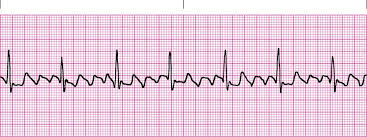
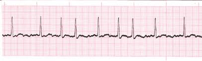

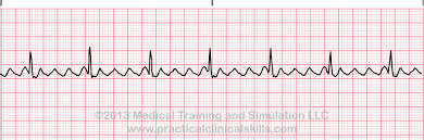
Heart rate over 100 beats per minutes is
tachycardia
bradycardia
depolarization
repolarization
Which of these is not considered when making a diagnosis?
P-R interval
rate
rhythm
T wave
When making a diagnosis in the classroom, we are looking at which view?
Lead I
Lead II
Lead III
V1
The outstanding characteristic of sinus arrest is
fast heart rate
a long pause with no heart beat
a long P-R interval
everything is normal
With sinus dysrhythmia
the rate is abnormal
the rhythm is abnormal
the p wave is abnormal
the t wave is abnormal
In wandering atrial pacemaker, the outstanding characteristic is
the P wave will have 3 or more shapes
the T wave is absent
the QRS repolarizes
the baseline shifts
What is the heart rate for multifocal atrial tachycardia?
60-100 bpm
over 100 bpm
below 60 bpm
20-40 bpm
In atrial flutter the atria will
not beat at all
beat as often as the ventricles beat
beat 3 or 4 more times than the ventricles
be normal
We see capital F waves in which dysrhythmia?
atrial fibrillation
premature atrial complexes
normal sinus rhythm
atrial flutter
Which of these is NOT true of atrial fibrillation?
the atria are quivering like a bowl of jello
the rhythm is irregular
p waves are not present
atrial rate is 60-100 bpm
MI stands for
myocardial infarction
muscle infarction
mycocordial infarction
muscle infraction
With premature atrial complexes, what is coming early?
P wave
ORS complex
U wave
T wave
If our patient's heart rate is 75bpm, rhythm is regular, P-R interval is normal, QRS is normal, then our diagnosis is
Sinus tachycardia
Sinus dysrhythmia
normal sinus rhythm
Sinus arrest
A common cardiac dysrhythmia characterized by premature heartbeats originating in the atria.
A-Fib
A-Flutter
PAC's
SA Block
Occurs when the natural pacemaker site shifts between SA node, the atria, and/or the AV node.
1st Degree AV Block
SA Block
Sinus Pause
Wandering Atrial Pacemaker
Pattern in which every third complex is a premature (PVC).
(a)
A cardiac cell or group of cells that produces an ectopic beat; cells that are notably different or abnormal in some manner
(a)
The extra blood that can be ejected into the ventricles by the atria as a result of the electrical impulse slowing as it enters the AV node.
(a)
A blood clot that forms on the wall in a diseased area of the endocardium or on the wall of an injured blood vessel
(a)

Determine which of the following electrocardiogram (ECG) tracings is missing P waves but is otherwise regular.
A
B
C
D
What causes heart sounds?
opening of heart valves
heart valve closure
pressure of blood in the ventricles
blood flowing from the atria into the ventricles
Increasing end-diastolic volume (EDV) and end-systolic volume (ESV) will increase stroke volume.
TRUE
FALSE
What does the QRS wave of the electrocardiogram (ECG) represent?
atrial depolarization
atrial repolarization
ventricular depolarization
ventricular repolarization
As soon as ventricular pressure falls below atrial pressure, the atrioventricular valve will open and ventricular filling will begin.
TRUE
FALSE
Which of the following terms is correctly matched to its description?
ventricular diastole: period of ventricular contraction
atrial systole: period of atrial relaxation
quiescent period: period of total heart relaxation
stroke volume: amount of blood pumped out by each ventricle in one minute

What separates the parietal and visceral pericardium?
epicardium
fibrous pericardium
myocardium
pericardial cavity
Which of the following descriptions of cardiac pacemaker cells is INCORRECT?
The rapid depolarization phase of the pacemaker cell action potential is due to Na+ influx through Na+ channels.
The pacemaker cells of the sinoatrial (SA) node exhibit the fastest rate of spontaneous depolarization.
The pacemaker cells of the atrioventricular (AV) node are smaller and have fewer gap junctions compared to the rest of the conduction system.
Cutting the vagus nerve would allow the pacemaker cells of the sinoatrial (SA) node to generate action potentials at a faster rate.
Electrical impulses pass through the conduction system of the heart in the following sequence:
SA node, AV node, bundle of His, bundle branches, and Purkinje fibers
SA node, bundle branches, AV node, bundle of His, and Purkin
SA node, bundle of His, AV node, bundle branches, and Purkinje fibers
SA node, bundle branches, bundle of His, AV node, and Purkinje fibers

Which portion of the electrocardiogram represents the wave-like change in charge in the positive direction received by the atria from the sinoatrial (SA) node?
T wave
S-T segment
P wave
QRS complex

Which of the following INCREASES stroke volume?
exercise
decrease in preload
decrease in end diastolic volume
severe blood loss
Which of the following would increase cardiac output?
parasympathetic stimulation
high blood pressure
decreased cytoplasmic calcium concentration during contraction
epinephrine
The right side of the heart is considered the systemic circuit pump.
true
false
Which heart chamber receives oxygenated blood from the lungs?
left ventricle
left atrium
right atrium
right ventricle

Why are gap junctions a vital part of the intercellular connection of cardiac muscles?
Gap junctions form strong intercellular connections
Gap junctions allow calcium to enter the cell, triggering calcium release from the SR.
Gap junctions contract when stimulated, strengthening the force of cardiac cell contraction.
Gap junctions allow action potentials to spread to connected cells
Which statement is correct regarding the ventricles?
The right ventricle empties into the pulmonary trunk.
The left ventricle empties into the pulmonary circuit.
The left ventricle has a thinner wall than the right ventricle.
The right ventricle forms a complete circle in cross section.

What heart problem would be experienced by an individual whose ECG is seen at C?
The heart would pump with too much pressure (hypertension).
The heart is not pumping any blood at all
The atria are not pumping blood.
The ventricles are not reaching systole in every cardiac cycle.
Which of the following structures receives the depolarization wave from the atria and passes impulses onto the ventricles?
SA node
atrioventricular bundle
bundle branches
AV node

Part A
Part complete
During which of these stages are the aortic and pulmonary valves open?
phase 1
phase 2a
phase 2b
phase 3

Which of these muscles is particularly associated with anchoring the right and left atrioventricular valves?
papillary muscles
pectinate muscles
trabeculae carneae
myocardium
Deoxygenated blood is pumped from the right side of the heart to the lungs in (a) circulation.
(a) circulation begins with blood being pumped out of the left side of the heart to the body.
The alternative name for the visceral pericardium is the
(a)
The condition when BP is decreased within three minutes of standing up is (a) hypotension
The area of the chest wall anterior to the heart and lower thorax is the (a) .
(a) are the upper chambers of the heart
(a) are the lower chambers of the heart.
Deoxygenated blood travels to the lungs through the (a) .
The heart muscle's dedicated system of blood supply are the (a) arteries.
The bicuspid valve is also called the (a) valve
The inner lining of the heart is called the (a) .
Blood return from the body through the inferior and superior
(a)
an abnormally slow heartbeat is called (a) .
Arteries carry blood ___ ___ the heart
(a)
Veins carry blood (a) the heart
The valve between the right atrium and right ventricle of the heart is the (a) valve
An unusually fast heartbeat is called (a) .
fainting is also called
diaphoresis
vertigo
cynosis
syncope
Profuse sweating is also called
diaphoresis
vertigo
cynosis
syncope
The condition of bluish skin is called
diaphoresis
vertigo
cynosis
syncope
_______ assessments can be done with an initial assessment. They identify new or overlooked problems. They are important because they can "flag" existing problems.
Initial
Focused
On-going
Emergency
Time lapsed assessments compare current status to the ______data
Subjective
Projected
Objective
Baseline
The E in the HELP mnemonic ensure systematic person-centered observation stand for
Environmental equipment
Essential equipment
Environmental essentials
_______ data is observable and measurable data that can be seen, heard, felt or measured by someone other than the person experiencing them
Objective
Subjective
_______ is the conscious and deliberate use of the five senses to gather data
Assessment
Interview
Observation
The act of confirming or verifying data is__________
Interview
Inference
Validation
The four methods used to collect data during an assessment (select all that apply)
Inspection
Palpitation
Percussion
Clarify
Auscultation
The _____ step of the nursing process interprets and analyzes data gathered
Assessment
Diagnosis
Implementation
Evaluation
Alfaro-LaFevre in her book identifies the shift from diagnose and treat to _______, prevent, manage and promote (PPMP)
Preside
Propose
Pattern
Predict
The formulation of nursing diagnosis statements include
(select all that apply)
Problem
Defining characteristics
Related factors
Medical diagnosis
Etiology
Caring for a patient who presents with labored respirations, productive cough, and fever. What would be an appropriate nursing diagnosis for this patient? (select all that apply)
Bronchial pneumonia
Impaired gas exchange
Ineffective airway clearance
Potential complications: sepsis
Risk for septic shock
Select The best description of the tricuspid valve.
left semilunar valve
right atrioventricular valve
left atrioventricular valve
right semilunar valve
The function of the pulmonic valve is to:
divide the left atrium and left ventricle
guard the opening between the right atrium and right ventricle
protect the orifice between the right ventricle and the pulmonary artery
guard the entrance to the aorta from the left ventricle
The second heart sound is the result of:
opening of the mitral and tricuspid valves
closing of the mitral and tricuspid valves
opening of the aortic and pulmonic valves
closing of the aortic and pulmonic valves
The student nurse wishes to listen in the pulmonic valve area. To do this, the stethescope would be placed at the:
second right interspace
second left interspace
left lower sternal border
fifth interspace, left midclavicular line
When auscultating the heart, your first step is to:
identify s1 and s2
listen for s3 and s4
listen for murmurs
identify all four sounds on the first round
The stethoscope bell should be pressed lightly against the skin so that:
chest hair doesn't simulate crackles
high-pitched sounds can be heard better
it does not act as a diaphragm
it does not interfere with amplification of heart sounds
While reviewing a medical record, a notation of 4+ edema of the right leg was noted. The best description of this type of edema is:
mild pitting, no perceptible swelling of the leg
moderate pitting, indentation subsides rapidly
deep pitting, leg looks swollen
deep pitting, indentation lasts lover than 3 seconds
The function of the venous system is:
to hold more blood when blood volume increases
to conserve fluid and plasma proteins that leak out of the capillaries
to form a major part of the immune system that dfends the body against disease
to absorb lipids from the intestinal tract
During a cardiovascular assessment, the nurse finds a bluish tinge on the client's lips, fingers, and toes. Wheat is the appropriate documentation for this finding.
Central cyanosis
Peripheral cyanosis
Central and peripheral cyanosis
cyanosis
When performing a dietary history on a client with a cardiovascular history, the nurse should obtain information related to which items?
Dairy consumption
Sodium intake
Whole grain intake
Vitamin supplements
When palpating the carotid arteries, it is essential that the nurse do which of the following?
Palpate the carotids while the client is supine
Avoid palpating the carotids simultaneously
Determine if there is a heave
Palpate for an enlarged heart
In preparing to perform a cardiovascular assessment, the nurse should initially place the client in which position?
Supine
Lithotomy
Sitting upright
Prone
The nurse is performing a cardiovascular assessment. To evaluate the client of pulmonary edema, the nurse would assess the client for which manifestation?
Edema in the lower extremities
Shortness of breath
A thrill
Splinter hemorrhages
During inspection of the carotid arteries, the nurse assessed a bounding pulse. The nurse should evaluate the client for which additional finding?
Fever
Bruits
Chest pain
Cyanosis
These are the smallest blood vessels in the cardiovascular system
veins
capillaries
arteries
atrium
These prevent blood from flowing back into the atria when the ventricles contract
semilunar valves
capillaries
tricuspid & bicuspid valves
arteries
This blood vessel splits in two and sends oxygen-poor blood to the lungs
pulmonary veins
inferior vena cava
pulmonary trunk
superior vena cava
This area of the heart initiates the action potential that causes the heart beat
SA node
Right Atrium
AV node
Left Atrium
One of the largest blood vessels that transports blood from the body region below the diaphragm to the heart
Pulmonary veins
Superior Vena Cava
Aorta
Inferior Vena Cava
Chamber of the heart that receives oxygenated blood from the lungs
right ventricle
left atrium
left ventricle
right atrium
These carry blood away from the heart
veins
capillaries
arteries
When blood leaves this chamber of the heart, it heads back to the lungs
right atrium
left atrium
right ventricle
left ventricle
Stops blood from flowing back into the ventricles each time the contract
tricuspid valve
semilunar valve
bicuspid valve
none of these
When blood leaves this chamber of the heart, it heads out to the body
Left Ventricle
Right Atrium
Left Atrium
Left Ventricle
When this chamber contracts it pushes blood through the tricuspid valve
Left Ventricle
Right Atrium
Left Atrium
Right Ventricle
These carry blood towards the heart
Veins
Capillaries
Arteries
These allow the heart to make its own electrical impulses
Veins
Arteries
Pacemaker cells
Pulmonary circuit blood travels through the _______ side of the heart and out to the _________.
left
body
right
body
left
lungs
right
lungs
This is responsible for distributing electical impulses throughout the heart
AV node
Arteries
SA node
Capillaries
Coronary circulation provides nourishment to
lungs
body
heart
none
Blood traveling in the systemic circuit goes through the _______ side of the heart and out to the _________
right
body
left
body
right
lungs
left
lungs
These small blood vessels have the thinnest walls in order maximize diffusion of nutrients in and out of interstitial fluid
arteries
capillaries
veins
none

Which of the following best describes this strip?
Tachycardia
Normal Sinus Rhythm
Bradycardia
Asystole
Which of the following best describes this strip?
Tachycardia
Normal Sinus Rhythm
Bradycardia
Asystole

Which of the following best describes this strip?
Tachycardia
Normal Sinus Rhythm
Bradycardia
Asystole

How many cardiac cycle shown in the diagram
2
4
5
6
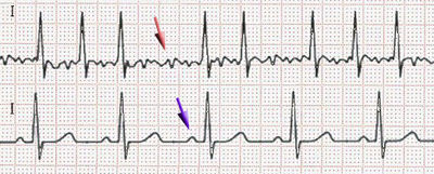
What is the ECG showing (Red Arrow)
Atrial Fibrillation
Ventricular Fibrillation
Asystole
Premature Ventricular Contraction
Heart Block

What is the ECG showing
Atrial Fibrillation
Ventricular Fibrillation
Asystole
Premature Ventricular Contraction
Heart Block

What is the ECG showing
Atrial Fibrillation
Ventricular Fibrillation
normal
Premature Ventricular Contraction
Heart Block
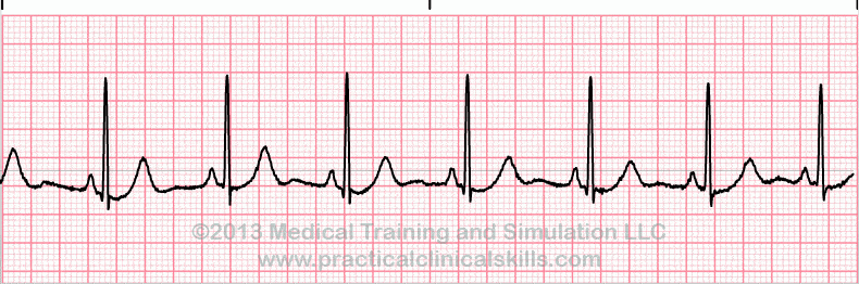
What is the ECG showing
Atrial Fibrillation
Ventricular Fibrillation
normal
Premature Ventricular Contraction
Heart Block
What is the ECG showing
Atrial Fibrillation
Ventricular Fibrillation
normal
Premature Ventricular Contraction
Heart Block

What is the ECG showing (arrow)
Atrial Fibrillation
Ventricular Fibrillation
normal
Premature Ventricular Contraction
Heart Block

Identify the ECG tracing.
Normal Sinus Rhythm
Atrial Fibrillation
Atrial Flutter
Ventricular tachycardia

Identify the ECG tracing.
Ventricular fibrillation
Ventricular tachycardia
Torsades de pointes
Asystole

What is the heart rate in this ECG tracing?
60 bpm
75 bpm
90 bpm
130 bpm

Identify the heart block, if any.
First-degree AV block
Second-degree AV block (type I)
Second-degree AV block (type II)
Third-degree AV block
Normal Sinus Rhythm
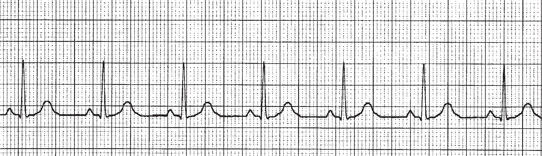
What is the hear rate of this ECG rhytm?
40
70
100
130
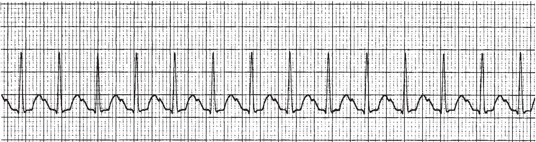
This ECG strip illustrates ____________.
Premature Atrial Contraction
Premature Ventricular Contraction
Atrial Fibrulation
Tachycardia

This ECG wave illustrates ____________.
An MI that occurred years ago
ST segment depression
An acute MI
Normal ECG wave
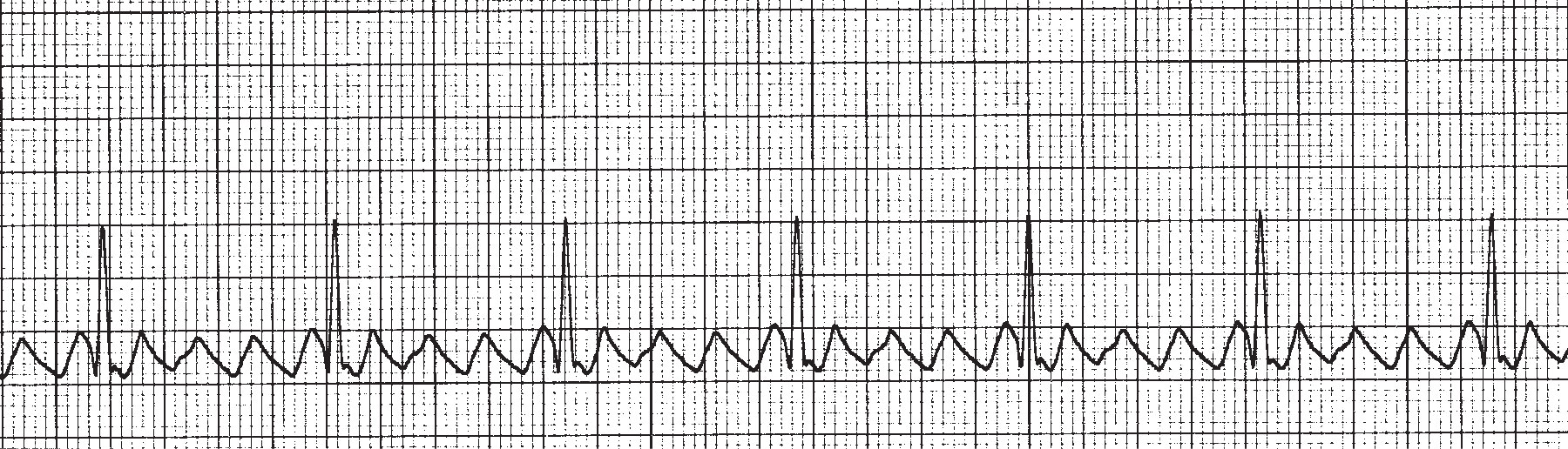
This ECG illustrates _______________.
Premature Atrial Contraction
Atrial Flutter
Atrial Fibrillation
Ventricular Fibrillation
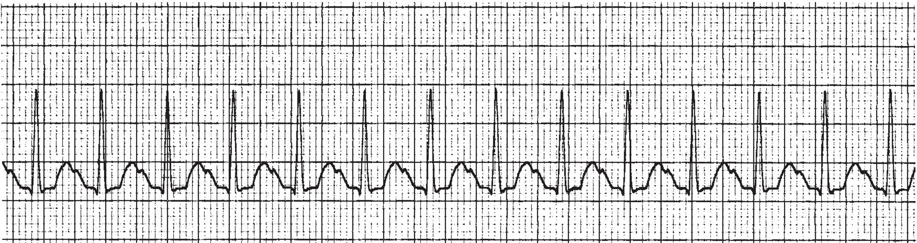
What is the HR of this ECG?
105pbm
135bpm
75bpm
165bpm
What is the ECG showing?
Atrial Fibrillation
Ventricular Fibrillation
normal
Premature Ventricular Contraction
Heart Block
What is the ECG showing?
Atrial Fibrillation
Ventricular Fibrillation
normal
Premature Ventricular Contraction
Heart Block

What is the ECG showing?
Atrial Fibrillation
Ventricular Fibrillation
normal
Premature Ventricular Contraction
Heart Block
What is the ECG showing ?
Atrial Fibrillation
Ventricular Fibrillation
normal
Premature Ventricular Contraction
Heart Block

your correct diagnosis is
Atrila Fibrillation
Ventricular fibriilation
Sinus Tacchycardia
Abnormal ECG

