
NEW
Font size
 Worksheets
MCQS
Total questions: 26
Worksheet time: 13mins
The partial fusion of two ventral aortae is called……………
Aortic sac
Pulmonary sac
Aortic arch
Left horn
Where is Ostium primum formed
Between RV & LV
Between RA & LA
Between RA & Ventricle
Within Coronary sinus
Which of the following arch arteries doesn’t disappear
First arch artery
Second arch artery
Fifth arch artery
Sixth arch artery
Pulmonary trunk develops from……………..arch artery
Third
Fourth
Fifth
Sixth
The descending aorta is derived from ……………………
Right Dorsal aorta and fused dorsal aorta
Left dorsal aorta and fused dorsal aorta
Right horn
Left horn
Connection between aorta and pulmonary artery is known as……………
Patent Ductus arteriosus
AV canal defect
Atrial septal defect
Ventricular septal defect
Following are the branches of aortic arch except
Brachiocephalic artery
Left common carotid artery
Left subclavian artery
Radial artery
The common carotid artery is derived from the………..
Proximal part of 3rd arch artery
Distal part of 3rd arch artery
Proximal part of 4th arch artery
Distal part of 4th arch artery
The communication between common atrial chamber and ventricle is called as
Atrioventricular canal
Septum primum
Septum secundum
Ostium primum
Fossa ovalis represents.........................
Septum primum
Septum secundum
Ostium primum
Ostium secundum
Abnormal communication between interatrial septum is called as ……
ASD
VSD
PDA
TOF
The left atrium is derived from the following three
Components except:
Left half of primitive atrial chambers
Left half of AV canal
Absorbed proximal part of pulmonary vein
Absorption of right half of AV canal
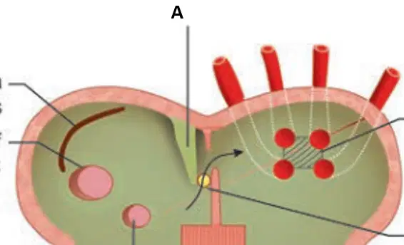
‘A’ represents ……..
Septum primum
Septum secundum
Fossa ovalis
Foramen ovale
Arterial end of the heart tube is…..
Right sinus venosus
Sinus venosus
Bulbus cordis
Left sinus venosus
Persistence of foramen ovale is called as
Patent ductus arteriosus
Patent foramen ovale
Ventricular septal defect
Atrial septal defect
The heart lies exposed, on the front of the chest,
and can be seen from the outside, due to defective development of the chest wall is called as
Mesocardia
Levocardia
Ectopia cordis
Dextrocardia
The septum that divide the truncus arteriosus into pulmonary trunk and ascending aorta is
Muscular part of septum
Bulbar septum
Spiral septum
All of the above
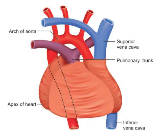
The figure represents……….
Dextrocardia
LEVOCARDIA
MESOCARDIA
ECTOPIA CORDIS
The condition in which the defect is located at the lower half of the IAS
Ostium primum ASD
Ostium secundum ASD
PFO
Sinus venosus type
Which of the following is not a component of TOF?
Pulmonary stenosis
Ventricular septal defect
Right Ventricular Hypertrophy
Co-arctation of aorta
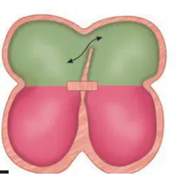
The diagram represents ….
Patent foramen ovale
Septum primum defect
Septum secundum defect
Interventricular septum defect
Truncus arteriosus is an embryological structure that give rise to………………..
Arch of Aorta
Thoracic aorta
descending aorta
Ascending aorta
Right horn of aortic sac forms……………….
Pulmonary vein
Pulmonary artery
Brachiocephalic artery
Common carotid artery
Liver is normally located in………..
Right
Left
Inferior
Superior
The position of right atrium depends on ……
Tricuspid valve
Right ventricle
Aorta
Liver
Tricuspid valve Is apical compared to the mitral valve
True
False

