
NEW
Font size
 Worksheets
Post-test Imaging-HF
Total questions: 15
Worksheet time: 11mins
A 42-year-old woman presents with acute heart failure and is diagnosed with suspected fulminant myocarditis. She undergoes a CMR.
What is the most appropriate timing for performing a CMR to evaluate myocarditis in this patient?
Within the first 24 hours of symptom onset
1-2 weeks after symptom onset
2-3 weeks after symptom onset
4-6 weeks after symptom onset
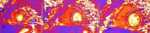
A patient with suspected myocarditis undergoes a CMR which shows an increase in T2 mapping values as shown.
Question: What do these CMR findings indicate in the context of myocarditis?
Presence of myocardial ischemia
Presence of myocardial inflammation and fibrosis
Presence of myocardial injury
Presence of myocardial edema
A 28-year-old male presents with palpitations and chest pain. ECG shows non-specific ST changes, and echocardiography is normal. CMR is performed to investigate myocarditis.
Question: Which CMR sequence is most useful to detect myocardial edema in this patient?
T1 mapping
T2 STIR imaging
Late gadolinium enchancement
Diffusion-weighted imaging
A 60-year-old man with a history of acute myocarditis undergoes CMR to monitor his condition.
Question: Which CMR parameter is most predictive of poor long-term outcomes in myocarditis?
Increased T2 mapping values
Increased T1 mapping values
Presence of late gadolinium enhancement (LGE) in the septal region
Normal extracellular-volume
A 72-year-old male whose planned to undergo coronary interventions presents with dyspnea on exertion. His echocardiogram showed severely reduced LVEF, and a CMR is ordered.
Question: Why would a CMR be indicated in this patient?
To evaluate the severity of coronary artery stenosis
To assess myocardial viability and scar burden
To measure renal artery stenosis
To detect pulmonary embolism
A 58-year-old female with a known ischemic cardiomyopathy is referred for cardiac magnetic resonance (CMR) imaging. The CMR reveals subendocardial late gadolinium enhancement (LGE) and thinned myocardiumin LAD territory.
Question: What does the presence of LGE on CMR indicate in this patient?
Active inflammation
Myocardial fibrosis and scar
Coronary artery stenosis
Increased myocardial perfusion
A 62-year-old male with a history of ischemic cardiomyopathy and persistent symptoms despite optimal medical therapy is evaluated for the possibility of revascularization. CMR is performed to assess myocardial viability.
Question: Which CMR finding would most likely support the decision to proceed with revascularization in this patient?
Extensive areas of transmural LGE
Preserved myocardial thickness with subendocardial infarction <50%
Decreased left ventricular ejection fraction
High extracellular volume (ECV) throughout the myocardium
What is the primary advantage of using MRI in the diagnosis of pericarditis?
MRI is less expensive than other imaging techniques.
MRI provides detailed images of the pericardial layers and can assess inflammation and pericardial effusion.
MRI uses ionizing radiation.
MRI is faster than CT scans.
Which of the following MRI sequences is most useful for detecting inflammation in pericarditis?
T1-weighted imaging
T2-weighted imaging
Late gadolinium enhancement (LGE)
Diffusion-weighted imaging (DWI)
What is a characteristic MRI finding in acute pericarditis?
Thickening of the pericardium without enhancement
Pericardial effusion with diffuse pericardial enhancement
Calcification of the pericardium
Reduced pericardial thickness
Which MRI feature can help differentiate constrictive pericarditis from restrictive cardiomyopathy?
Presence of pericardial thickening
Absence of pericardial effusion
Septal bounce and ventricular interdependence
Decreased left ventricular ejection fraction

The T2-weighted images and LGE in the following figures were obtained from a young man with dyspnea. What is the likely diagnosis?
Cardiac tumor
Pulmonary embolism
Pericarditis
Arrhythmogenic right ventricular dysplasia
Constrictive pericarditis
Which one this following FALSE regarding Dilated Cardiomyopathy (DCM) ?
The prevalence of Dilated Cardiomyopathy in adults was 0.036-0.4% whereas in children the prevalence more lower
According to etiology about half patient had history of premature death (sudden cardiac death, accidental death, still birth or sudden infant death syndrome) and must be further evaluation
Gene linked Dilated Cardiomyopathy of Dystrophin are caused by Autosomal Dominant, meawhile LMNA (Lamin A/C) Dilated Cardiomyopathy are caused by X-linked inheritance
Role of Cardiac Magnetic Resonance Imaging in DCM as risk stratification, detection of inflammation and exclude ischemia
Concomitant Atrioventricular block with dilated cardiomyopathy suggest caused by myocarditis or LMNA
What is the factor that influenced outcome in patient with dilated cardiomyopathy ?
Location and Pattern of LGE
Value of ejection fraction
History of atrial fibrillation
Increased of LA volume index
Transient LV dilatation
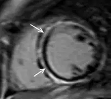
Which one is TRUE regarding this picture ?
The risk of Sudden Cardiac Death (SCD) in this patient was lower
Family history must be explored because related to X-linked inheritance
Ring-like pattern of LGE associated with DSP mutation
Arrythmogenic and restrictive cardiomyopathy rarely found in these case
This patient may possible had extensive myocardial infarction

