
 Worksheets
Lead Morphology
Total questions: 15
Worksheet time: 15mins

What is the name of the elongated, tightly packed cells (part E) directly under the upper epidermis?
Palisade mesophyll
Guard cell
Spongy mesophyll
Lower epidermis
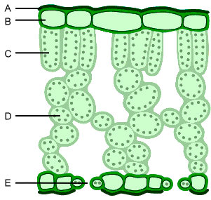
What is letter "D" pointing to?
upper epidermis
palisade mesophyll
spongy mesophyll
bundle sheath
What is the primary function of lead in electrocardiography (ECG)?
To measure blood pressure
To record electrical activity of the heart
To monitor brain waves
To assess lung function
Which of the following best describes the term 'lead morphology' in the context of ECG?
The shape and size of the ECG leads
The arrangement of leads on the patient's body
The pattern of electrical activity recorded by the leads
The material composition of the leads
In an ECG, which lead is typically used to provide a view of the heart's electrical activity from the right arm to the left leg?
Lead I
Lead II
Lead III
aVR
What does a normal P wave morphology indicate in an ECG reading?
Normal atrial depolarization
Ventricular hypertrophy
Myocardial infarction
Atrial fibrillation
Which lead configuration is used to obtain a standard 12-lead ECG?
3 limb leads and 3 chest leads
6 limb leads and 6 chest leads
4 limb leads and 8 chest leads
5 limb leads and 7 chest leads
What is the significance of the QRS complex morphology in an ECG?
It indicates atrial repolarization
It represents ventricular depolarization
It shows the heart rate
It measures blood oxygen levels
Which of the following changes in lead morphology might indicate a myocardial infarction?
Inverted T waves
Prolonged PR interval
Shortened QT interval
Increased P wave amplitude
How does the morphology of the T wave in an ECG help in diagnosing heart conditions?
It indicates the heart's electrical axis
It shows ventricular repolarization
It measures atrial contraction
It assesses blood flow
What is the role of augmented leads in an ECG?
To provide additional views of the heart's electrical activity
To measure the heart's mechanical function
To assess the heart's blood supply
To monitor respiratory rate
Which lead morphology is most commonly associated with left ventricular hypertrophy?
Tall R waves in V1
Deep S waves in V6
Tall R waves in V5
Inverted T waves in aVL
In an ECG, what does the morphology of the ST segment indicate?
Atrial depolarization
Ventricular repolarization
Ventricular depolarization
Atrial repolarization
What is the importance of lead placement in determining ECG morphology?
It affects the amplitude of the waves
It changes the heart rate
It alters the blood pressure readings
It modifies the respiratory rate
Which of the following best describes the morphology of a normal sinus rhythm in an ECG?
Irregular P waves and QRS complexes
Regular P waves followed by QRS complexes
Absent P waves and irregular QRS complexes
Inverted T waves and prolonged QT interval

