
 Worksheets
CMA EKG Quiz
Total questions: 29
Worksheet time: 58mins
What is the purpose of the p wave in an EKG?
To indicate ventricular depolarization
To indicate atrial depolarization
To indicate atrial repolarization
To indicate ventricular repolarization
Where is the SA node located?
Right atrium near the entry of the superior vena cava
Left atrium
Right ventricle
Top of the interventricular septum
A patient is experiencing electrocardiogram changes. Which wave should the nurse recognize as representing ventricular repolarization?
Q wave
R wave
S wave
T wave
A patient is admitted for diagnosis of an electrocardiogram abnormality. Which wave should the nurse plan care based upon changes in the patient’s ventricular depolarization?
P wave
QRS complex
U wave
T wave
The nurse is determining the functioning of a patient’s sinoatrial node. How many times per minute should the nurse expect the node to normally fire?
20 to 40
25 to 55
40 to 85
60 to 100

Where is the P wave located?
A
B
C
D
E

What is considered the QRS complex?
A, B, & C
C, D, & E
B, C, & D
D & E

What is the T wave located?
A
B
C
D
E
What does depolarization result in?
relaxation
contraction
repulsion
attraction
What does re-polarization result in?
relaxation
contraction
attraction
repulsion
Blood enters the heart from the _______?
Superior and inferior vena cava
Bicuspid and Tricuspid
aorta
pulmonary artery
After the blood returns from the lungs, it is now____?
deoxygenated
oxygenated
purple
deoxygenated blood coming from____?
body to lungs
head to body
lungs to body
lungs to head
The SA Node is known as?
Pacemaker
gatekeeper
ventricle
cardiac muscle
how many chambers of the heart are there
3
4
5
6
IF a doctor wants information regarding a patients heart over an extended period of time he might use a:
Holter monitor
12-lead EKG
Nothing allows him to do this
Echocardiogram
Why do we use 12 leads for an EKG?
So we can see fewer views of the hearts electrical activity
So we can see more views of the hearts mechanical activity
To provide the MD with different views of the heart's electrical activity
To provide the MD with different views of the heart's mechanical function
Where would you place the electrode for V3?
5th intercostal space left midclavicular line
Between electrodes V2 and V4
4th intercostal space just to the right of the sternum
Horizontally even with V4 and V5 in the left midaxillary line
In which location would you place the electrode for V1?
Horizontally even with V4 and V5 in the left midaxillary line
5th intercostal space left midclavicular line
Right leg
4th intercostal space just to the right of the sternum
What is the paper speed most commonly used during an EKG?
5mm/mV
10mm/mV
25mm/sec
50mm/sec
Which color is the V1 lead?
White
Green
Purple
Brown

Which of the following best describes this strip?
Tachycardia
Normal Sinus Rhythm
Bradycardia
Asystole

Which of the following best describes this strip?
Tachycardia
Normal Sinus Rhythm
Bradycardia
Asystole

Which of the following best describes this strip?
Tachycardia
Normal Sinus Rhythm
Bradycardia
Asystole

What is the ECG showing
Atrial Fibrillation
Ventricular Fibrillation
Asystole
Premature Ventricular Contraction
Heart Block

Identify the ECG tracing.
Normal Sinus Rhythm
Atrial Fibrillation
Atrial Flutter
Ventricular tachycardia
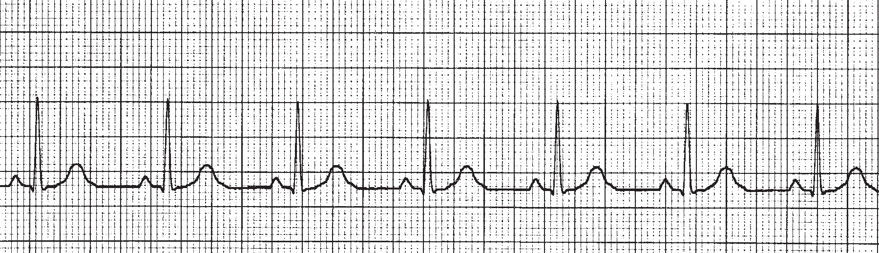
What is the hear rate of this ECG rhytm?
40
70
100
130
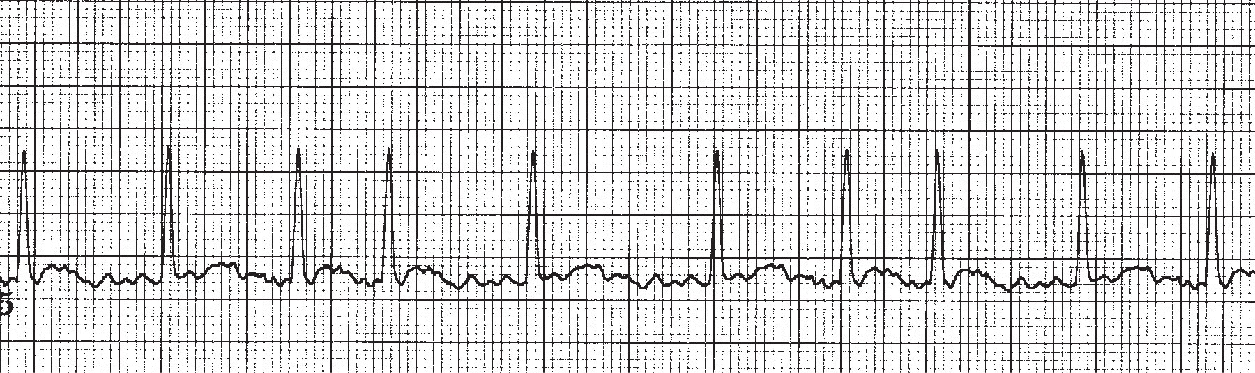
This ECG illustrates ____________________.
Atrial Flutter
Bradycardia
Atrial Fibrillation
Myocardial Infarct
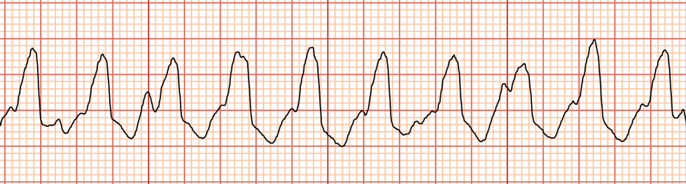
Identify the tracing
Atrial Fibrillation
Sinus Tachycardia
Ventricular tachycardia
Supraventricular tachycardia

