
 Worksheets
LP1- ch.19: Heart
Total questions: 181
Worksheet time: 2hrs 24mins
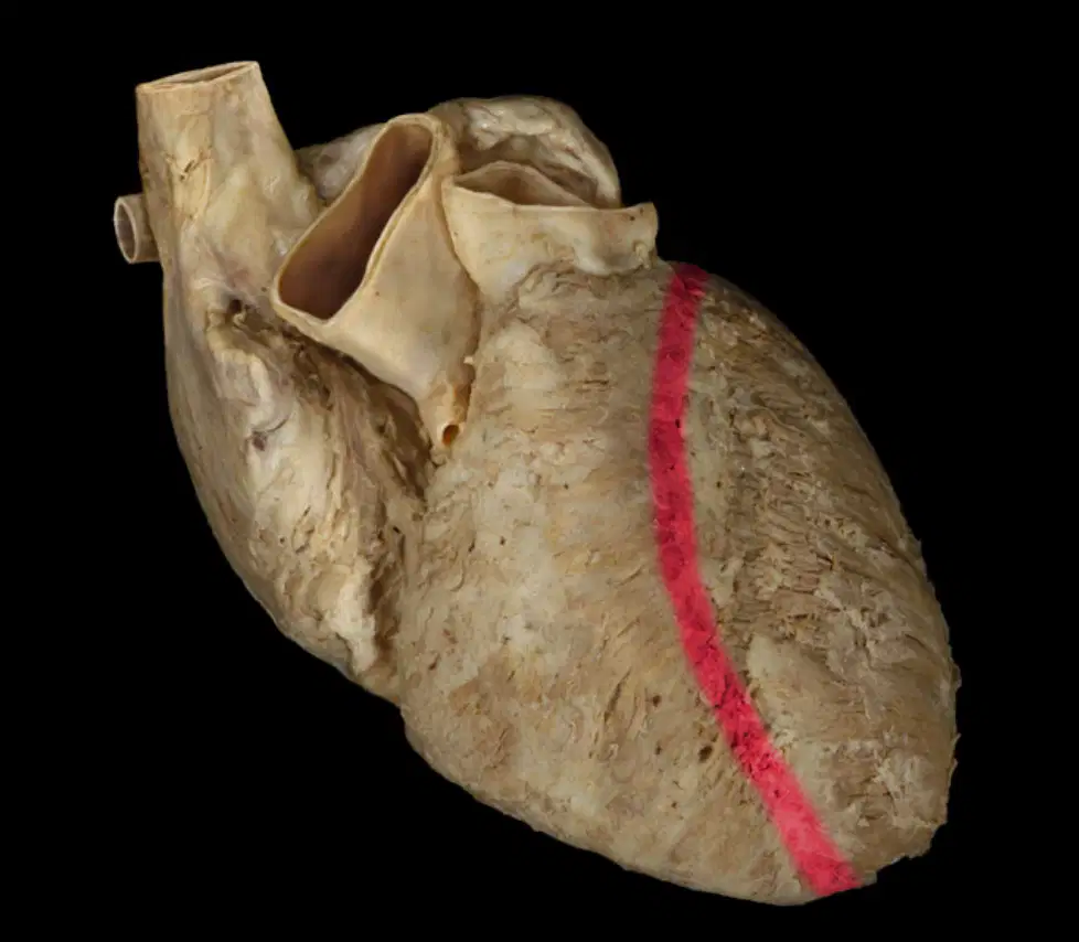
What is the name of the following structure?
(a)
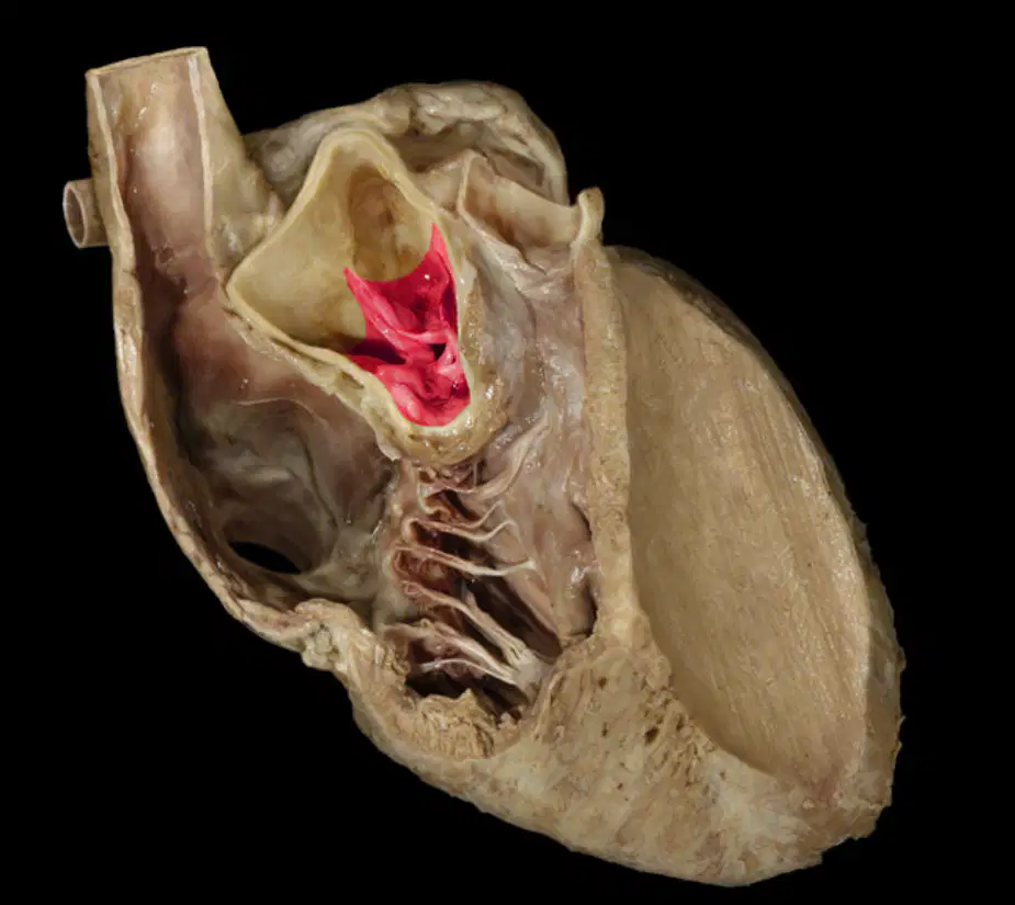
What is the name of the following structure?
(a)
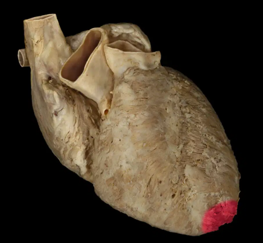
What is the name of the following structure?
(a)
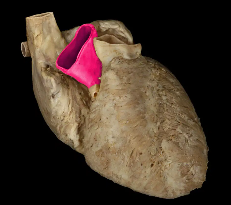
What is the name of the following structure?
(a)
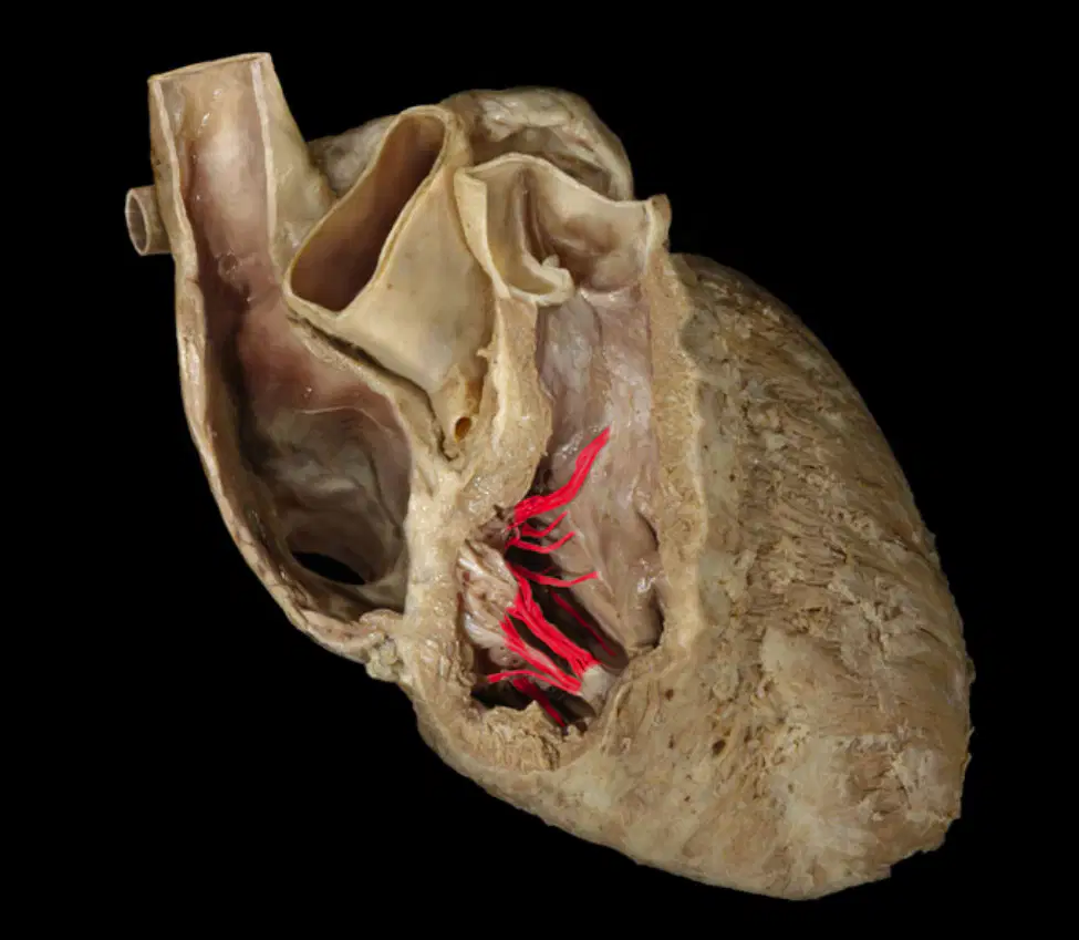
What is the name of the following structure?
(a)
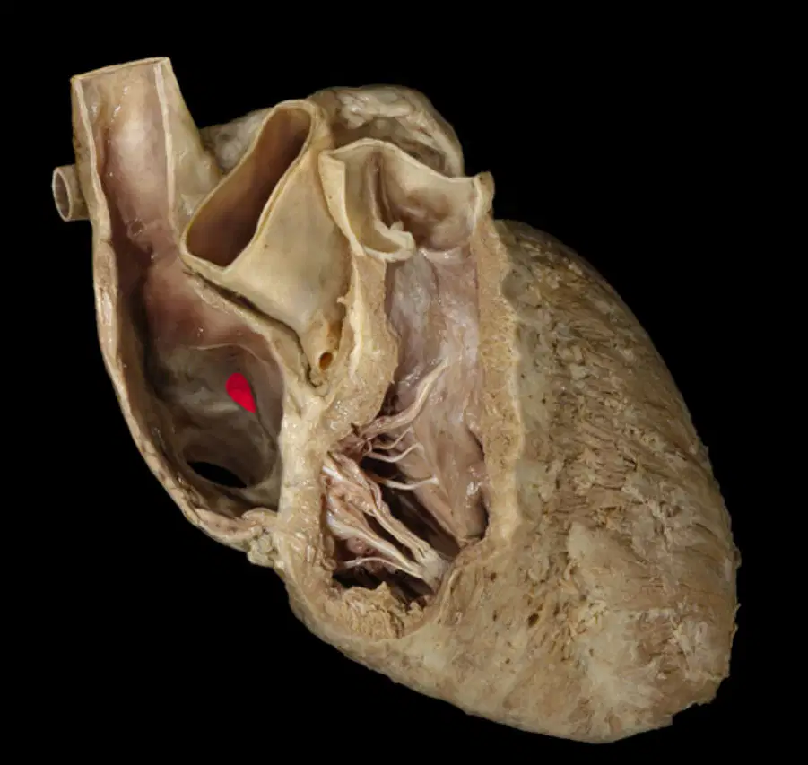
What is the name of the following structure?
(a)
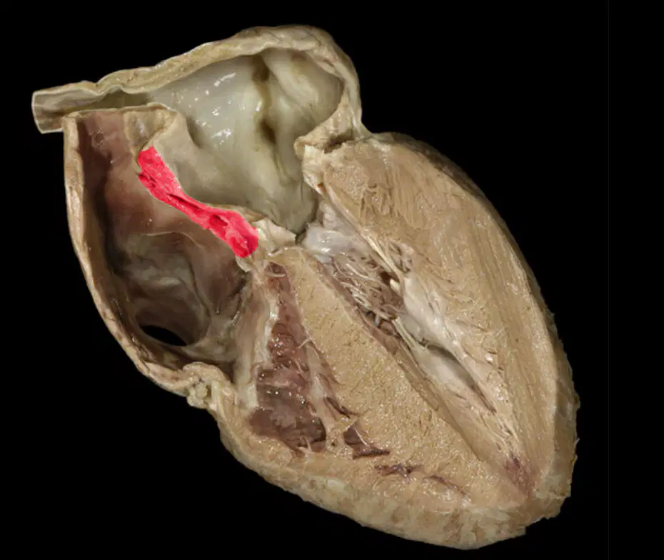
What is the name of the following structure?
(a)
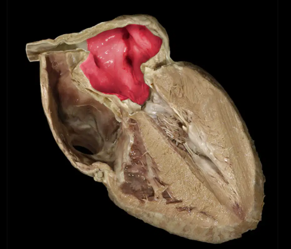
What is the name of the following structure?
(a)
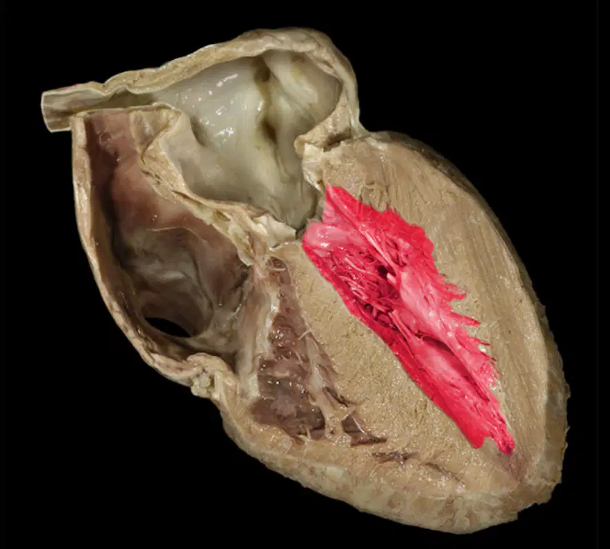
What is the name of the following structure?
(a)

What is the name of the following structure?
(a)
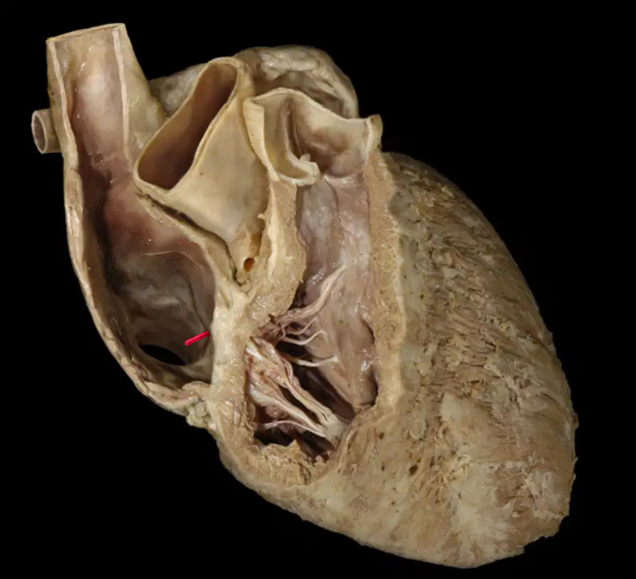
What is the name of the following structure?
(a)
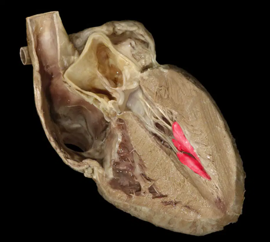
What is the name of the following structure?
(a)
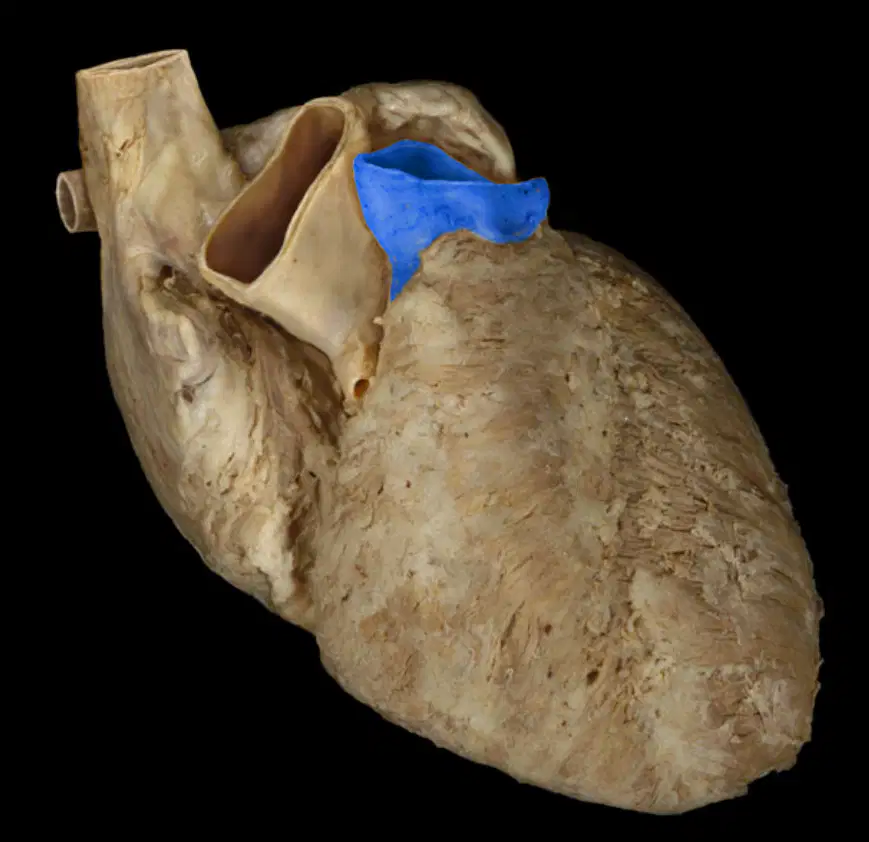
What is the name of the following structure?
(a)

What is the name of the following structure?
(a)
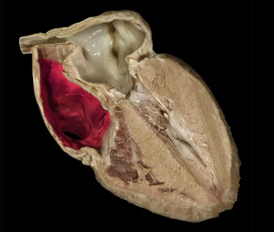
What is the name of the following structure?
(a)
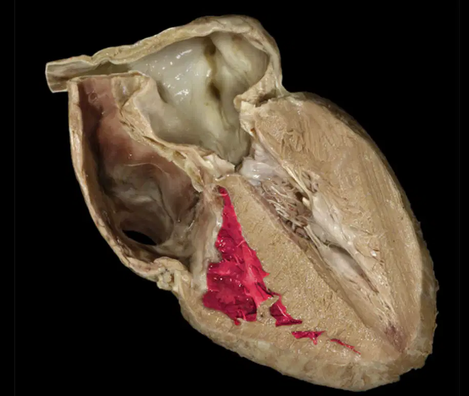
What is the name of the following structure?
(a)
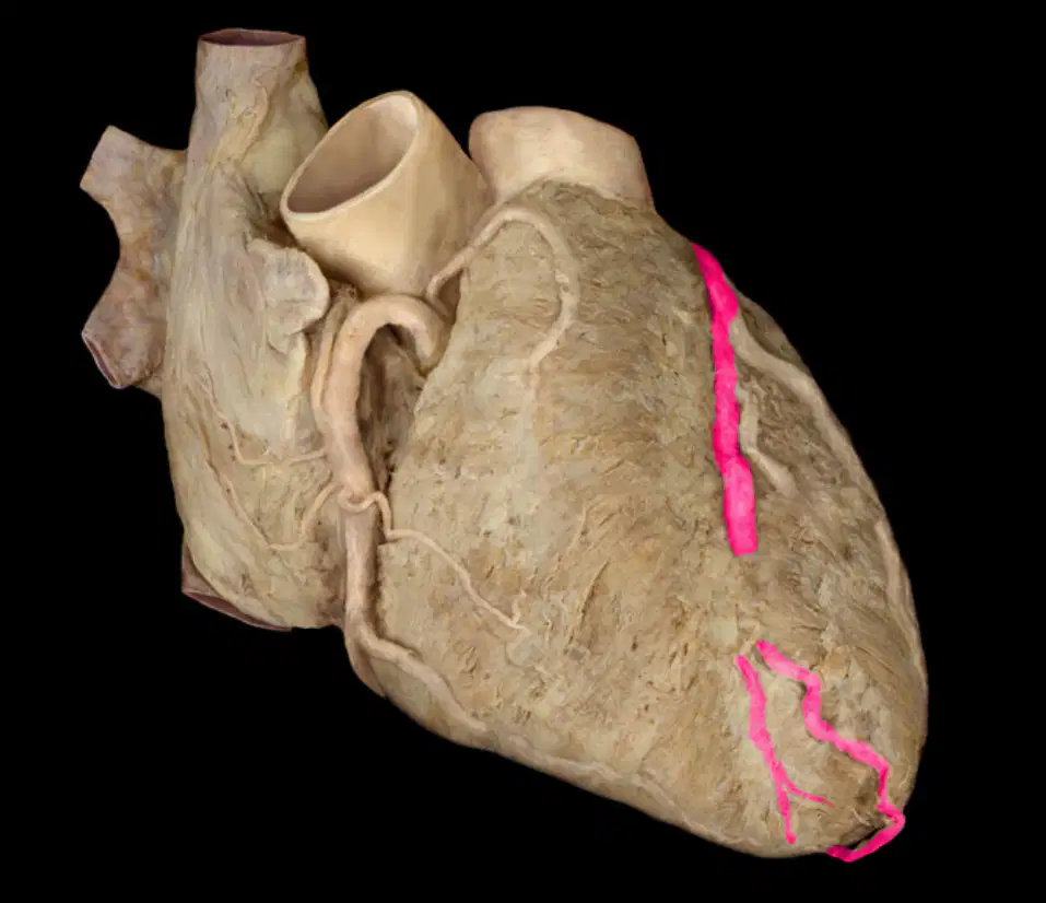
What is the name of the following structure?
(a)
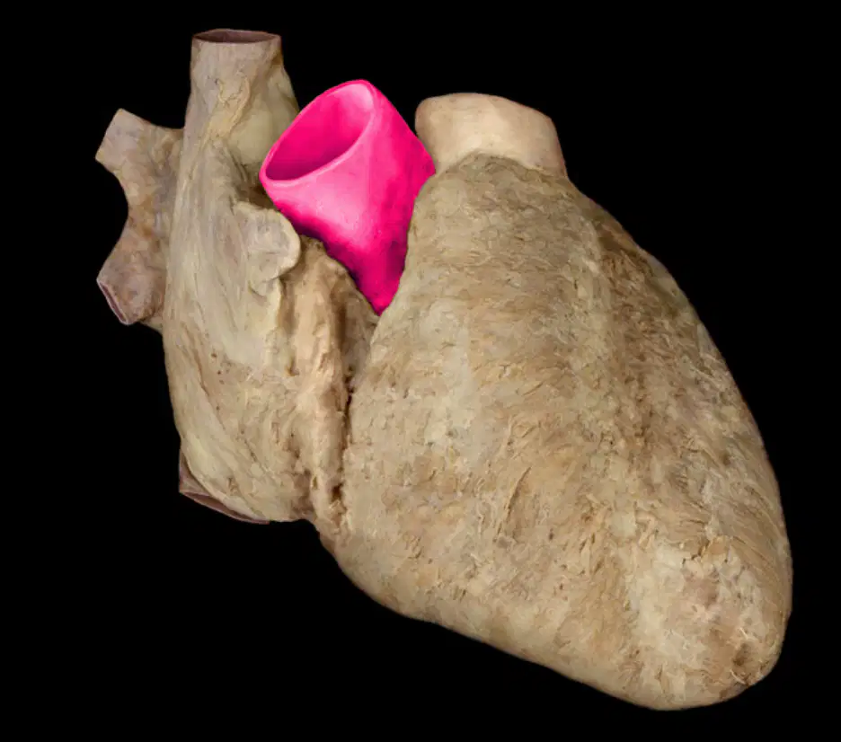
What is the name of the following structure?
(a)
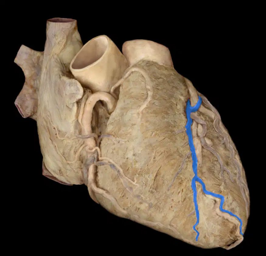
What is the name of the following structure?
(a)

What is the name of the following structure?
(a)
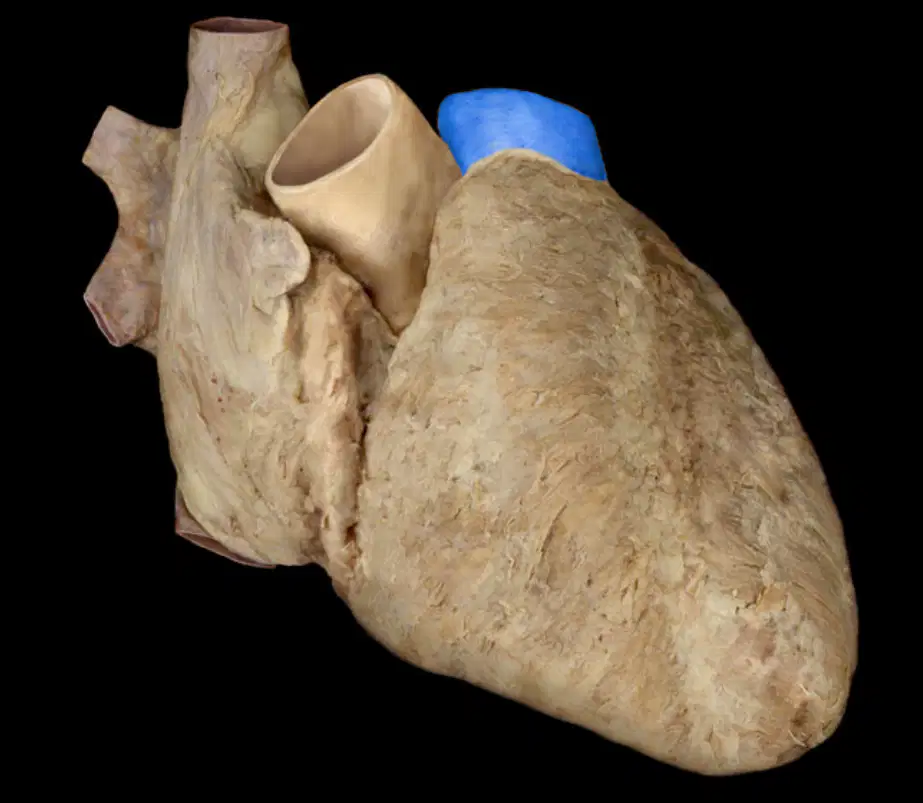
What is the name of the following structure?
(a)
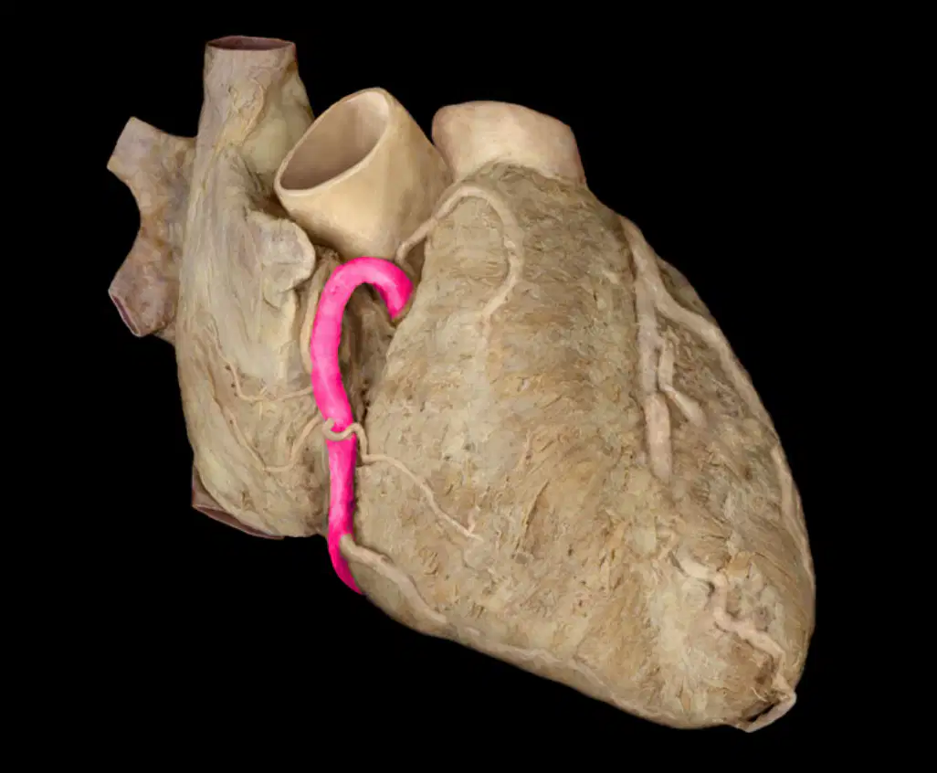
What is the name of the following structure?
(a)

What is the name of the following structure?
(a)
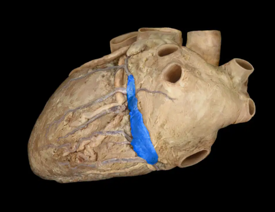
What is the name of the following structure?
(a)


What is the most muscular chamber of the heart?
left ventricle
left atrium
right atrium
right ventricle


Valve that blood is forced through from right ventricle into pulmonary arteries.
pulmonary semi lunar valve
aortic valve
mitral valve
In the walls of the heart, there are two thin layers around a thick layer of muscle called the
epithelial tissue.
pericardium.
connective tissue.
myocardium.


The heart layer that keeps blood from sticking to the inside of the heart made up of squamous epithelial cells
endocardium
pericardium
myocardium
epicardium
Pericardium is a serous membrane it has two layers. The parietal layers of the membranes line the walls of the body cavity. The inner visceral layer covers organ (viscera).
True
False
Aorta
The largest arterial trunk that carries blood from the heart to be distributed by branch arteries through the body
a vessel that returns blood to the heart
a body structure that is temporarily closes a passage or orifice, or permits movement of fluid in only one direction.
An integrated group of cells with a common function, structure, or both
Artery
A valve in the heart that guards the opening between the left atrium and the left ventricle; prevents the blood in the ventricle from returning to the atrium. Alternative name is bicuspid valve.
A vein that is the largest vein in the human body and returns blood to the right atrium of the heart from bodily parts below the diaphragm.
The body structure that temporarily closes a passage or orifice, or permits movement of fluid in only one direction.
Any of the tubular, branching, muscular, and elastic-walled vessels that carry blood from the heart through the body.
Mitral Valve
The semilunar valve separating the aorta from the left ventricle that prevents blood from flowing back into the left ventricle.
A vein that is the largest vein in the human body and returns blood to the right atrium of the heart from bodily parts below the diaphragm.
An anatomical cavity or passage especially a chamber of the heart that receives blood from the veins and forces it into a ventricle or ventricles
A valve in the heart that guards the opening between the left atrium and the left ventricle; prevents the blood in the ventricle from returning to the atrium. Alternative name is bicuspid valve.


What is the function of structure labelled 5?
carry deoxygenated blood to the lungs
carry oxygenated blood to all parts of the body
carry deoxygenated blood from all parts of the body to the heart
carry oxygenated blood from the lungs
Which structure of the heart prevents the mixing of oxygen-riched blood and oxygen-poor blood?
Aorta
Atrium
Valve
Septum


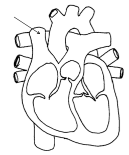


The largest artery in the body. Carries oxygenated blood from the left ventricle to the rest of the body.
aorta
apex
pulmonary artery
vena cava
The blood vessels that supply oxygen to the heart's tissues.
cornary artery
apex
pulmonary artery
vena cava
Chamber that receives oxygenated blood from the lungs.
left atrium
right atrium
left ventricle
right ventricle
Chamber that receives deoxygenated blood from the vena cava.
left atrium
right atrium
left ventricle
right ventricle
The chamber that receives deoxygenated blood from the right atrium and pumps it to the lungs.
left atrium
right atrium
left ventricle
right ventricle
The thickest chamber and is responsible for pumping oxygenated blood to tissues all over the body.
left atrium
right atrium
left ventricle
right ventricle
The blood vessels that carry oxygenated blood from the lungs to the left atrium.
pulmonary vein
pulmonary artery
aorta
vena cava
Separates the right atrium from the right ventricle.
tricuspid valve
bicuspid valve
aortic valve
mitral valve
The second largest vein in the human body. Returns blood to the right atrium from the upper half of the body.
superior vena cava
right ventricle
chordae tendinae
apex

What is this? (lower case)
(a)

interventricular septum
vena cava
pulmonary artery
apex
coronary artery
What is the purpose of a valve?
prevents the retroprofusion of blood
provides the heart tissue with oxygen
provides electrical stimulation
oxygenates the blood
The pumping of blood from the body to the lungs and back to the heart for the purpose of oxgenating the blood is called (a) circulation.
(a) circulation pumps oxygen-rich blood to the body and returns deoxygenated blood back to the heart.
In most of the body, the arteries carry oxygenated blood and the veins carry deoxygenated blood. Which of the following are exceptions
pulmonary artery
pulmonary vein
aorta
vena cava
renal artery
P Wave
SA Nodes contracts
Both atria contracts
Impulses reach the av node 0.10 delay allows
Repolarization or recovery of the ventricles
Impulses hit av bundle branch off into two bundles down front of heart into purkinje fibers
QRS
Repolarization or recovery of the ventricles
Impulses hit av bundle branch off into two bundles down front of heart into purkinje fibers
SA Nodes contracts
Both atria contracts
Impulses reach the av node 0.10 delay allows atria


The nurse is reviewing a patient’s electrocardiogram. Which wave should the nurse recognize as being the first positive wave in a normal cardiac cycle?
P wave
Q wave
R wave
S wave
A patient is experiencing electrocardiogram changes. Which wave should the nurse recognize as representing ventricular repolarization?
Q wave
R wave
S wave
T wave
A patient is admitted for diagnosis of an electrocardiogram abnormality. Which wave should the nurse plan care based upon changes in the patient’s ventricular depolarization?
P wave
QRS complex
U wave
T wave

(a)

(a)

(a)

(a)

(a)
The correct sequence of the three events in ECG
P wave , T wave , QRS complex
QRS complex , P wave , T wave
T wave , QRS complex , P wave
P wave, QRS complex, T wave
P wave is
Repolarization of atria
Repolarization of ventricles
Depolarization of atria
Depolarization of ventricles
T wave is
Repolarization of atria
Repolarization of ventricles
Repolarization of ventricles and atria
Depolarization of atria and ventricles
QRS complex is
Repolarization of atria
Repolarization of ventricles
Depolarization of atria
Depolarization of ventricles

Which one is represent P wave
A
B
C
E
Which one is represent T wave
A
B
C
E
Which one is represent QRS complex
A
B
C
E
The ECG is the most commonly used clinical tool for diagnosing what?
Cardiac dysfunction
Cardiac Failure
Congestive Heart Failure
COPD

Name the EKG
Normal Sinus Rhythm
Sinus Tachycardia
Sinus Bradycardia
Atrial fibrillation

Name the EKG
Normal Sinus Rhythm
Sinus Tachycardia
Sinus Bradycardia
Atrial fibrillation

Name the EKG
Atrial fibrillation
Atrial flutter
Super ventricular tachycardia
Asystole
Everything is normal, rate is 60 -100
Sinus tachycardia
Sinus bradycardia
Sinus arrhythmia
Normal sinus rhythm
Atria are irritable and quivering, causing fibrillatory waves between QRS complex
asystole
atrial flutter
A fib
V fib
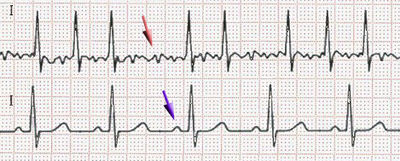
What is the ECG showing (Red Arrow)
Atrial Fibrillation
Ventricular Fibrillation
Asystole
Premature Ventricular Contraction
Heart Block

What is the ECG showing
Atrial Fibrillation
Ventricular Fibrillation
normal
Premature Ventricular Contraction
Heart Block
Heart rate over 100 beats per minutes is
tachycardia
bradycardia
depolarization
repolarization
Which of these is not considered when making a diagnosis?
P-R interval
rate
rhythm
T wave
MI stands for
myocardial infarction
muscle infarction
mycocordial infarction
muscle infraction
If our patient's heart rate is 75bpm, rhythm is regular, P-R interval is normal, QRS is normal, then our diagnosis is
Sinus tachycardia
Sinus dysrhythmia
normal sinus rhythm
Sinus arrest

Interpret this Rhythm:
Normal sinus rhythm
Ventricular fibrillation
Sinus Brady with PAC's
Sinus Bradycardia

Identify the cardiac rhythm strip pictured above
Ventricular Tachycardia
Normal Sinus Rhythm
Atrial Fibrillation
Sinus Tachycardia

Identify the cardiac rhythm strip pictured above
Atrial Flutter
Normal Sinus Rhythm
Atrial Fibrillation
Sinus Tachycardia

Which of the following best describes this strip?
Tachycardia
Normal Sinus Rhythm
Bradycardia
Asystole
Name the EKG
Normal Sinus Rhythm
Sinus Tachycardia
Sinus Bradycardia
Atrial fibrillation

Which best describes this rhythm?
Atrial Fibrillation
Atrial Flutter
Asystole
PEA

Which best describes this rhythm?
Asystole
PEA
Pulseless Ventricular Tachycardia
Ventricular Fibrillation

Identify the cardiac rhythm seen above
Ventricular tachycardia
Sinus bradycardia
Atrial Fibrillation
Atrial Flutter
What does the term 'ST segment' indicate in an ECG interpretation?
It indicates the blood pressure of the patient
It indicates the oxygen level in the blood
It indicates the period between ventricular depolarization and repolarization.
It indicates the heart rate during ECG
Explain the significance of the 'P wave' in an ECG interpretation.
It has no significance in ECG interpretation.
It represents ventricular depolarization.
It represents atrial depolarization.
It indicates atrial repolarization.
What is the medical term for an irregular heartbeat?
arrhythmia
hypertension
tachycardia
dyspnea
Explain the difference between 'bradycardia' and 'tachycardia' in heart rhythm terminology.
Bradycardia is a slower than normal heart rate, while tachycardia is a faster than normal heart rate.
Bradycardia is a condition of irregular heart rhythm, while tachycardia is a normal heart rhythm.
Bradycardia and tachycardia are the same thing.
Bradycardia is a faster than normal heart rate, while tachycardia is a slower than normal heart rate.
What is the function of 'beta-blockers' in cardiac medication terminology?
They help to increase blood pressure and heart rate
They have no effect on blood pressure or heart rate
They are used to treat respiratory conditions
They help to lower blood pressure, slow down the heart rate, and reduce the workload on the heart.
What does this ECG show?
Normal sinus rhythm
frequent atrial ectopics

What is the rate and rhythm?
Ventricular tachycardia
140-200
Sinus tachycardia
100-150
What is the normal heart rate range for a sinus rhythm in adults?
40-60 beats per minute
60-100 beats per minute
100-120 beats per minute
120-140 beats per minute
Which of the following is a characteristic feature of atrial fibrillation on an ECG?
Regular P waves
Absence of P waves
Inverted P waves
Tall, peaked P waves
In a junctional rhythm, where does the electrical impulse originate?
Sinoatrial node
Atrioventricular node
Purkinje fibers
Bundle of His
What is the primary difference between sinus tachycardia and sinus bradycardia?
The presence of P waves
The rate of the heartbeats
The shape of the QRS complex
The length of the PR interval
How can you differentiate between ventricular tachycardia and ventricular fibrillation on an ECG?
Ventricular tachycardia has a regular rhythm, while ventricular fibrillation does not
Ventricular fibrillation has a regular rhythm, while ventricular tachycardia does not
Both have regular rhythms
Both have irregular rhythms
Which of the following arrhythmias is most likely to cause a sudden drop in blood pressure?
Sinus arrhythmia
Atrial flutter
Ventricular fibrillation
Junctional rhythm
What is the typical treatment for a patient with atrial fibrillation to prevent stroke?
Beta-blockers
Anticoagulants
Calcium channel blockers
Diuretics
What is the main difference in the ECG appearance of a junctional rhythm compared to a normal sinus rhythm?
Absence of QRS complexes
Inverted or absent P waves
Prolonged QT interval
Elevated ST segment
If the P wave on an EKG looks abnormal, which structures in the heart are likely damaged?
SA/AV node
Aortic valve
Bundle of His
Purkinje fibers
The p-wave correlates with which event?
atrial depolarization
ventricle depolarization
ventricle repolarization
atrial repolarization
What does depolarization result in?
relaxation
contraction
repulsion
attraction

Intrinsic electrical impulses start at which of the following locations?
AV Node
Bundle of His
Purkinje fibers
SA Node
The pacemaker of the heart
AV node
SA node
Bundle of His
Bundle branches
A cardiac cycle is:
ventricular relaxation
ventricular contraction
equivalent to 1 complete heart beat
a heart attack
What is the second step to heart contraction?
Atria contract
ventricles contract
depolarization of SA node
Impulse spreads throughout the atria
A heart rate 100 bpm or above is called _______.
bradycardia
tachycardia
arrhythmia
fibrillation
The heart layer that keeps blood from sticking to the inside of the heart and forming potentially deadly blood clots is called
endocardium
pericardium
myocardium
epicardium
The SA node fires at a rate of
40-60 bpm
80-90 bpm
60-80 bpm
60-100 bpm
What does the first heart sound (Lubb) correspond with?
Ventricular diastole
Ventricular systole
AV Valve closure
Pulmonary Valve closure
What does the second heart sound (dubb/dupp) correspond with?
Ventricular diastole
Ventricular systole
AV valve closure
Semi-lunar valve closure
Which valve's closure is responsible for the "lub" sound?
Aortic and Pulmonary valves
Tricuspid and Mitral valves
Aortic and Mitral valves
Tricuspid and Pulmonary valves
Which valve's closure is responsible for the "dub" sound?
Aortic and Pulmonary valves
Tricuspid and Mitral valves
Aortic and Mitral valves
Tricuspid and Pulmonary valves
Which heart sound marks the beginning of ventricular systole?
S1 ("lub")
S2 ("dub")
S3
S4
Which heart sound marks the end of ventricular systole?
S1 ("lub")
S2 ("dub")
S3
S4
Which heart sound is usually louder and longer?
S1 ("lub")
S2 ("dub")
S3
S4
What causes the "dub" sound in the heart?
Opening of the atrioventricular valves
Closing of the atrioventricular valves
Opening of the semilunar valves
Closing of the semilunar valves
What causes the "lub" sound in the heart?
Opening of the atrioventricular valves
Closing of the atrioventricular valves
Opening of the semilunar valves
Closing of the semilunar valves
What instrument is used to listen to heart sounds?
Thermometer
Sphygmomanometer
Stethoscope
Electrocardiogram (ECG)
What is the primary purpose of listening to heart sounds?
To measure blood pressure
To diagnose lung diseases
To assess heart function and detect abnormalities
To measure body temperature
Which heart condition might cause a "murmur" sound?
Heart attack
Valve abnormalities
High cholesterol
Diabetes