
 Worksheets
Intro the Foot
Total questions: 38
Worksheet time: 26mins
How many tarsal bones are there in the foot?
5
6
7
8
Which of the following is not a type of bone in the foot?
Tarsal
Metatarsal
Phalanges
Femur
Which bone is located above the Calcaneus?
Talus
Navicular
Cuboid
1st Cuneiform
What are the bones called that are located between the Tarsal bones and the Phalanges?
Metatarsals
Carpals
Metacarpals
Tarsals
Identify the type of arch labeled as 'A' in the diagram.
A. Anterior Metatarsal & Transverse Arches
B. Medial Longitudinal Arch
C. Lateral Longitudinal Arch
What is the plantar fascia?
Where does the plantar fascia originate and end?
It originates from the calcaneus and ends at the metatarsal heads.
It originates from the tibia and ends at the fibula.
It originates from the femur and ends at the patella.
It originates from the humerus and ends at the ulna.
What is the function of the plantar fascia?
The function of the Tibialis Posterior muscle in the foot is:
Plantarflexion and inversion of the foot
Dorsiflexion of the foot
Eversion of the foot
Flexion of the toes
What is the function of the 'Flexor hallucis longus'?
Flexes the big toe
Extends the big toe
Abducts the big toe
Adducts the big toe
The function of the 'Tibialis anterior' is:
Dorsiflexion of the foot
Plantarflexion of the foot
Inversion of the foot
Eversion of the foot
Which artery gives off a number of branches to supply the dorsum of the foot?
Anterior Tibial Artery
Posterior Tibial Artery
Sural Artery
Deep Fibular Artery
Which artery provides the branches for the plantar portion of the foot?
Anterior Tibial Artery
Posterior Tibial Artery
Sural Artery
Deep Fibular Artery
Fill in the blank: The five major nerves that provide innervation to the foot are the Tibial nerve, sural nerve, deep fibular nerve, superficial fibular nerve, and the ________ nerve.
saphenous
ulnar
radial
median
The functional anatomy of the foot includes which of the following movements?
Flexion and extension
Abduction and adduction
Inversion and eversion
All of the above
The foot serves as a shock absorber during stance by:
distributing weight evenly across the foot
increasing the speed of walking
reducing the need for footwear
enhancing the flexibility of the toes
Improper footwear and poor hygiene are associated with which foot problems?
Blisters and calluses
Headaches and dizziness
Back pain and neck strain
Vision problems
What type of surfaces may overstress joints and soft tissue?
Are the majority of skin conditions like blisters, corn, calluses, and ingrown toenails preventable?
Yes
No
The foot is a highly vulnerable area to a variety of injuries because:
it is frequently used and bears the body's weight.
it is protected by strong muscles and bones.
it is rarely exposed to external forces.
it has a limited range of motion.
What are some methods to prevent foot injuries?
Selecting appropriate footwear
Correcting biomechanical structural deficiencies
Paying attention to hygiene
All of the above
The sesamoid bones in the foot are found within which tendon?
Achilles tendon
Flexor hallucis longus tendon
Tibialis posterior tendon
Flexor digitorum brevis tendon
Which joint in the foot is primarily responsible for the movement of toe flexion?
Metatarsophalangeal (MTP) joints
Interphalangeal (IP) joints
Tarsometatarsal (TMT) joints
Subtalar joint

Identify the muscle
peroneus longus
tibialis anterior
rectus femoris
vastus muscles
What is the primary action of the flexor digitorum longus muscle?
Dorsiflexion of the foot
Plantar flexion and flexion of the toes
Eversion of the foot
Inversion of the foot
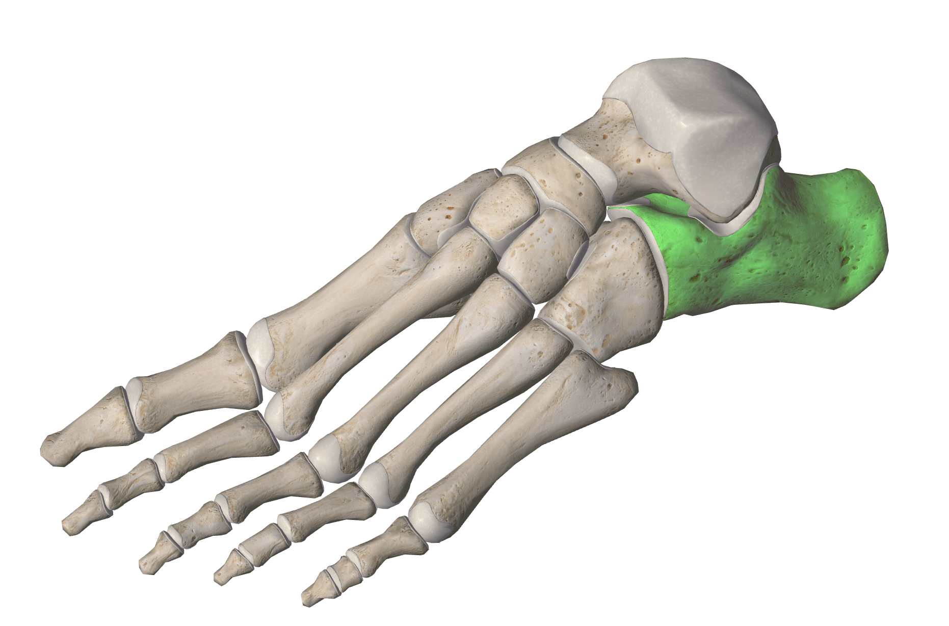


tarsals
phalanges
tibula
metatarsals

Anterior Talofibular Ligament

Calcaneofibular Ligament

Posterior Talofibular Ligament

__________ tendon sits on the posterior of the lower leg and ankle
Soleus
Achilles
Gastrocnemius
Tibialis Anterior

Which ligament is highlighted in red
posterior talofibular ligament
anterior talofibular ligament
calcaneofibular ligament
deltoid

What is the name of this ankle ligament, pointed at by the Red arrow
Anterior Talofibular Ligament
Anterior Tibiofibular Ligament
Calcaneofibular Ligament
Posterior Talofibular Ligament
Deltoid Ligament

What is the name of this ankle ligament, pointed at by the Red arrow
Anterior Talofibular Ligament
Anterior Tibiofibular Ligament
Calcaneofibular Ligament
Posterior Talofibular Ligament
Deltoid Ligament

What is the name of this ankle ligament, pointed at by the red arrow
Anterior Talofibular Ligament
Anterior Tibiofibular Ligament
Calcaneofibular Ligament
Posterior Talofibular Ligament
Deltoid Ligament

