
NEW
Font size
 Worksheets
Heme Cell Morphology Practice #2
Total questions: 20
Worksheet time: 10mins
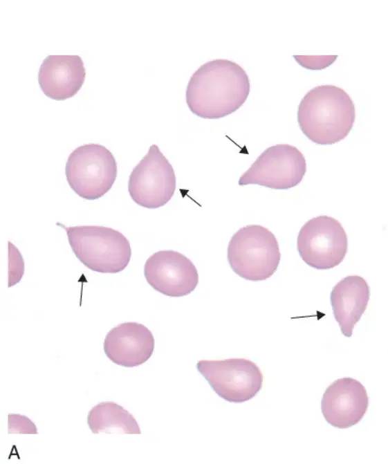
What are dacrocytes also known as?
Round cells
Teardrop cells
Square cells
Oval cells
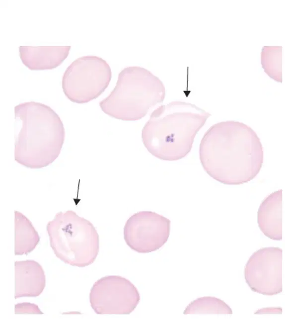
What does the Wright-Giemsa stain in the image show?
Pappenheimer bodies
Howell-Jolly bodies
Heinz bodies
Basophilic stippling

What type of cell is shown in the image?
Neutrophil
Monocyte
Eosinophil
Basophil
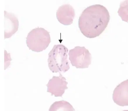
What is the structure indicated by the arrow in the diagram?
Howell-Jolly body
Cabot ring
Heinz body
Siderotic granules
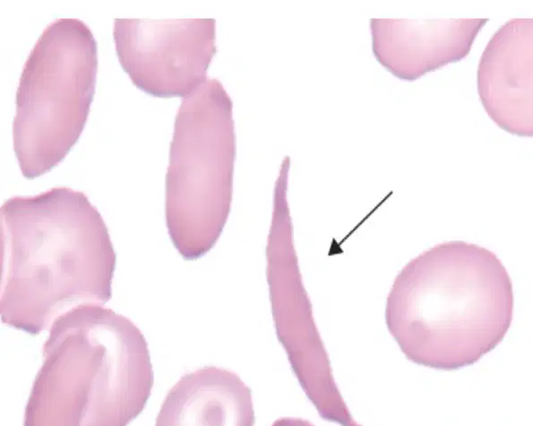
What type of cell is shown in the diagram?
Red blood cell
White blood cell
Sickle cell
Platelet
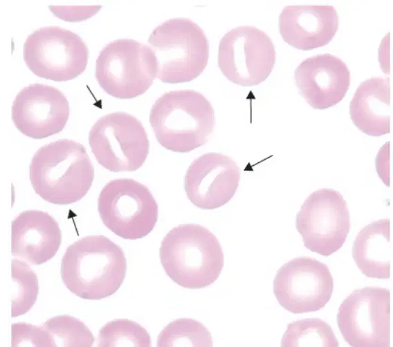
What type of cells are shown in the image?
Erythrocytes
Stomatocytes
Leukocytes
Thrombocytes

What type of stain is shown in the image?
A) Gram stain
B) Sudan Black B stain
C) Hematoxylin and Eosin stain
D) Wright's stain
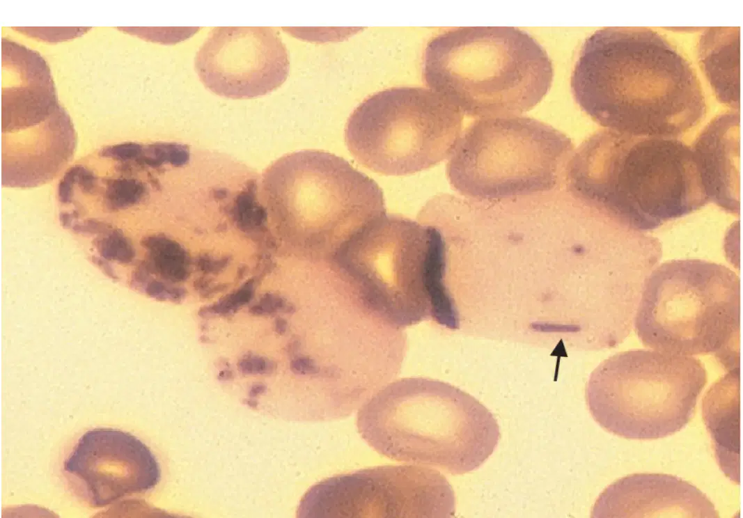
What does the image depict?
Negative myeloperoxidase stain
Positive myeloperoxidase stain
Positive SBB stain
Negative SBB stain
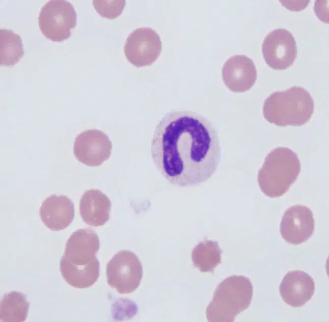
What type of cell is shown in the image?
Red Blood Cell
Band Neutrophil
Platelet
Lymphocyte
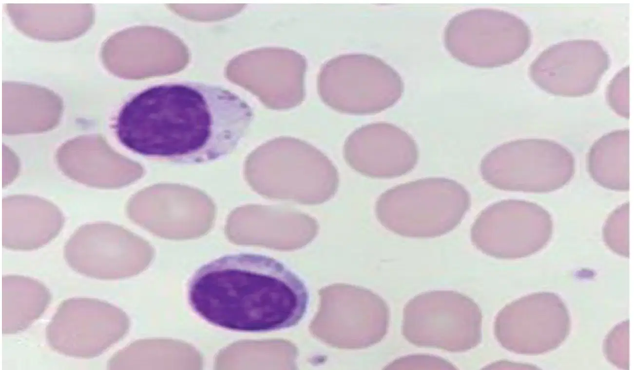
The image shows what type of cell?
Lymphocyte
Monocyte
Lymphocytic blast
Basophil
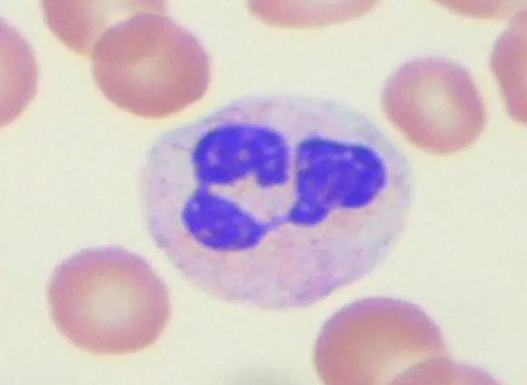
What type of cell is shown in the image?
Red blood cell
Segmented neutrophil
Platelet
Lymphocyte
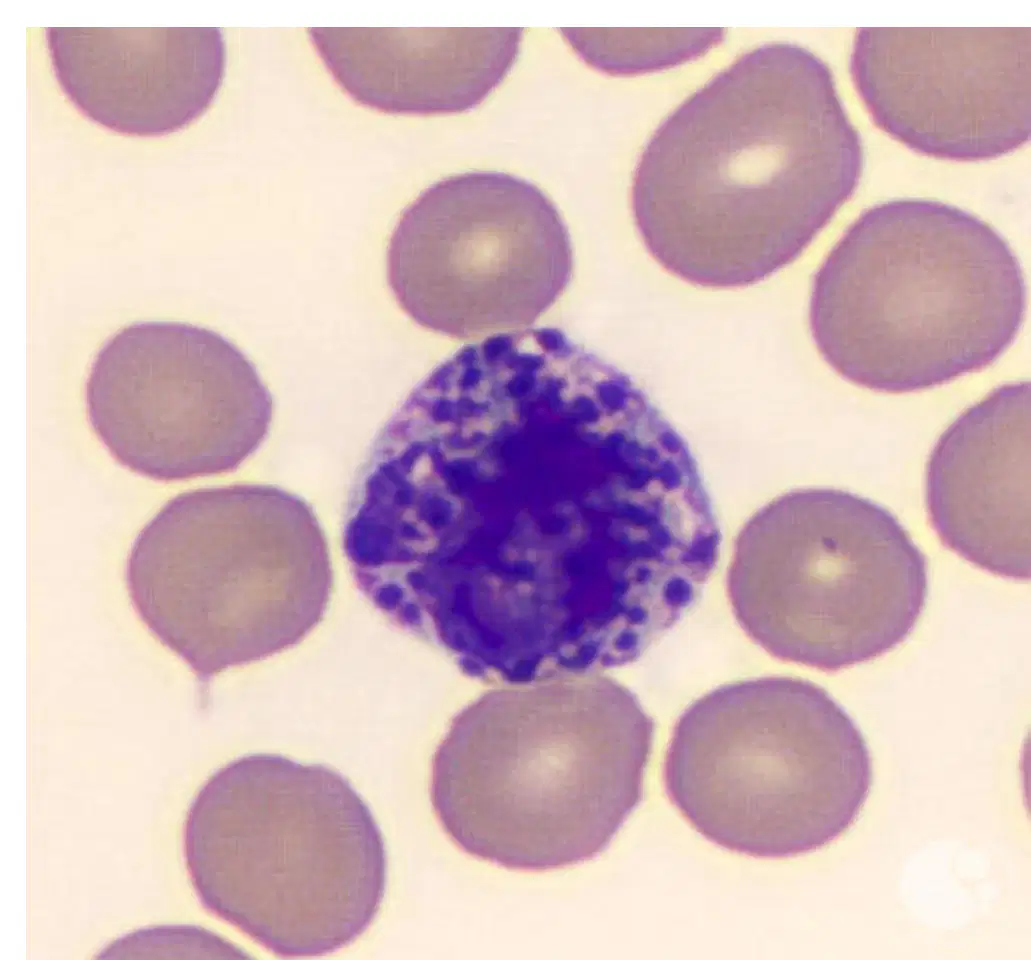
What type of cell is shown in the diagram?
Neutrophil
Basophil
Eosinophil
Lymphocyte
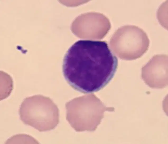
Identify the type of cell shown in the image.
Neutrophil
Lymphocyte
Eosinophil
Basophil

What morphology is being shown by the WBC's in the image?
Alder-Reilly anomaly
Chediak-Higashi syndrome
May-Hegglin anomaly
Toxic granulation
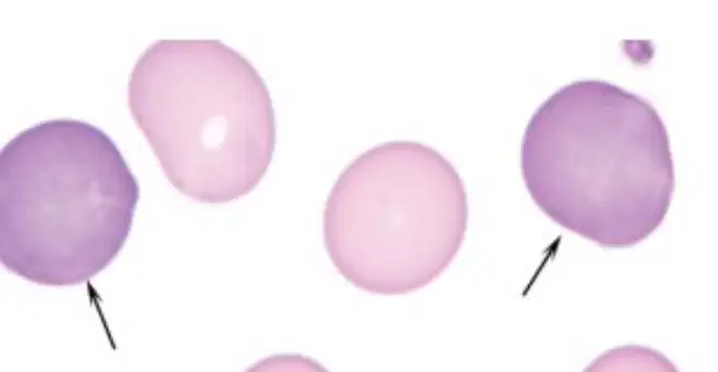
What is shown in the image?
Polychromatic Erythrocyte (Reticulocyte)
Neutrophil
Lymphocyte
Monocyte
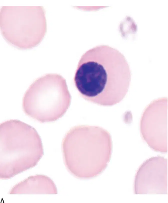
What does this image depict?
Nucleated Red Blood Cells
Neutrophils
Platelets
Lymphocytes
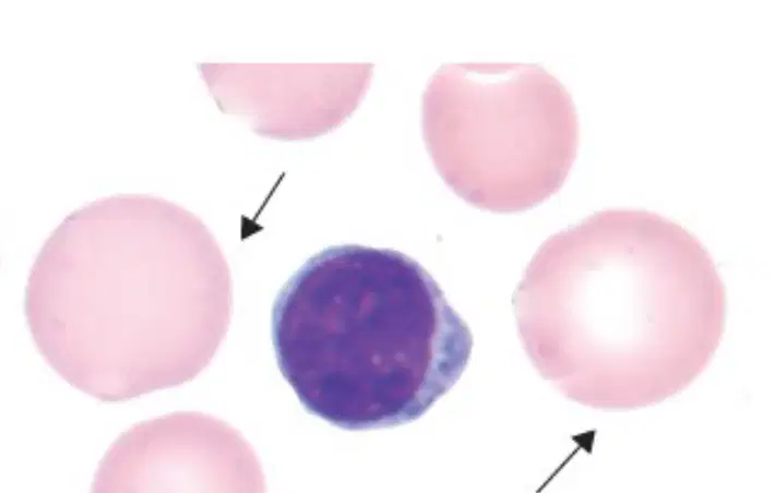
What does the image depict?
Normal RBCs
Microcytic RBCs
Macrocytic RBCs
Sickle cell RBCs
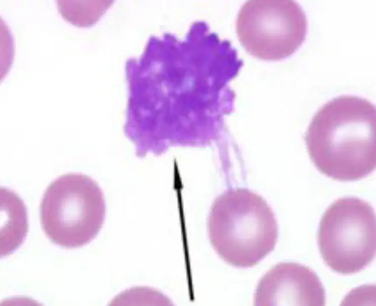
What is shown in the image?
Red Blood Cell
Smudge Cell
White Blood Cell
Platelet
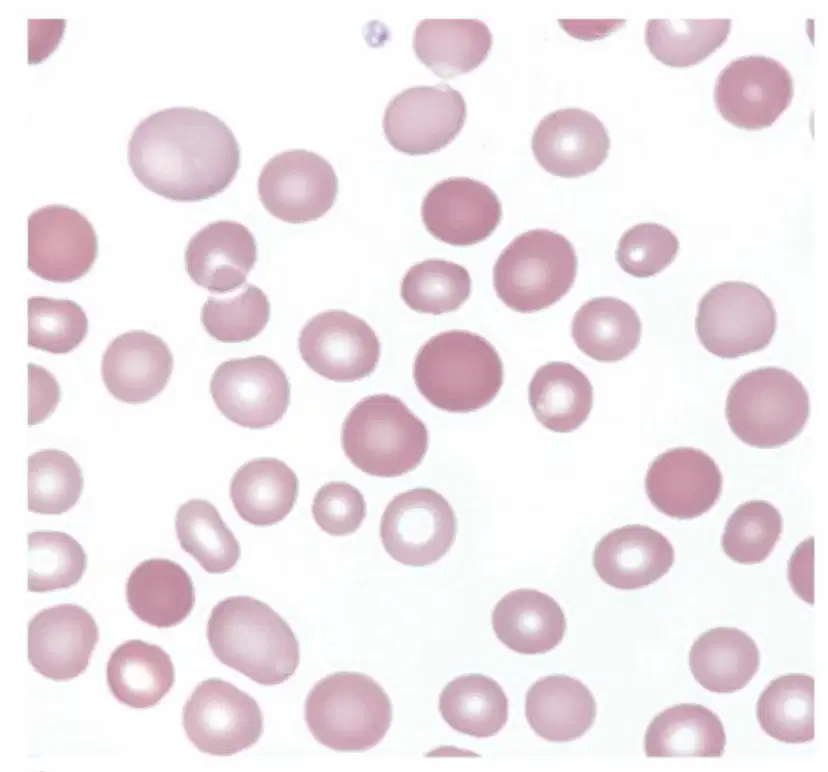
What condition is characterized by red blood cells of unequal sizes?
Anisocytosis
Leukocytosis
Thrombocytosis
Poikilocytosis
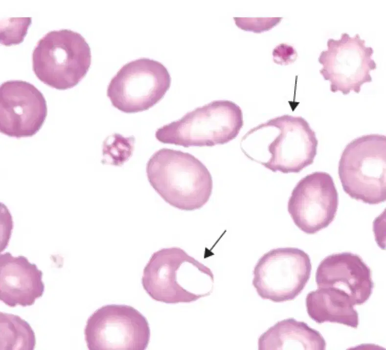
What type of cells are shown in the image?
Red blood cells
White blood cells
Blister cells (Schistocytes)
Platelets

