
 Worksheets
Heme Cell Morphology Practice #3
Total questions: 17
Worksheet time: 9mins

What type of cells are shown in the image?
Blister cells
Bite cells
Target cells
Acanthocyte cells

What does the image depict?
Normal RBCs
Hypochromic RBCs
Sickle Cell RBCs
Macrocytic RBCs

What type of cells are shown in the image?
Neutrophils
Spherocytes
Lymphocytes
Erythrocytes

What does the image titled 'Iron stain' show?
Siderotic granules
Pappenheimer bodies
Dhole bodies
Lipid granules

What is shown in the diagram labeled 'New Methylene Blue Stain'?
Reticulocytes
Heinze Bodies
Pappenheimer bodies
Siderotic grahules

What is shown in the image labeled 'A'?
A) Basophilic Stippling
B) Eosinophilic Granules
C) Neutrophilic Bands
D) Lymphocytes

What is shown in the image?
Rouleaux
Autoagglutination
Dacrocytes
Cold agglutinins
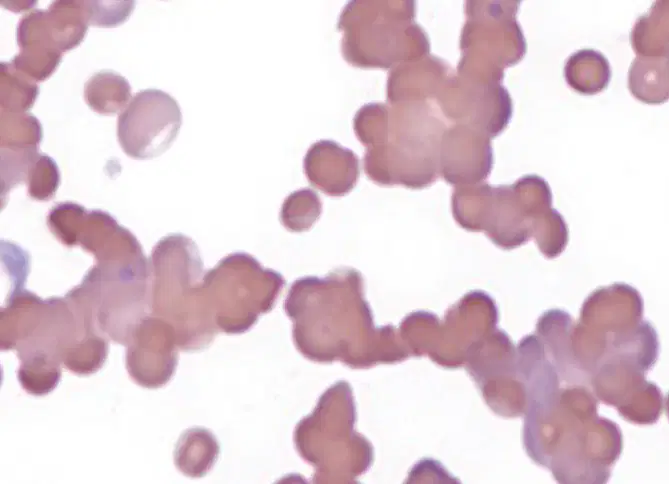
What does the red cell morphology depict in this image?
Normal red blood cells
Autoagglutination
Bacterial infection
Viral infection
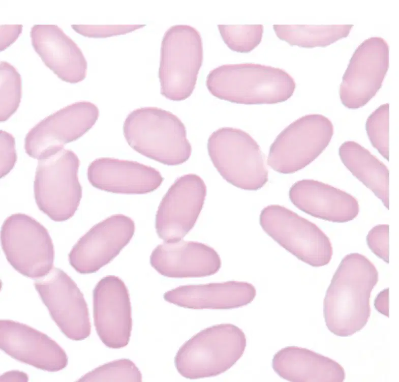
What type of cells are shown in the image?
Spherocytes
Elliptocytes
Erythrocytes
Leukocytes

What are the RBC inclusions shown?
Howell Jolly Bodies
Pappenheimer bodies
Dohle bodies
Siderotic granules

What type of cells are shown in the image?
Spherocytes
Ovalocytes
Elliptocytes
Stomatocytes

What type of cell is shown in the image?
Lymphocyte
Hairy Cell Lymphocyte
Platelet
Neutrophil
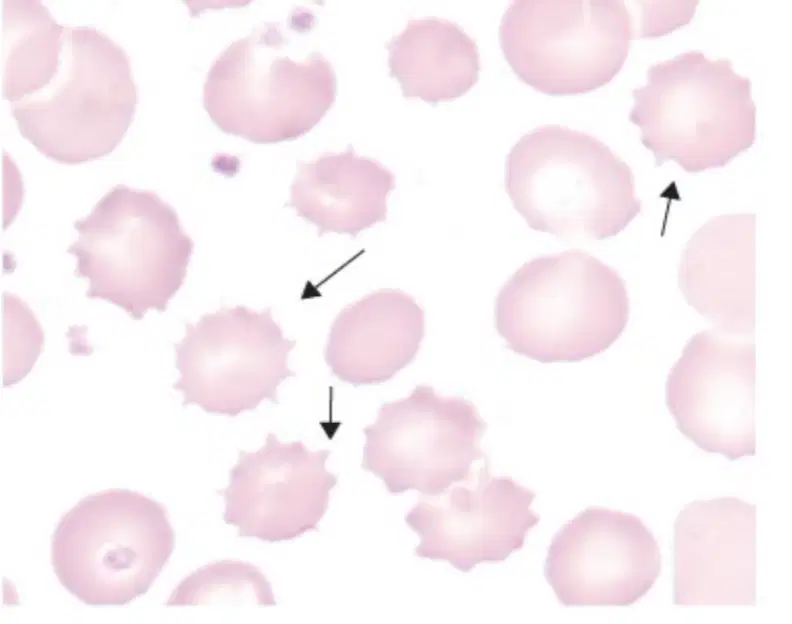
What is another name for Echinocyte?
Burr Cell
Sickle Cell
Red Blood Cell
White Blood Cell

What is the pattern shown in the image associated with Burkitt Lymphoma?
A) Starry Sky Pattern
B) Cloudy Pattern
C) Striped Pattern
D) Dotted Pattern
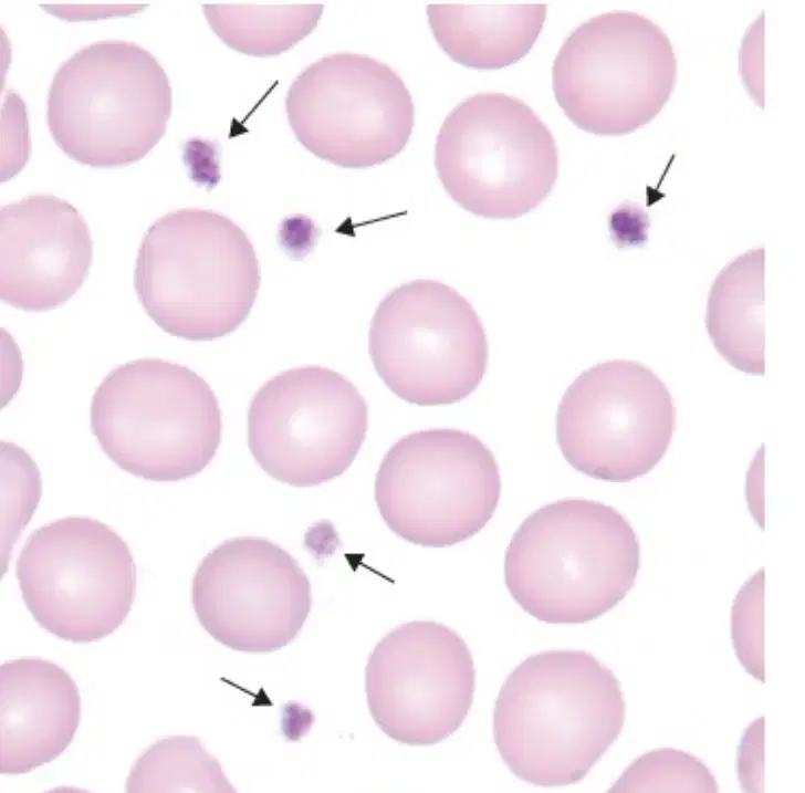
Identify the cells shown in the diagram.
Codocytes
Thrombocytes
Dacrocytes
Fragments

What is shown in the image?
Hemoglobin 'A' Crystal
Hemoglobin 'B' Crystal
Hemoglobin 'C' Crystal
Hemoglobin 'D' Crystal

What type of cell is shown in the image?
Neuron
Megakaryocyte
Erythrocyte
Lymphocyte

