
NEW
Font size
 Worksheets
Heme Cell Morphology Practice #4
Total questions: 20
Worksheet time: 10mins

What is seen in the image related to May-Hegglin anomaly?
Normal Platelet
Giant Platelet
Red Blood Cell
White Blood Cell

What anomaly is shown in the image, 71% of these cells exhibited this morphology?
Pelger-Huet
May-Hegglin
Chediak-Higashi
Alder-Reilly

What is shown in the diagram?
Hyposegmentation
Hypersegmented
Lymphocyte
Monocyte
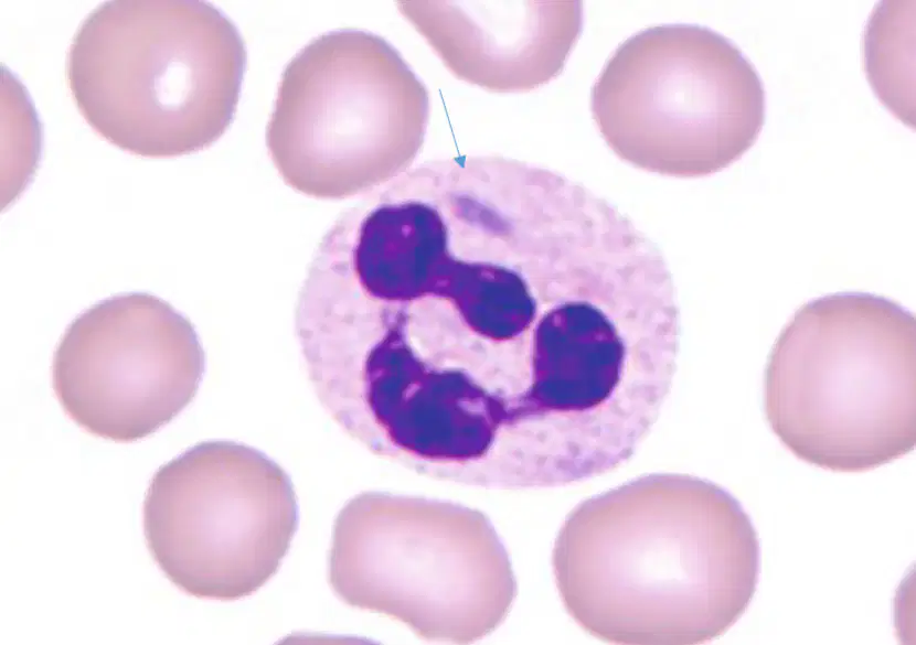
What is the inclusion shown in the image?
May-Hegglin
Dohle Body
Howell-Jolly Body
Pappenheimer body

What type of WBC is shown in the image?
Normal Cell
Pyknotic Cell
Toxic Cell
Reactive Cell
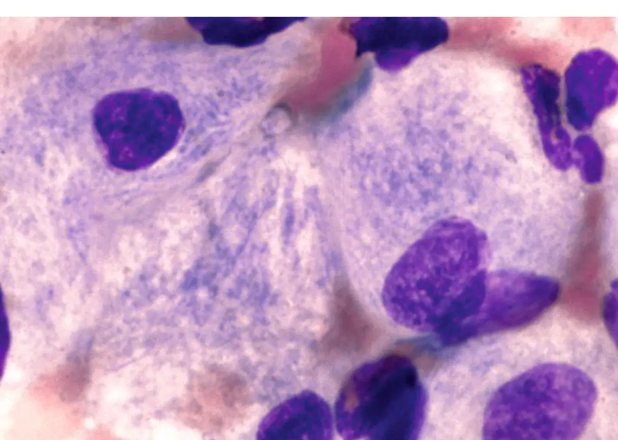
What type of cell is shown in the image?
Plasma cell
Gaucher Cell
Megakaryocyte
Niemann-Pick Cell
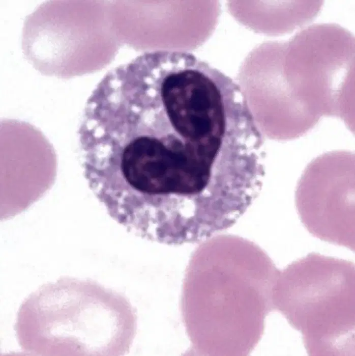
What is the morphology shown with this WBC?
Cell division
Vacuolation
Pyknosis
Reactive
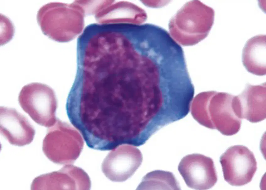
The image shows what type of cell.
Reactive lymph
Plasmacytoid lymph
Lymphoblast
Monocyte

What type of stain is being shown here?
LAP
SBB
Oil red O
Grams

A marked reduction seen in the LAP score would be seen in what Leukemic condition?
CML
Leukemoid reaction
ALL
AML

What WBC morphology is shown in the image?
Alder-Reilly anomaly
Toxic granulation
May-Hegglin
Dhole-bodies

What does the nucleated cell in the image depict?
White Blood Cell
Nucleated Red Blood Cell
Platelet
Plasma
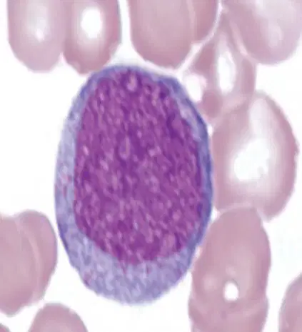
Identify the cell type shown in the image.
Promyelocyte
Myeloblast
Lymphoblast
Monocyte

What type of cell is shown in the image?
Red blood cell
Segmented Neutrophil
Platelet
Lymphocyte

The WBC image shows:
Reactive Segmented Neutrophil
Reactive Lymphocyte
Reactive Monocyte
Vacuolated Neutrophil
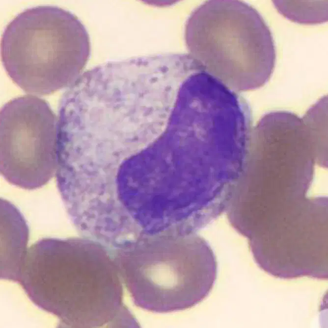
What type of cell is shown in the image?
Promyelocyte
Metamyelocyte
Myelocyte
Band
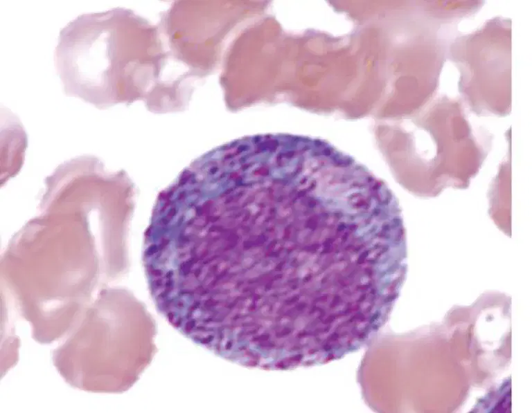
What is shown in the image?
Metamyelocyte
Promyelocyte
Myelocyte
Monocyte
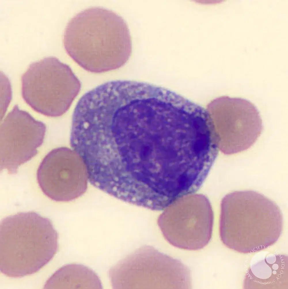
Identify the type of cell shown in the image.
Myelocyte
Promyelocyte
Metamyelocyte
Myeloblast
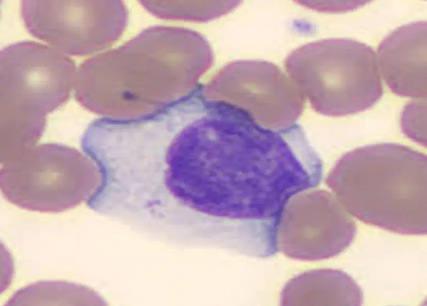
What is shown in the image?
Lymphoblast
Reactive Lymphocyte
Lymphocyte
Monocyte

Identify the WBC shown.
Plasma cell
Plasmacytoid cell
Monocyte
lymphocyte

