
 Worksheets
Liver
Total questions: 144
Worksheet time: 2hrs 11mins
Is a continuous transparent layered membrane
(a)
Lines the abdominal cavity and covers the abdominal organs (or viscera).
(a)
Provides a pathway for blood vessels
and lymph
(a)
(secrets a small amount of serous fluid for lubrication)
o Covers the organs
(a)
o Lines the abdominal wall
(a)
Encloses most of the abdominal organs; enclosed organs called “intraperitoneal
(a)
Small sac bordered anteriorly by the stomach, posteriorly by the pancreas and a portion of the transverse colon
(a)
Passageway between greater and lesser sacs just inferior to the liver
(a)
Double peritoneum extends from liver to lesser curvature of stomach and beginning of duodenum
(a)
Large fold of peritoneum that extends from stomach, passes anteriorly to the colon and small intestine
(a)
Space within the abdomen that houses most
of the organs (liver, stomach, intestines)
(a)
Liver is considered an ___ organ
Intraperitoneal
Retroperitoneal
Located within the peritoneal cavity and covered by the visceral peritoneum
(a)
The area on the liver that is not covered by peritoneum is called the (a)
The (a) area is located on the posterior superior surface of the right of lobe the liver
The (a) area lies in direct contact with the diaphragm
The right sub hepatic space is also called __
Morisons Pouch
The place
Hepatorenal recess
Pancreatic recess
(a) is anatomical space located beneath the RT lobe of the liver, adjacent to the right kidney
The (a) space is an area between the liver or spleen and diaphragm
Which space is a common site for abscess formation?
Right hepatic space
Bare Area
Liver
Subphrenic space
The (a) is located beneath the right costal margin.
Which abdominal regions does the liver fall into?
Right Hypochondrium
Hypogastrium
Epigastrium
Left hypochondrium
What lies superiorly to the liver?
The right kidney
Navel region
Dome of diaphragm
Bony lumbar region
What lies posteriorly to the liver?
Bony lumbar region
Dome of diaphragm
Right kidney
Caudal region
The following measurement applies to which lobe of the liver:
Midclavicular line measurement 13cm to 17cm
Left lobe
Which lobe of the liver varies greatly in size?
Left lobe
Right lobe
Which of the following best describes the liver's weight in adult males?
3-3.9 pounds
4.5 pounds
1400g to 1800g
1.8 mg
Which of the following best describes the liver's weight in adult females?
30 kg
1200-1400 g
2.6-3 pounds
40-80mg
The liver occupies almost all of the (a)
The liver occupies the greater part of the (a)
The liver occupies the (a) as far as the mammary line
Which of the following can vary greatly in the liver
Placement
Contour
Shape
Location
The liver lies (a) to the diaphragm
What covers the greater portion of the right lobe?
Ribs
Lungs
Heart
Brain
Which lobe extends several centimeters below the xiphoid process?
Right lobe
Left lobe
The fundus of the stomach lies _ and _ to the LT lobe of the liver
(a)
Duodenum lies __ to the RT and __ segment of the left lobe
(a)
Body of pancreas is usually just (a) to the left lobe
The (a) border of the liver contacts the RT kidney, IVC, and AO
The diaphragm covers the (a) border of the liver
Which of the following does NOT lie on the posterior borders of the liver?
IVC
Splenic artery
Hepatic artery
Cystic duct
Which of the following does NOT lie on the posterior borders of the liver?
Gallbladder
Confluence
RT Kidney
Colon
LT kidney
What alters the projections of the liver?
(a)
(a) tumors can elevate the liver
Right hepatic flexure of colon
(a)
Right kidney/adrenal gland
(a)
First portion of duodenum
(a)
Gallbladder
(a)
Gastric indentation
(a)
IVC groove (posterior)
(a)
Anterior mid portion (medial portion of let lobe) → quadrate lobe
(a)
Left lateral boundary:falciform ligament
(a)
Posterior midportion (below porta hepatis) → caudate lobe
(a)
How many lobes is the liver divided into?
1
2
3
4
Which of the following are the names for the lobes of the liver?
Left
Right
Upper
Caudate
Quadrate
How many times larger is the right lobe than the left lobe?
4
3
6
5
The right lobe has two segments the __ and __ segements
(a)
The right lobe is separated horizontally by the (a)
The right lobe of the liver appears to be separated from the left lobe by the __ and __
Falciform ligament
Teres hepatis
Brain
Main lobar fissure - anterior surface
The (a) divides the medial segment of the LT lobe from the anterior segment of the RT lobe
extension of the posterior segment of RT lobe that can extend to iliac crest
(a)
divides Left lobe into medial and lateral segments
(a)
Intersegmental fissure is marked on external liver margin by the (a)
Runs horizontally between the medial and lateral segments
(a)
Left lobe is bound (a) by the Porta Hepatis
Left lobe is bound (a) by the GB fossa
Left lobe is bound (a) by the fossa for umbilical vein
Which of the following are the two fissures of the left lobe?
Falciform ligament
Ligamentum venosum
Ligamentum teres hepatis
Ductus venous
Round ligament
(a)
fibrous remnant of fetal circulation in which the umbilical vein is connected to IVC, in adulthood it is usually seen attached to Left branch of Portal vein within Porta hepatis (gateway to liver
(a)
The caudate lobe is bound (a) by the port hepatis
(a) , the caudate lobe is separated from the RT lobe by IVC
The (a) part of the caudate lobe is bounded by the fissure for the Ligamentum Venosum
The caudate lobe is (a) Connected to the RT lobe of the liver by the caudate process, which passes laterally between the Portal vein and the IVC at the Porta Hepatis.
Which of the following are also names for the caudate lobe?
lower segment
Spiegels lobe
Segment 2
Segment 1
Located on the visceral surface of the liver, specifically between the IVC and the Ligamentum venosum.
(a)
The (a) lobe is the largest lobe
Close to the anterolateral abdominal wall
Rt lateral diaphragm undersurface
Subdivided into anterior and posterior segments
(a)
Close to undersurface of the diaphragm
Varies in size and shape
Inferior margin is close to then gastric body and antrum
(a)
Anterior to:
o Pancreas body
o Splenic vein
o Splenica rterySubdivided into lateral and medial segments
(a)
Located on the inferior surface of the RT lobe.
Sits on the inferior surface of the RT lobe, between the anterior margin of the liver and the Porta hepatis
(a)
The (a) lobe Is the smallest lobe
Related to the lumbar region (posterior abdominal and thoracic walls)
Covered by the lesser sac
Separated from LT lobe by LHV and the fissure of the Ligamentum Venosum
(a)
Liver covered by thin connective tissue layer called (a)
(a) is boundary between RT and LT lobes of liver
(a) appears as a hyper echoic line extending from portal vein to neck of GB
Sonographer uses this ligament as a landmark to find GB if diseased
(a)
(a) extends from umbilicus to diaphragm and contains ligamentum teres
appears as a bright echogenic focus with ultrasound and is seen as rounded termination of falciform ligament
(a)
separates theLeft lobe from caudate lobe
• Seen on ultrasound as a thin, horizontal line just anterior to caudate lobe
(a)
Which lobe of the liver is 1?
(a)
Can you usually see the caudal lobe on a scan?
yes
No
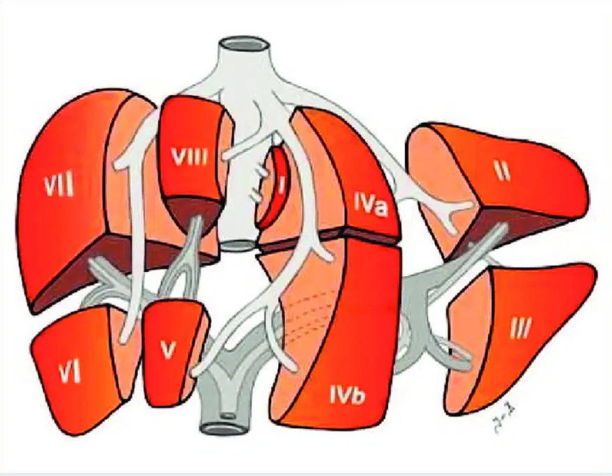
Which lobe is labeled 1?
(a)
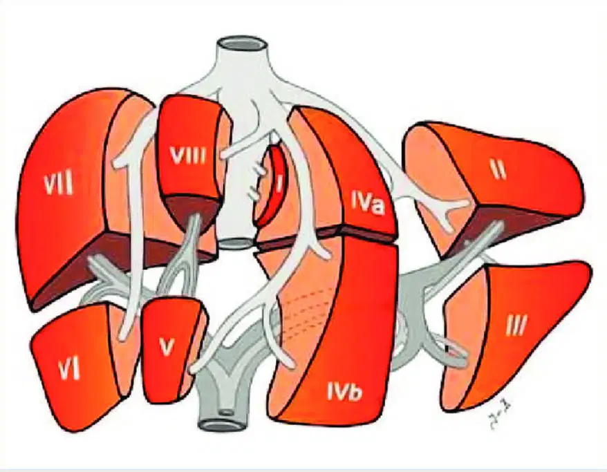
Which lobe is labeled 2?
(a)
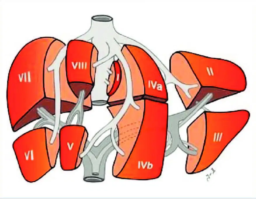
Which lobe is labeled 3?
(a)
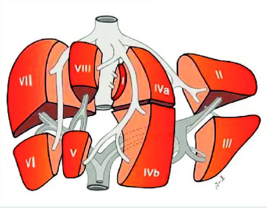
Which lobe is labeled 4a?
(a)
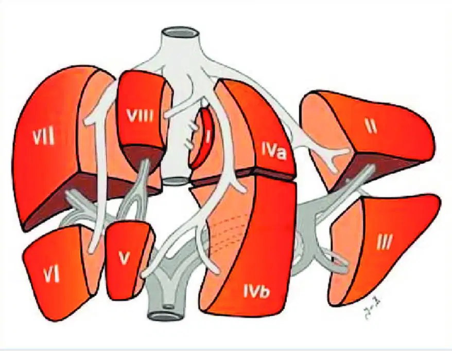
Which lobe is labeled 4b?
(a)
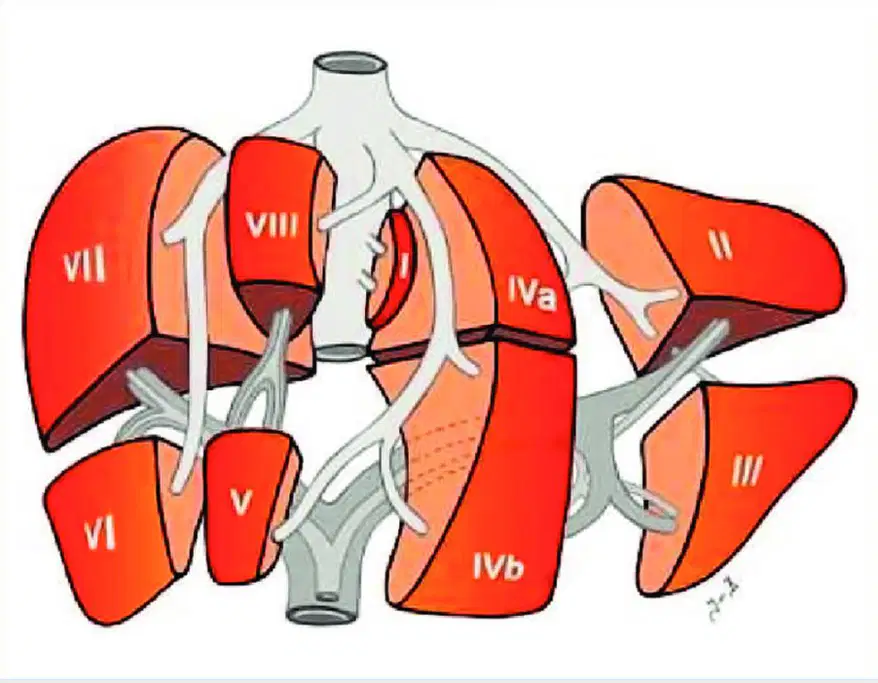
Which lobe is labeled 5?
(a)
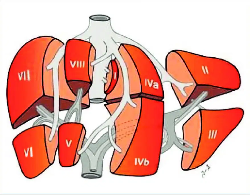
Which lobe is labeled 6?
(a)
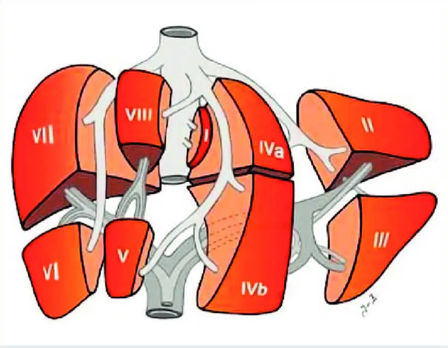
Which lobe is labeled 7?
(a)
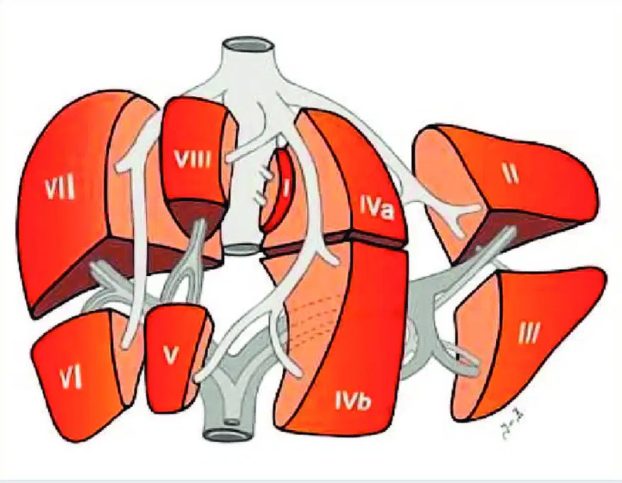
Which lobe is labeled 8?
(a)
Runs from galbladder fossa to inferior vena cava and passes through portion of caudate lobe; this is the plane we refer to for the main lobar fissure
(a)
Divides liver into right and left hemiliver (liver halves); hemilivers further divided into anterior and posterior segments
(a)
Anterior and posterior segments divided into inferior and superior segments
(a)
Anterior segment only divided into superior and inferior segments
(a)
Liver Portion: caudate, bordered posteriorly by inferior vena cava and anteriorly by LPV
(a)
Liver portion: left hemiliver, segment 2 and 3 found to the left of the ligamentum venosum
(a)
Right hemiliver, segments 5 and 8 are anterior; segments 6 and 7 are posterior
(a)
Hepatic Artery
Portal venous system
Hepatic veins
(a)
(a) hepatic vein is the largest
(a) hepatic vein is the smallest
What is the difference between the portal and hepatic veins is there:
Intensity of blood flow within the liver
Direction of blood flow within the liver
Portal flow within the liver
Hepatic vein
(a) formed by the union of the SMV (Superior Mesenteric vein) & SV (Splenic vein)
- delivers blood from the digestive organs to the liver for processing
- enters the liver
(a)
- drain the liver and return the blood to the heart
- exit the liver
(a)
The portal triad consists of:
Portal vein
Hepatic artery
Hepatic vein
Bile duct
These structures travel together, throughout liver, to support physiological function of liver
(a)
The portal vein is contained within a connective tissue sheath called ___ ___, which contributes to its echogenicity
(a)
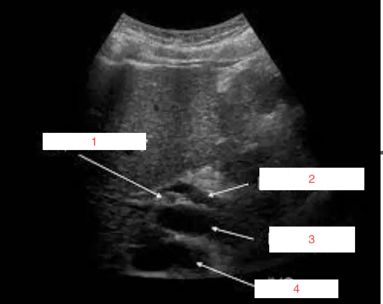
What is labeled 1
IVC
Bile duct
Hepatic vein
Hepatic artery
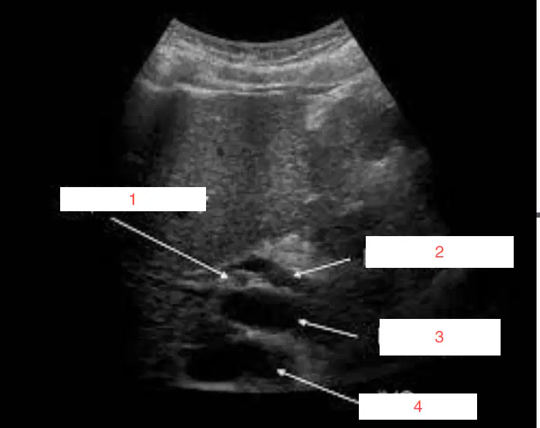
What is labeled 2 in this image?
Hepatic artery
Bile duct
Main PV
IVC
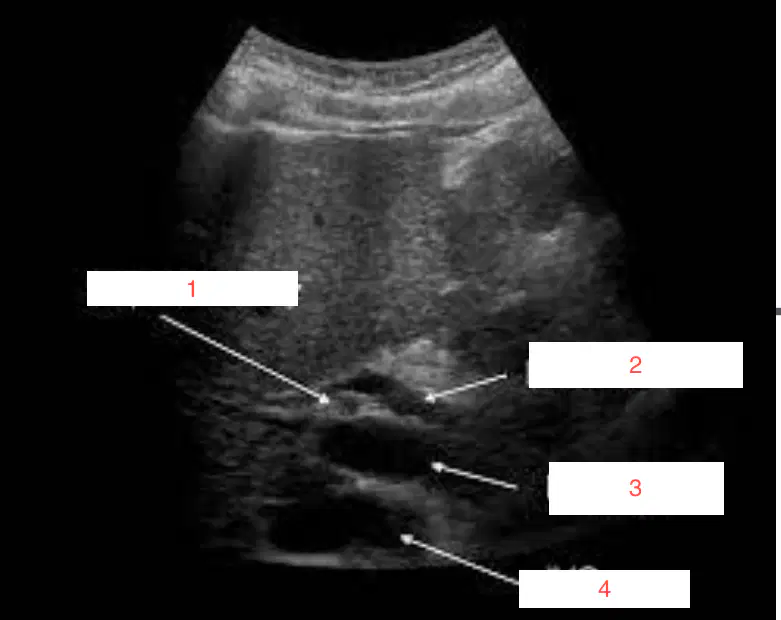
What is labeled 3?
Main PV
IVC
Hepatic artery
Bile duct
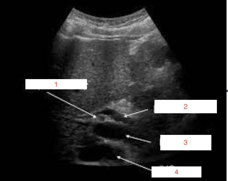
What is labeled 4?
Hepatic Artery
Bile duct
Main PV
IVC
Left hepatic vein divides LT lobe of the liver into its __ and __ segments.
(a)
Left portal vein divides LT lobe of the liver into ___ and __ segments based on its transverse plane
(a)
Drains into the anterior and posterior segments of the right lobe
(a)
Medial and lateral segments of the left lobe
(a)
Drains into the right lobe and medial left lobe
(a)
(a) is drained by veins that directly connect to the inferior vena cava
Drained by veins that directly connect to the inferior vena cava (IVC):
Long hepatic veins
Emissary hepatic veins
Spieghel veins (type of extrahepatic veins)
Short hepatic veins
(a) supplied by multiple arteries, including branches of the RHA and LHA
Parenchyma of (a) is used to sonographically evaluate other organs.
(a) equal to or less echogenic than the liver.
(a) isoechoic or hyperechoic to liver
(a) is used as a window to visualize abdominal structures such as pancreas, gallbladder
The liver is a primary center of (a)
Typically, there are approximately _ million lobules within the liver
(a)
The liver uses (a) to metabolize carbohydrates.
(a) is a major component of the bile that is secreted by the liver and serves to emulsify fats
The liver synthesizes the (a) albumin, fibrinogen, prothrombin, and globulins
The liver (a) vitamins and other metabolic substances
Has a tip that can extend left laterally, all the way to the spleen. The sonographic appearance is the same as the normal liver.
(a)
Which structure is located on the inferior surface of the RT lobe between the anterior margin of the liver and the Porta hepatis?
Quadrate lobe
Caudate lobe
Right lobe
Left lobe
Caudate lobe is connected to the Rt lobe of liver
True
False
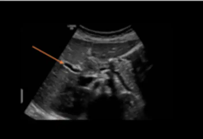
What is red arrow pointing to?
(a)