
 Worksheets
CSE Prep
Total questions: 68
Worksheet time: 1hrs 21mins
Pathology
Weakened, enlarged airspace’s often along with an increase in goblet cells and mucus gland
COPD
Chronic inflammatory/obstructive disease known for exacerbations of wheezing and coughing
Asthma
Thick tenacious mucus accumulation in the lungs, blocks passageway of the pancreas and prohibits enzymes from reaching intestine.
Cystic fibrosis
Gas or free air in the pleural space
Pathology of pneumothorax
Abnormal condition that affects cardiac pumping caused by MI, ischemic heart disease or cardiomyopathy
CHF/pulmonary edema
Pathology
An increase in mean pulmonary artery pressure greater than 25 at rest usually due to COPD or ILD
Pulmonary hypertension, PH
Interruption of coronary blood flow for an extended period causing potentially irreversible damage to the heart muscles
Myocardial infarction/ischemia
Chronic disorder of neuromuscular junction resulting in descending paralysis
Myasthenia gravis
Autoimmune disorder of the peripheral nervous system
Guillian-barre syndrome
Portion of brain loses blood supply as a result of a vascular occlusion or hemorrhage
Stroke/cerebral infrc, CVA
Pathology
Acute illness or injury to the lungs that results in reduced lung compliance, diffuse Atelectasis, and refractory hypoxemia
ARDS
Failure of the cardiovascular system to adequately perfume tissues resulting in an impairment of cellular metabolism: reduction in blood flow to the tissues
Shock
Decrease renal function secondary to diabetes Mellitus or renal insufficiency
Diabetes/renal failure
Not enough surfactant or depressed surfactant leading to Atelectasis and hypoxemia
Infant RDS, hyaline membrane disease
Overuse of medication that exceeds the recommended dose
Drug overdose
Pathology: Muscular Dystrophy
A group of genetic disorders that cause progressive weakness and degeneration of the muscles that control movement.
Bacterial infection that affects the nervous system, causes muscle stiffness and spasms (jaw/neck muscles)
Tetanus
Viral infection, genetics, puncture wound
Etiology
History of present illness, current medications
Assessment
Establish airway, monitor VT, VC, MIP, O2,
Treat
Pneumonia
CXR, CBC, sputum culture
Diagnostic
O2, pulmonary hygiene therapy, hyperinflation (IS/SMI, IPPB), MV, VAP protocol
Treat
Penicillin G, amoxicillin
Drugs
S. Aureus (MRSA), treat with vancomycin . Cefotaxime, ceftriaxone, ciprofloxacin
Penicillin resistant pathogen
Acquired Immunodeficiency Syndrome, AIDS
Virus attacks the immune system, gradually weakening it over time and making it difficult for the body to fight infections
Pathology
Frequent pneumonia, bronchitis, sinus/ear infections, Meningitis, low platelets counts or anemia
History
Drug cocktails, Antiretroviral, high activity antiretroviral therapy
Treatment
Trimethoprim and sulfamethoxazole, corticosteroids within 72 hrs
Pneumocystis carinii pneumonia, PCP
Accumulation of blood in the pleural space
Pathology
Hemothorax
General appearance
Cyanosis, tracheal shift away from from affected side, productive cough (hemoptysis), dull percussion
COPD Medications
SABA:
Albuterol, xopenex, ipratropium
LABA:
Salmeterol, spirivia
Steroids:
Fluticasone, budesonide
Combination MDIs:
Advair, symbicort
First line treatment: especially in exacerbations
LAMAs, tiotropium (spirivia), Glycopyrrolate (Seebril), umeclidinium (incruse), aclidinium.
Outpatient COPD
Spirometry, ABG, review therapy (meds/techniques), psychological (support system, depression), cessation of smoking (done prior to rehab), avoid secondhand smoke
F/U exam: 3-4 weeks
SABA, LABA, Steroids, Combo MDI
Medications
Controlled lip pursed breathing, I/S, coughing, diet and exercise
Other

GOLD

COPD classification
Pathology: CBABE
You know this
CF
Obstructive condition p, irreversible damage of bronchial walls (excessive inflammation) , copious amounts of bronchial secretions, commonly limited to lobe or segment
Bronchiectasis
Reoccurring episodes of dyspnea, wheezing, chest tightness
Asthma
Increased mucus production in trachea and bronchi, productive cough that lasts at least 3 months of the year
(Chronic) bronchitis
Increased lung expansion due to air trapping, irreversible damage to alveolar walls, enlargement of air spaces
Emphysema
Pathology: Adult Cardiovascular
Sudden, rapid decline in heart functioning and the amount of blood your heart can pump to the rest of the body. (Lower left chamber, left ventricle)
Acute heart failure, AHF
Occurs as a result of damage to the right side of heart , resulting from pulmonary hypertension. Increased right ventricular workload that causes hypertrophy of the RV. Often caused by COPD
Cor pulmonale
When the arteries that supply blood to the heart become narrowed or blocked, which can restrict blood flow and oxygen to the heart muscle. Caused by atherosclerosis
Coronary artery disease
Pathology: Adult Cardiovascular
Pulmonary Embolus
Ventilation without perfusion
Pulmonary Embolus
Anxious, diaphoretic, cyanotic, cool/clammy, SOB, tachypnea, wheezing/crackles/pleural friction rub, possible hemoptysis, sudden onset of signs/symotoms
Assessment
CXR: increased density in infarcted area, wedge shape infiltrate. Hemodynamic: increased PAP. Capnography: decreased PeCO2, normal PaCO2. VD/VT: increased.
Diagnostic testing
1)Spiral CT -most sensitive,
2)V/Q scan, 3)Pulmonary angiogram
Special tests

Treatment
Thermal Lung Injury
Tachypnea and cough may be productive of thick black sooty secretions
Respiratory pattern
Normal in early stages, may present with wheezes, crackles, or rhonchi, inspiratory strider
Breath sounds
100% NRB, IF severe, establish an airway FIRST. CPAP to minimize pulmonary edema. Burn injuries determined by bronchoscopy,
Treatment
Pediatric Pathology
A viral infection characterized by inflammation of the bronchioles that results in wheezing and difficulty breathing in children
Bronchiolitis
Viral infection of the upper airways that results in subglottic swelling and an obstruction below the vocal cords. Results in stridor.
Croup
Acute upper airway infection that causes severe supraglottic swelling.
Epiglottitis
Chronic respiratory disorder in premature and low birth weight infants characterized by a reduction in the overall surface area for gas exchange (results from the treatment of RDS with MV and high O2 over a prolonged period of time >28 days)
Bronchopulmonary dysplasia
Pediatric Pathology: Croup
Cough-barking, stridor, hoarse voice, nasal flaring, rhinorrhea, substernal/intercostal retractions, cyanosis
Assessment
O2 (24-28% initially), ABG
Diagnostic
Racemic via SVN, corticosteroids (decadron) when unresponsive to epi, MV
Drugs
Pediatric Pathology: Epiglottitis
2-6yrs, pale, cyanotic, lifeless, drooling, hoarseness, dysphagia, tongue thrust forward during inspiration, diminished BS with inspiratory stridor.
Assessment
Visualization of pharynx, oral temp, ABG, IV, Neck X-ray
DO NOT DO-avoid stimulation
24-36hr initially
Intubation
Lateral neck X-ray (after sedation) will show thumb sign
Confirmation of diagnosis
Pediatric Pathology: Bronchiolitis
Nasal discharge, lethargic, nasal flaring, cyanosis
Assessment
Tachypnea, apnea in severe cases, grunting, retractions, wheeze, upper airway noise from secretions
Respiratory
Plenty of fluids, oral decongestants, clearance of secretions from nasal passages
Treatment: Home
(Severe symptoms: apnea, CHD, CF, BPD, asthma)
O2 with humidity, aerosolized bronchodilators, IV fluids, bronchial hygiene
Treatment: Hospital
Pediatric Pathology: BPD
Early surfactant replacement therapy
PRIORITY
Prevents atelectasis and intubation
Non invasive CPAP
Bronchodilators, diuretics or fluid management (monitor input) vaccines for RSV and influenza
Drugs
To prevent infection
Radiant warmer
Treatment for adult cardiovascular
O2, CPAP for pulmonary edema/hypoxemia, NPPV for impending respiratory failure: first line For persistent hypoxia, unresponsive to O2, increased WOB (hallmark signs)
AHF
O2, monitor vital signs, treat underlying cause, decrease workload by lowering PAP. Digitalis, diuretics, nitric oxide
Cor Pulmonale
Minimize risk factors, if angina: O2, nitroglycerin, angioplasty/stent, CABG
Coronary artery disease, CAD
Cessation of smoking, limit cholesterol/fats, exercise, Control BP and aspirin for blood thinner
Minimize risk factors for CAD
Treatment of Emphysema / Chronic bronchitis
SABA, LABA, anticholingeric, LAMA, ICS, Antibitoics (indicated by sputum)
Medication
Low flow O2, NPPV for acute exacerbations of ventilator failure (ph: <7.35, CO2: >45)
General Management
Nutrition, exercise, method to aid in secretions, home O2 and aerosol therapy.
Outpatient rehab/home care
What patient type is characterized by:
Chronic lung disease, dyspnea, increased AP with obstructive lung disease, distended neck veins, chest pain, peripheral edema, and clinical manifestation appear during exercise testing
Cor pulmonale
CHF
Coronary artery disease
What patient type is characterized by:
Barrel chest, clubbing of fingers, cyanosis, use of accessory muscles, and expiratory wheeze only
COPD
ARDS
Asthma
CF
What patient type is characterized by:
Pursed lip breathing, prolonged exhalation, chest tightness, use of accessory muscles, increased AP during episode, pulses paradoxes (severe)
Asthma
COPD
Pneumothorax
What patient type is characterized by:
Barrel chest, cyanosis, clubbing, thick viscous mucus accumulation
CF
Thermal lung injury
CHF
What patient type is characterized by:
cyanosis, tracheal or mediastinal shift away from affected side, bruising over affected area
Pneumothorax
COPD
CHF
What patient type is characterized by:
Anxious, surface burns, black soot marks, cyanosis (cherry red CO2)
TLI/Smoke inhalation/CO poisoning
PH
COPD
What patient type is characterized by:
Peripheral/pedal edema, diaphoresis, cyanosis, tachyonea, orthopnea, crackles and wheezing, pink frothy secretions
CHF/pulmonary edema
pulmonary Hypertension, PH
MI
What patient type is characterized by:
Anxious, diaphoretic, cyanosis, peripheral edema, jugular vein distension
CHF
Pulmonary hypertension
Myocardial infarction/ischemia
What patient type is characterized by:
Diaphoretic, anxious, chest pain
Myaocardial ischemia/infarction
Pulmonary hypertension
Pulmonary embolism
What patient type is characterized by:
General weakness at rest/drooping eyes, double vision (diplopia) difficulty swallowing (dysphagia)
Myasthenia gravis
Guillian barre
What patient type is characterized by:
Autoimmune weakness specially in legs, cyanosis, shallow breathing
Myasthenia gravis
Guillian barre
What patient type is characterized by:
Slow shallow, respiration/diminished breath sounds, altered level of consciousness, euphoria
Drug overdose
Stroke, acute brain attack, cerebral infarction, cerebral vascular accident
(CVA)

What patient type is characterized by:
Motor/speech loss, Cheyne-stokes
Drug overdose
Stroke, acute brain attack, cerebral infarction, cerebrovascular accident(CVA)
What patient type is characterized by:
Cyanotic, tachypnea, substernal/intercostal retraction
ARDS
IRDS
What patient type is characterized by:
Pale or cyanotic, cold, clammy, lethargic, unresponsive, diaphoretic, poor capillary refill
Shock
Diabetes/renal failure
What patient type is characterized by:
Kussmauls, rales if CHF is present
Diabetes/renal failure
Drug overdose
What patient type is characterized by:
cyanosis, GA <38, low APGAR scores, L:S <2:1
IRDS/ Hyaline membrane disease (HMD)
ARDS
Initial settings for NPPV with AHF
If in cardiogenic shock: warfarin, thrombolytics, diuretics
3-5 To get VT 6-8ml.. Lower volumes prevent compromise of venous return
IPAP
3-5, can increase 8-12 as tolersted
EPAP
Titrate to maintain >92%
FiO2
pH: <7.30, CO2: >50
Intubation
Nitride, diuretics, vaspressors (milrinonr)
Drugs, fluid resuscitation
Diagnostic Testing
CXR: hyperinflation/increased AP
PFT: decrease volumes
ABG
COPD
Reduced peak flow, post-bronchodilator spirometry (increased by 12% or 200ml), bronchial provocation test (methacholine), significant decrease in FEV1 confirms this
Asthma
Atelectasis/fibrosis, staphyloccocus/haemophilus, sweat chloride test (>60)
CF, mucoviscidosis
CXR: Hyperlucensy, no vascular markings on affected side, ABG: Acute hyperventilation with hypoxemia
Pneumothorax
CXR: increased radiodensity, tracheal shift to the away. CBC: reduced RBC/Hb/Hct
Hemothorax
Diagnostic Testing
CoHb levels measure with Co-oximetry or hemoximeter
TLI, smoke inhalation, CO
CXR: butterfly/bat wings, hemodynamics: increased PCWP and PAP, cardiac enzymes: high BNP
CHF, Pulmonary edema
Increased PAP, respiratory alkalosis w hypoxemia, CXR: enlarge pulmonary arteries
Pulmonary Hypertension
ECG: elevated T waves, elevated S-T segment, Cardiac enzymes: elevated troponin level
Myocardial ischemia, infarction
Tensilon test, electromyography, blood test for Ach receptor antibodies
Myasthenia gravis
Diagnostic Testing
Lumbar puncture: high protein in CSF >500, abnormal electromyograph, elevated IgM immunoglobain leve,s
Guillian Barre
Drug toxicity
Drug overdose
CT/MRI, cerebral angiogram, elevated ICP (5-10 normal, treat >20)
Stroke, CVA
CXR: increased opacity/honeycomb or ground glass. PAP elevated with normal PCWP
ARDS
ABG: hypoxemia, hemodynamics: decreased CVP, PAP, PCWP, QT. Decreased urine output
Shock
Diagnostic Testing
ABG: metabolic acidosis, Urine output: Less than 0.5L a day
Renal failure/diabetes
CXR: increased opacity/ground glass
IRDS/hyaline membrane disease
.
.
Outpatient Rehab/Home care: Asthma
Developed asthma plan, and scheduled
F/U exam
ICS (fluticasone, pulmicort, azmacort, singulair) - most effective long-term therapy. Albuterol, servant (LABA only with ICS), advair/symbicort as combo MDI
Medications
Physical activity encouraged, temp/humidity control in home
Environment
Lung expansion therapy, PEFR daily: pre/post bronchodilators, PT education
Other
Treatment
Low O2, bronchodilators (SABA, LABA, anticholingeric, LAMA) bronchial hygiene as needed, NPPV for acute exacerbation of ventilatory failure, rehab (if smoker), increased fluids
COPD
O2, duoneb, oral or IV corticosteroid, intubation for acute hypercapnic respiratory failure (pH:<7.30). With severe asthma: initial 4mL/kg VT
Asthma, Acute
Eliminate/minimize triggers, LABA, ICS, mast cell stabilizers (cromolyn sodium), leukotriene inhibitors (montelukadt, zafirlukast, zileuton)
Asthma, Long-term
Heliox, magnesium sulfate, subcutaneous epinephrine
Adjunct therapy for asthma
20% or less: bedrest 30 days, 20% or more: chest tube, unstable pt: needle aspiration
Pneumothorax
Treatment
thoracentesis/chest tube drain, O2, hyperinflation therapy (IS/IPPB) after chest tube, MV with PEEP for ventilatory failure.
Hemothorax
100% O2, hyperbaric therapy, hyperinflation, aerosolized meds (bronchodilators, mucolytics, corticosteroids), monitor ABG, electrolytes, fluids, bronchoscopy,
TLI/CO
High flow O2 with NRB, Fowler position, hyperinflation (IPPB, IPV), diutrietcs, analgesic morphine
CHF/pulmonary edema
O: oxygen
M: morphine
A: aspirin
N: nitroglycerin
Myocardial Ischemia, infarction
Monitor: Vt, VC, MIP.
MV as indicated, hyperinflation therapy, pulmonary hygiene
Myasthenia Gravis
treatment: myasthenia gravis
Anticholinesterase (mestinon or prostigmine)
Improve muscle strength
Anticholinesterase - short duration: more suitable for diagnosis
Tensilon
Suctioning, intubation with MV with acute ventilatory failure
Myasthenia crisis management
Stimines (mestinon)
Treatment
Treatment of CHF
Preload reduction agents: nitroglycerin, nitroprusside, morphine
Antidysrhythmic agents:
-bradycardia: atropine
-tachycardia: procainamide, metoprolol
Treatment of Myocardial infarction
Antiarrhythmic drugs: amiodarone, procainamide, atropine
Nitrates for chest pain
Defibrillate: Pulseless ventricular tachycardia or ventricular fibrillation
Treatment
O2, IS/IPPB, pulmonary hygiene, deep breathing/coughing, CPT,
Guillian barre
100% O2 first, establish airway, Narcan, flumazenil (romazicon) for sedative (benzo), acetylcysteine for acetaminophen OD
Drug overdose
Anticoagulation therapy, vasodilators, thrombolytic therapy for acute ischemic stroke
Stroke/ CVA
O2, CPAP/PEEP, VT 6ml/plat less than 30, recruitment measures
PEEP can go as high as 30 or until cardiopulmonary compromise is seen (Optimal PEEP)
ARDS
MV, vasopressors (dopamine/dobutamine), inotropic agents for HF (digitalis), antibiotics, IV fluids for hypovolemia
Shock
Treatment
O2, CPAP, neutral thermal environment, administer surfactant (calfactant, beractant)
IRDS
Bronchial hygiene therapy, O2, bronchodilators, mucolytic (dornase Alpha), inhaled antibiotics (TOBI)
CF
.
.
What are the six key points we like to discuss when it comes to educational program for families and close friends of patients with COPD? 1) (a) 2) (b) 3) (c) 4) (d) 5) (e)

Asthma action plan
GREEN: 80-100%
YELLOW: 50-79%
RED: < 50%
Neonatal Resuscitation
Meconium Aspiration
If meconium (first poop) is present, suction mouth and nose with a bulb syringe if the baby is____.
Vigorous
If meconium (first poop) is present, DO NOT suction mouth and nose if the baby is ___.
Non-vigorous
Initiate PPV if infant is not breathing OR
If HR is less than 100
HFOV, iNO, ECMO
Other ventilation/oxygen therapies
Antibiotics, steroids
Drug therapy
Neonatal Resuscitation
Meconium Aspiration

FiO2 target pre-ductal spO2 aft. Birth
Start chest compressions when the HR remains at ___ despite of 30 seconds of effective PPV.
Less than 60bpm
3:1
Compression/ventilation ratio
Deliver___ compressions and ___ ventilation each minute.
90/30
Normal Values: CBC
4-6
RBC
12-16
Hb
40-50%
Hct
5-10 (thousand)
WBC
Electrolytes
135-145
Na+, sodium
3.4-4.5
K+, potassium
90-100
Cl-, chloride
Infant (Term)
110-160
HR
60/40
BP
30-60
RR
3000g (3kg)
Weight
Normal: 7-10
Poor: 4-6
Emergency: 0-3
APGAR (1 & 5 mins)
Calculations
20-40% (up to 60% on vent)
VD/VT
17-20%
CaO2
4-5%
C(a-v)O2
3-5% normal,
<20 acceptable, >20 bad
Qs/Qt
Hemodynamics
93-94mmhg
MAP
2-6mm Hg
(4-12 cm h2O)
CVP
25/8 (mean: 13-14)
PAP
4-12
PWP/PCWP
1) 4-8
2) 2.5-4.0
1) QT
2) CI
Prevention

VAP

Infection control: Home care

Pulmonary Rehab
Special procedures
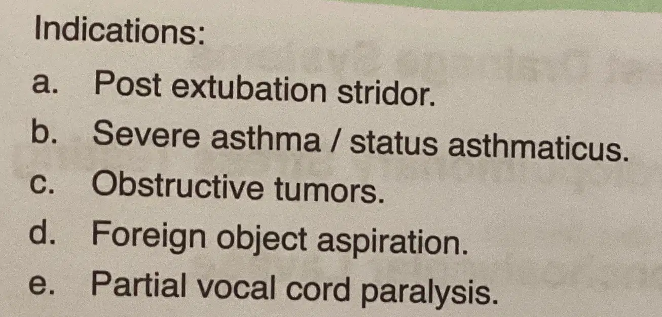
Heliox

HBO

iNO

Appearance of pleural fluid

Pleural effusion
Special procedures

BAL

Cardiopulmonary stress test

Cardioversion

Defibrillation

Chest tubes
Mechanical Ventilation

Initial settings for infant

Vent patient monitoring

Spontaneous monitoring

Normal loop graphics

weaning
NPPV

Indications
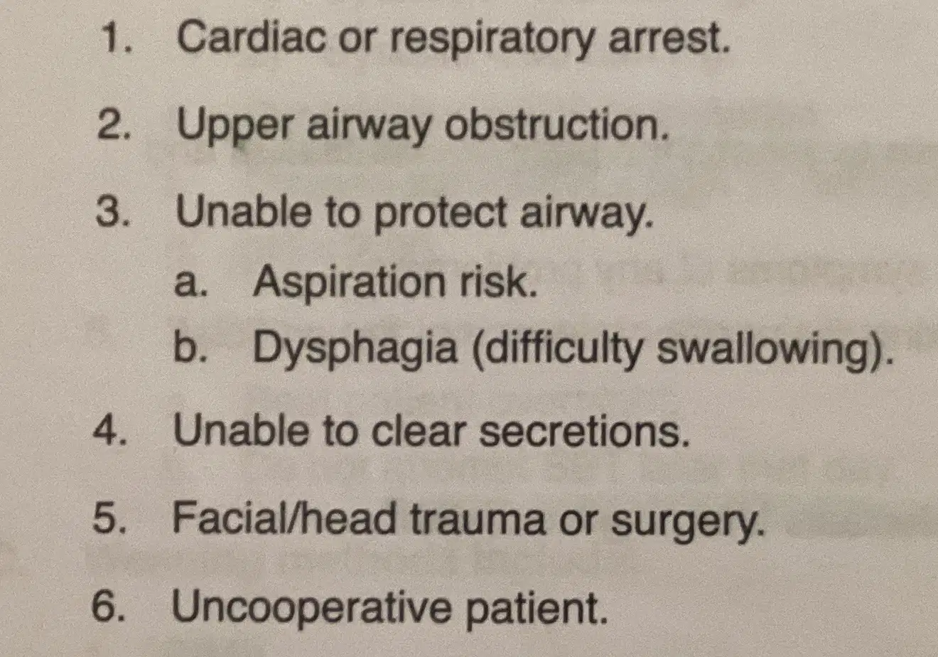
Contraindications

Initial settings
Breathing Techniques

SMI, IPPB, aerosol therapy, etc
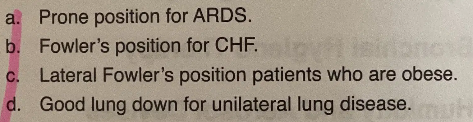
Patient position, oxygenation

IPPB

Bronchial hygiene therapy

PEP
Breathing techniques with medication devices

MDI

DPI

BNP level in CHF, <100 normal
ECG reading and treatment

A-Flutter

A-Fib

PVC

V-tach

V-flutter/Fib
ECG reading and treatment

Asystole

Ischemia

Injury

Infarction