
Font size
 Worksheets
First week of cardiology
Total questions: 101
Worksheet time: 58mins
The truncus arteriosus will become the _______
ascending aorta and pulmonary trunk
smooth portion of ventricles
trabeculated portion of the ventricles
trabeculated part of the atria
The primitive atrium of the heart tube will become
ascending aorta and pulmonary trunk
smooth portion of ventricles
trabeculated portion of the ventricles
trabeculated part of the atria
The primitive ventricle of the heart tube will become the
ascending aorta and pulmonary trunk
smooth portion of ventricles
trabeculated portion of the ventricles
trabeculated part of the atria
The bulbus cordis will become
ascending aorta and pulmonary trunk
smooth portion of ventricles
trabeculated portion of the ventricles
trabeculated part of the atria
A newborn is diagnosed with transposition of the great vessels shortly after birth. Which of the following maternal conditions is most commonly associated with an increased risk of this congenital heart defect?
gestational hypertension
gestational diabetes
maternal hypothyroidism
maternal anemia
A term newborn presents with central cyanosis and minimal response to oxygen therapy. Echocardiography confirms transposition of the great vessels (TGV). Which of the following is the most appropriate immediate pharmacologic intervention to stabilize the infant?
Administer indomethacin to close the ductus arteriosus
Begin a dopamine infusion to improve cardiac output
Administer prostaglandin E1 to maintain ductal patency
Initiate furosemide to reduce pulmonary congestion
Match the following
ascending aorta and pulmonary trunk
Truncus arteriosus
ventricular septum and av valves
endocardial cushions
Superior vena cava
common cardinal veins
portal and mesenteric veins
vitelline veins
coronary sinus and posterior RA
Sinus venosus
Which of the following best explains the functional closure of the foramen ovale in a healthy newborn shortly after birth?
Decreased systemic vascular resistance increases right atrial pressure, sealing the foramen ovale
increased pulmonary vascular resistance reidrects blood through the foramen ovale, closing it
Increased pulmonary venous return raises left atrial pressure, functionally closing the foramen ovale
closure of the ductus arteriosus increases right atrial pressure, forcing the foramen ovale shut
Which of the following is a potential complication of a patent foramen ovale (PFO) in an adult?
volume overload of the right atrium leading ot heart failure
right to left shunting of oxygenated blood causing systemic hypoxia
passage of venous thrombi to the left atrium, increasing the risk of systemic embolism
increasing pulmonary circulation pressure leading to pulmonary edema
How will an atrial septal defect present on auscultation
Fixed wide split of S2 and possible systolic ejection murmur at the left upper sternal border.
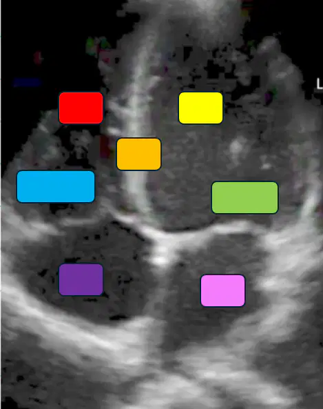
Match the following
Blue
Tricuspid
Green
mitral valve
Orange
Septal wall
Red
Right ventricle
Purple
Left atrium
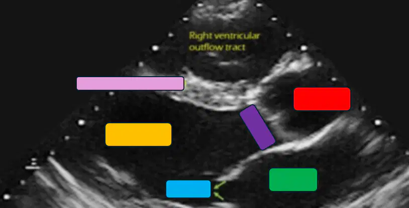
Match the following
Blue
mitral valve
green
Left atrium
Blue
mitral valve
Red
Aorta
Purple
aortic valve
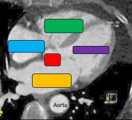
Match the following
blue
right atrium
Green
right ventricle
Orange
Left atrium
red
aorta
purple
Left ventricle
S1 heart sound is produced by the closure of
pulmonary and aortic valves
tricuspid and mitral valves (AV)
S1 heart sound is produced by the closure of
pulmonary and aortic valves
tricuspid and mitral valves (AV)
S2 heart sound is produced by the closure of
pulmonary and aortic valves
tricuspid and mitral valves (AV)
S1 heart sound is best appreciated at the _____ and is accentuated by placing patient in the ______ position
What can pause pathological S2 splitting that is wide and fixed
severe aortic stenosis; left bundle branch block
atrial septal defect
pulmonary stenosis; right bundle branch block
What can pause pathological S2 splitting that is wide (but not fixed)
severe aortic stenosis; left bundle branch block
atrial septal defect
pulmonary stenosis; right bundle branch block
What can pause pathological S2 splitting that is paradoxical
severe aortic stenosis; left bundle branch block
atrial septal defect
pulmonary stenosis; right bundle branch block
Where is the P2 component of S2 best appreciated
erbs point
Cardiac apex
2nd R parasternal ICS
2nd L parasternal ICS
Where is the A2 component of S2 best appreciated
erbs point
Cardiac apex
2nd R parasternal ICS
2nd L parasternal ICS
Physiologic S2 splitting will cause a ______ of A2 and P2 during ________
wide fixed splitting; expiration
What is a paradoxical S2 split (seen in severe aortic stenosis and left bundle branch block)
A paradoxical S2 split occurs when P2 closes before A2
Narrowing of S2 split in inspiration is indicative of ______ splitting
wide fixed splitting
In a patient with excessive blood volume you will hear what heart sound early in diastole, after S2 (aka gallop)
S3 heart sound is physiologic in what conditions
S4 heart sound is physiologic in what conditions
S3 heart sounds are pathologic in what conditions
Young and fit, pregnancy
systolic heart failure, mitral regurgitation, aortic regurgitation
Elderly individuals
diastolic heart failure, chronic hypertension, aortic stenosis
S4 heart sounds are pathologic in what conditions
Young and fit, pregnancy
systolic heart failure, mitral regurgitation, aortic regurgitation
Elderly individuals
diastolic heart failure, chronic hypertension, aortic stenosis
____ is produced by atrial kick due to high left ventricular pressures
S1
S2
S3
S4
S1 results from the closure of the AV valves. A softer/absent S1 can result from an immobile mitral valve. This can be seen in ________
Severe/late mitral stenosis
Early mitral stenosis
Systole starts when the _____ close
______ begins after the closing of the mitral valve and is characterized by the highest oxygen consumption
During the cardiac cycle, the highest amount of oxygen consumption is during ________
isovolumetric contraction
The T wave in the cardiac cycle represents what event
The ______ of the aortic pressure demarcates the end of systole and the onset of diastole, just after the aortic valve closes.
systolic peak
diastolic minimum
pulse pressure
Which part of the cardiac cycle is associated with the QRS complex on ECG
Isovolumetric contraction contraction
The _____ phase of the cardiac cycle occurs after the opening to the aortic valve
Once left ventricular pressure is lower than the aortic pressure, _____
mitral valve opens
Aortic valve closes
tricuspid valve closes
aortic valve closes
Pulmonary valve closes
Coronary blood flow is going to peak during
early systole
early diastole
late systole
late diastole
Which heart sound occurs due to rapid filling into a volume overloaded left ventricle
S1
S2
S3
S4
What heart sound is heard during atrial systole at the conclusion of diastole
S1
S2
S3
S4
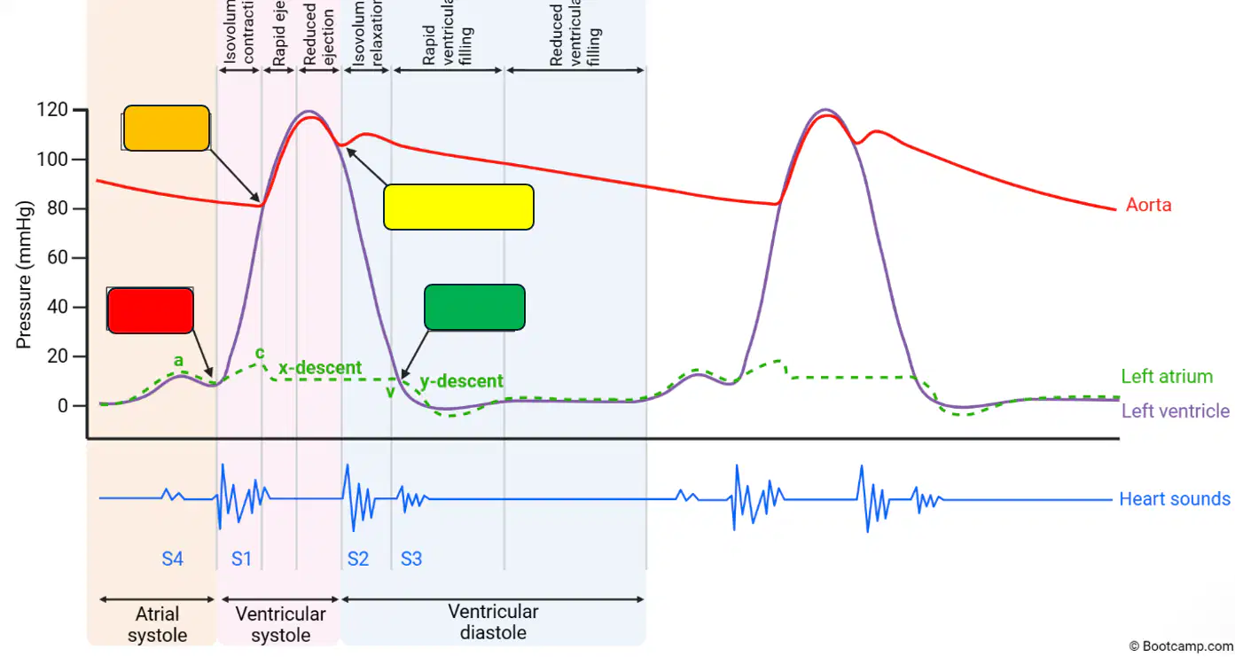
Red
Mitral valve closes
Aortic valve opens
Aortic valve closes
mitral valve opens
Orange
Mitral valve closes
Aortic valve opens
Aortic valve closes
mitral valve opens
Yellow
Mitral valve closes
Aortic valve opens
Aortic valve closes
mitral valve opens
Green
Mitral valve closes
Aortic valve opens
Aortic valve closes
mitral valve opens
Generally speaking, the jugular venous waveform focuses on
right side of the heart
left side of the heart
systole only
diastole only
The P wave on an ecg represents ______
If a p wave was absent from an ecg, what would be affected in the jugular venous wave form
A wave
C wave
X descent
V wave
Y descent
Which component of the jugular venous waveform corresponds to ventricular contraction
A wave
C wave
X descent
V wave
Y descent
Which component of the jugular venous waveform corresponds to atrial relaxation
A wave
C wave
X descent
V wave
Y descent
Which component of the jugular venous waveform corresponds to atrial filling
A wave
C wave
X descent
V wave
Y descent
Which component of the jugular venous waveform corresponds to ventricular filling
A wave
C wave
X descent
V wave
Y descent
How would tricuspid regurgitation present on a jugular venous wave form
Absent A-waves
Enhanced V wave
Increased Y descent
Blunting of Y descent
Cannon A-waves
How would atrial fibrillation present on a jugular venous wave form
Absent A-waves
Enhanced V wave
Increased Y descent
Blunting of Y descent
Cannon A-waves
How would tamponade present on a jugular venous wave form
Absent A-waves
Enhanced V wave
Increased Y descent
Blunting of Y descent
Cannon A-waves
How would constrictive pericarditis present on a jugular venous wave form
Absent A-waves
Enhanced V wave
Increased Y descent
Blunting of Y descent
Cannon A-waves
In aortic stenosis, the left ventricular pressure will be
unaffected
much greater than the aortic pressure
much lower than the aortic pressure
the same as the aortic pressure
Which of the following best describes the effect of chronic aortic regurgitation on pulse pressure
narrowed pulse pressure due to decreased stroke volume
widened pulse pressure due to increased stroke volume and decreased diastolic pressure
No change in pulse pressure because cardiac output remains constant
widened pulse pressure due to increased diastolic pressure
Which of the following will present with the loss of the dicrotic notch
Aortic stenosis
Aortic regurgitation
Mitral stenosis
Mitral regurgitation
Which of the following will present with a tall V-wave
Aortic stenosis
Aortic regurgitation
Mitral stenosis
Mitral regurgitation
Which of the following will present with much greater left atrial pressures (relative to left ventricular pressures) during diastole
Aortic stenosis
Aortic regurgitation
Mitral stenosis
Mitral regurgitation
Which of the following would not classically result from aortic regurgitation
Increased pulse pressure
Increased aortic pressure during diastole
Loss of dicrotic notch
increased aortic pressure during systole
Right sided heart failure often results from
Primary pulmonary problem (i.e. pulmonary hypertension)
Secondary systemic problem (i.e. peripheral edema)
Secondary pulmonary problem (i.e. pulmonary edema)
Primary systemic problem (i.e. hypertension, increased SVR)
Match the phases
Phase 0
Sodium rapidly enters cells; Ca slowly
Phase 1
Sodium channels close
Phase 2
K+ rapidly exits cell; Ca++ slowly enter
Phase 3
Ca++ channels close; K+ exits
Phase 4
Na+/K+ pump restores resting potential
Class 1 Arrhythmics
sodium channel blocker
beta blocker
potassium channel blocker
calcium channel blocker
Class 2 Antiarrhythmics
sodium channel blocker
beta blocker
potassium channel blocker
calcium channel blocker
Class 3 Antiarrhythmics
sodium channel blocker
beta blocker
potassium channel blocker
calcium channel blocker
Class 4 Antiarrhythmics
sodium channel blocker
beta blocker
potassium channel blocker
calcium channel blocker
The ______ represents the duration of the ventricular action potential
P wave
T wave
QT Interval
QRS Interval
PR Interval
The ____ represents the conduction time from atrium to ventricle
P wave
T wave
QT Interval
QRS Interval
PR Interval
The ______ represents the intraventricular conduction time (ventricular depolarization)
P wave
T wave
QT Interval
QRS Interval
PR Interval
The ______ represents the conduction time from atrium to ventricle
P wave
T wave
QT Interval
QRS Interval
PR Interval
Match the following antiarrhythmic drugs
Quinidine, Procainamide
1a
Lidocaine, phenytoin
1b
Verapamil, diltiazem
4
Amiodarone, sotalol
3
Propranalol, metaprolol
2
______ are used for refractory Vtac and WPW, as well as Afib without any structural cardiac or valvular changes.
Flecainide and propafenone
Quinidine and procainamide
lidocaine
propanolol and metaprolol
Which class of antiarrhythmics block voltage gated sodium channels, inhibiting depolarization (phase 0), as well as lower selectivity to block calcium and potassium channels
Class I antiarrythmics
Class II antiarrythmics
Class III antiarrythmics
Class IV antiarrythmics
Which group of antiarrhythmics elongate atrial refractory period, slow SA rate, and slow conduction through the AV node
Class I antiarrythmics
Class II antiarrythmics
Class III antiarrythmics
Class IV antiarrythmics
Non-selective Beta1 and Beta2 blocker.
Propranolol
Esmolol
Labetalol
Metaprolol
Which of the following is a side effect that is more strongly associated with/unique to propranolol amongst the other beta blockers
AV block
bradycardia
hypotension
exercise intolerance
nightmares
Lidocaine, a Class 1B antiarrhythmic, decreases the AP duration and is effective in treating ventricular arrhythmias post MI. Generally, class 1 antiarrhythmics decrease the conduction of depolarized cardiac cells. Which component of the ventricular action potential is influenced by Class 1 antiarrhythmics?
Phase 0
Phase 1
Phase 2
Phase 3
Phase 4
A 66 year old female presents to the emergency department with substernal chest pain. ECG reveals an ST elevation myocardial infarction (STEMI) and the patient undergoes percutaneous coronary intervention with successful stunting. The following day, she complains of palpitations and shortness of breath. Her blood pressure is 100/72 mmHg. The attending physician opts for medical management of this patient’s arrhythmia. Which of the following antiarrhythmics is contraindicated in this patient
Amiodarone
Mexiletine
Flecainide
Procainamide
Sotalol
A researcher is studying a new compound for use in the acute treatment of ischemic ventricular arrhythmia. When tested on myocardial cells, it causes a decrease in the slope of phase 0 depolarization and a decrease in the duration of the action potential. The compound exhibits increased activity when exposed to ischemic ventricular tissue. The experimental drug resembles which of the following
Disopyramide
Propafenone
Dofetilide
Carvedilol
Lidocaine
What is the most efficient class III antiarrhythmic drug
Two major drug interactions with amiodarone are with _____ and _____
aspirin, heparin
metformin, warfarin
lisinopril, heparin
warfarin, procainamide
Which class of antiarrhythmics can cause profound decreases in heart rate and contractility
Class I
Class II
Class III
Class IV
Side effects of _____ antiarrhythmics include gingival hyperplasia, peripheral edema, hypotension, dyspepsia, and hyperglycemia
Class I
Class II
Class III
Class IV
A 62 year old female presents to the emergency department with a 2 hour history of chest pain with ST-segment elevations in leads II, III, and aVF. She has a history of diabetes millitus. She receives emergent angioplasty and stunting but has recurrent runs of ventricular tachycardia after the procedure. She is then prescribed an antiarrhythmic drug that acts by slowing phase 3 depolarization of the action potential. Which of the following medications was she most likely prescribed.
Diltiazem
Procainamide
Esmolol
Quinidine
Sotalol
______ Inhibits the membrane of Na/K pump; enhancing myocardial contractility. Stimulates parasympathetic system via the vagus nerve; inhibits SAN rate and decrease conduction through AVN.
Digoxin/digitalis
adenosine
Mg++
Procainamide
K+
_____ is a cofactor of the membrane Na/K ATPase pump and a calcium ion antagonist; decreasing HR
Digoxin/digitalis
adenosine
Mg++
Procainamide
K+
________ stabilizes the membrane potential; increases the refractory period of the SA node and slows conduction through the AV node.
Digoxin/digitalis
adenosine
Mg++
Procainamide
K+
A 50 year old woman presents with a facial rash, arthralgia and muscle pain (myalgia) for the last 3 months. She has been treated with drug X for the past 6 months. Lab findings reveal elevated antinuclear antibodies (ANA) with homogenous patterns. Drug X is most likely which of the following?
Procainamide
Imipramine
Thioridazine
Tetracycline
Rifampin
Match the following following the physiological effects of adrenergic receptors
Alpha-1
NE>E
Alpha-2
E>NE
Beta-1
E=NE
Beta-2
E>>NE
Vasoconstriction of blood vessels of:
-Skin
-GIT
-Kidney
-Brain
Alpha-1; postsynaptic
Alpha-2;
Presynaptic
Beta-1;
Postsynaptic
Beta-2;
Postsynaptic
Inhibits release of norepinephrine; at skeletal muscle synapses inhibits alpha1-mediated constriction in skeletal muscle arteries
Alpha-1; postsynaptic
Alpha-2;
Presynaptic
Beta-1;
Postsynaptic
Beta-2;
Postsynaptic
Gq protein coupled activates phospholipase C; PIP2–>IP3 + DAG
Alpha-1; postsynaptic
Alpha-2;
Presynaptic
Beta-1;
Postsynaptic
Beta-2;
Postsynaptic
Gi protein coupled inhibits adenyl cyclase
ATP—> X —> cAMP
Alpha-1; postsynaptic
Alpha-2;
Presynaptic
Beta-1;
Postsynaptic
Beta-2;
Postsynaptic
FDA Indication:
-Bradyarrhythmias (heart blocks)
-Opthamology for retinal dilator
-Organophosphate poisoning
Atropine
Ipratropium/tiotropium
scopolamine
oxybutynin
Dicyclomine
FDA Indication:
-Bradyarrhythmias (heart blocks)
-Opthamology for retinal dilator
-Organophosphate poisoning
Atropine
Ipratropium/tiotropium
scopolamine
oxybutynin
Dicyclomine
FDA Indication:
Bronchiodilator COPD and asthma
Atropine
Ipratropium/tiotropium
scopolamine
oxybutynin
Dicyclomine
FDA Indication:
Motion sickness
Post-op nausea/vomiting
Atropine
Ipratropium/tiotropium
scopolamine
oxybutynin
Dicyclomine