
 Worksheets
Pathology Supple Set C
Total questions: 20
Worksheet time: 7mins
1-4 weeks after group A beta hemolytic nephritogenic streptococci infection of the pharynx or impetigo is etiology of.
Acute Proliferative GN
Chronic Proliferative GN
Acute Poststreptococcal GN
Chronic Poststreptococcal GN
Pathogenesis of APGN.
Glomerular deposition of immune complex from circulating or planted antigen
Proliferating & damage of glomerular cells
Infiltration of leucocytes, neutrophils
Hyper-complementemia
Focal proliferative GN.
Down syndrome
Goodpasture's syndrome
SLE
Diabetes Mellitus
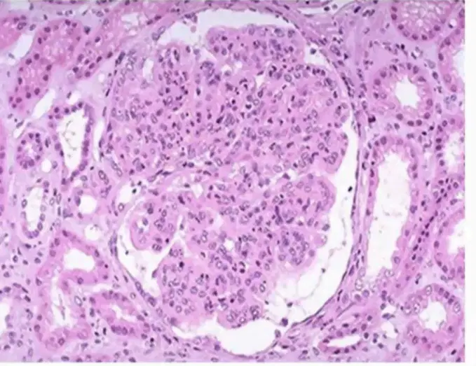
Microscopic changes in the glomeruli of APGN.
Glomerulus show diffuse hypercellularity
Glomerulus show diffuse hypocellularity
Caused by intracapillary luecocytes (neutrophils), and proliferation of intrinsic glomerular cells
Caused by extracapillary luecocytes (neutrophils), and proliferation of extrinsic glomerular cells
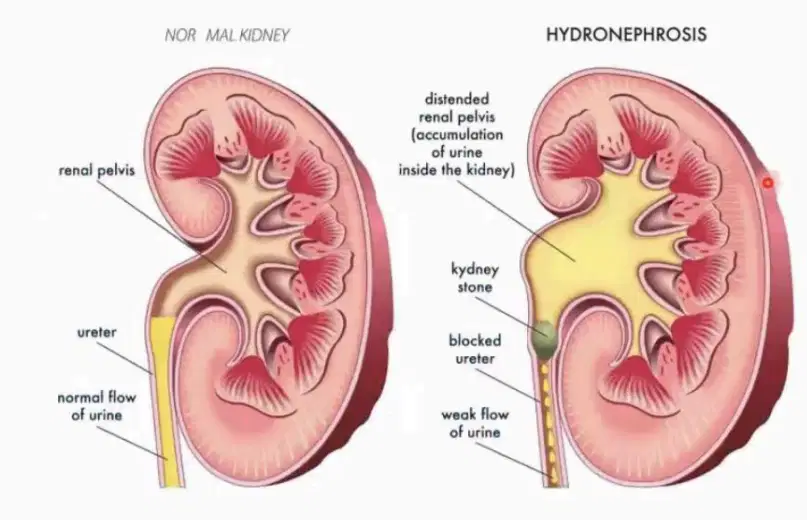
Definition of hydronephrosis.
Dilatation of renal pelvis and calyces associated with progressive hypertrophy of kidney due to obstruction to inflow of urine.
Dilatation of renal pelvis and calyces associated with progressive atrophy of kidney lead to obstruction to outflow of urine.
Dilatation of renal pelvis and calyces associated with progressive atrophy of kidney due to obstruction to inflow of urine.
Dilatation of renal pelvis and calyces associated with progressive atrophy of kidney due to obstruction to outflow of urine.
Pathogenesis of hydronephrosis.
1. partial obstruction decrease GFR
2. filtrate diffuse back affected pelvi-calyces
3. overwork and dilated causing renal atrophy
4. obstruction cause interstitial inf. reaction lead to fibrosis
1. Fully obstruction decrease GFR
2. filtrate diffuse back affected pelvi-calyces
3. overwork and dilated causing renal atrophy
4. obstruction cause interstitial inf. reaction lead to fibrosis
1. partial obstruction decrease GFR
2. filtrate diffuse back affected pelvi-calyces
3. overwork and dilated causing renal hypertrophy
4. obstruction cause interstitial inf. reaction lead to fibrosis
1. partial obstruction increase GFR
2. filtrate diffuse back affected pelvi-calyces
3. overwork and dilated causing renal atrophy
4. obstruction cause interstitial inf. reaction lead to fibrosis
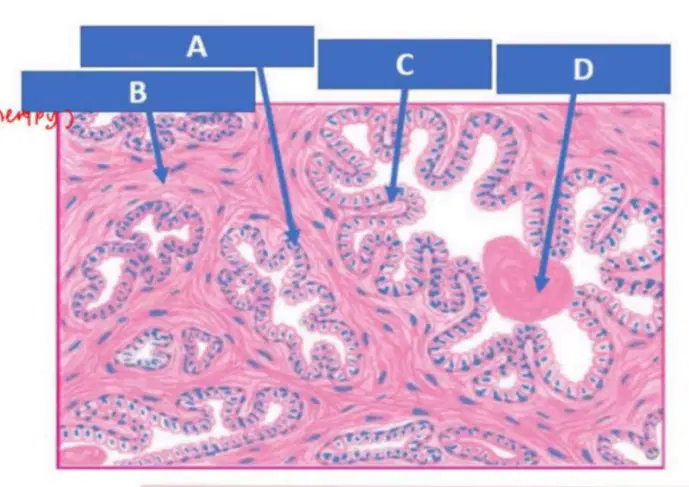
Label.
A : Double layered epithelium (tall columnar & basal cells)
B : Fibromuscular stroma
C : Papillary infolding
D : Corpora amylacea
A : Double layered epithelium (simple columnar & basal cells)
B : Corpora amylacea
C : Papillary infolding
D : Fibromuscular stroma
A : Double layered epithelium (simple columnar & basal cells)
B : Fibromuscular stroma
C : Papillary infolding
D : Corpora amylacea
A : Fibromuscular stroma
B : Double layered epithelium (simple columnar & basal cells)
C : Papillary infolding
D : Corpora amylacea
Method of microbial spreading.
Air-borne
Direct
Hematogenous
Local extension

Choose the correct.
Bacterial : Turbid
Viral : Clear
Fungal : Clear
Bacterial : Clear
Viral : Clear
Fungal : Turbid
Bacterial : > 40
Viral : Normal-Low
Fungal : Normal
Bacterial : < 40
Viral : Normal
Fungal : Normal-Low
Cloudy appearance showing.
Platelets
Neutrophils
Bacterial
Glucose
Increase opening pressure cause swelling of meninges and lead to.
(choose most correct)
haemorrhage
pus formation
Headache
Scars
Bacteria consume glucose, so glucose level will be?
increase
decrease
Immune response cause WBC's count?
increase
decrease
Elevated protein level due to?
Formation of BBB
Breakdown of BBB
Contiguos spread.
Otitis media
Sunisitis
Endocarditis
Dental infection
Hematogenous spread.
Pericarditis
Pulmonary embolism
Endocarditis
Lung abscess
Immunocompromised State.
HIV/AIDS
Chemotherapy
Pre-transplant patients
Diabetes Mellitus
Trauma or Neurosurgery.
Ventriculoperitoneal shunt
Penetrating head injuries
Craniotomy
Lobatomy
Complications of bacteria infection.
Hydronephrosis
Hydrocephalus
Cerebral abscess
Sensorineural hearing loss
How bacteria infection developed into an abscess.
1. Microb enter brain via (contigous/hematogenous spread)
2. Aided by patient increase immunity
3. Early stage (cerebritis)
4. Late stage (abscess formation)
1. Microb enter brain via (contigous/hematogenous spread)
2. Aided by patient reduce immunity
3. Early stage (abscess formation)
4. Late stage (cerebritis)
1. Microb enter brain via (contigous/hematogenous spread)
2. Aided by patient increase immunity
3. Early stage (abscess formation)
4. Late stage (cerebritis)
1. Microb enter brain via (contigous/hematogenous spread)
2. Aided by patient reduce immunity
3. Early stage (cerebritis)
4. Late stage (abscess formation)