
 Worksheets
ECG115 Exam 2 Review
Total questions: 58
Worksheet time: 35mins
Junctional rhythms have an intrinsic rate of:
40-60 bpm
20-40 bpm
10-20 bpm
60-100 bpm
Which of the following dysrhythmias is NOT considered part of the supraventricular tachycardia classification?
atrial fibrillation
sinus tachycardia
ventricular tachycardia
junctional tachycardia
In this rhythm, the SA node fails to initiate the electrical activity, and the rhythm is established by the backup pacemaker site.
sinus rhythm
junctional rhythm
atrial rhythm
ventricular rhythm
The (a) on the ECG is primarily affected by junctional rhythms.
Which type of heart block is sometimes referred to as Wenckebach.
1st degree HB
2nd degree type 1 HB
2nd degree type 2 HB
3rd degree HB
In a first-degree AV block, all sinus impulses get through to the ventricles.
True
False
Which of the following AV blocks have consistent PR intervals? Select all that apply.
1st degree HB
2nd degree type 1 HB
2nd degree type 2 HB
3rd degree HB
In Mobitz II second-degree AV block, where do the P waves originate?
SA node
AV node
Bundle of His
Purkinje fibers
A third degree heart block is best characterized by:
no relationship between P waves & QRS complexes
a steady relationship between the P waves & QRS complexes
a prolonged but regular PR interval
a progressively increasing PR interval
Wenckebach is characterized by:
consistent PR intervals
wide bizarre QRS complexes
absent P waves
gradually prolonging PR intervals
A first-degree heart block is best characterized by:
a completely normal EKG
no relationship between QRS complexes & P waves
a progressively increased PR interval, then a missing QRS, in a cyclical pattern
a prolonged PR interval
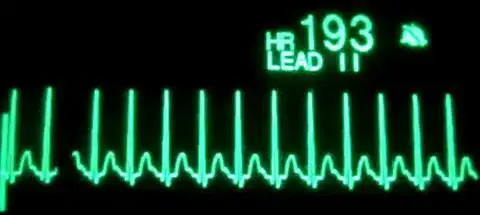
Identify the following rhythm.
Supraventricular Tachycardia (SVT)
Junctional Tachycardia
Sinus Tachycardia
Ventricular Fibrillation
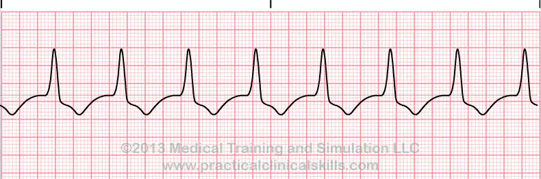
Identify the following rhythm.
Junctional bradycardia
Premature junctional contractions
Junctional escape rhythm
Accelerated junctional rhythm
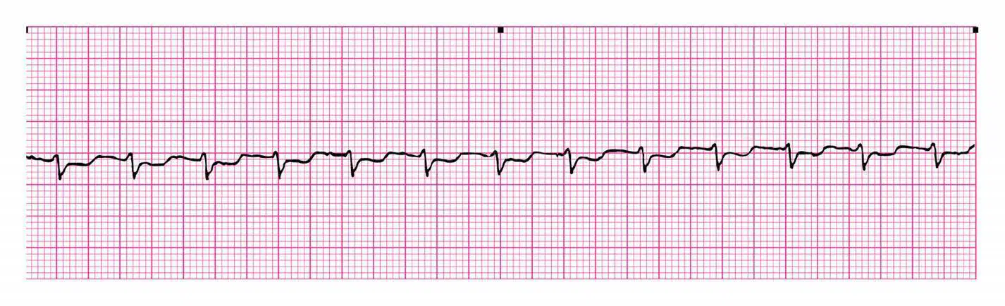
Identify the following rhythm.
Accelerated junctional rhythm
Junctional bradycardia
Premature junctional contractions
Junctional tachycardia
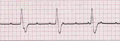
Identify the following rhythm.
Third-degree AV block
Second-degree Type I AV block
Second-degree Type II AV block
First-degree AV block

Identify the following rhythm.
Third-degree AV block
Second-degree Type I AV block
Second-degree Type II AV block
First-degree AV block
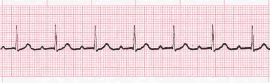
Identify the following rhythm.
Third-degree AV block
Second-degree Type I AV block
Second-degree Type II AV block
First-degree AV block
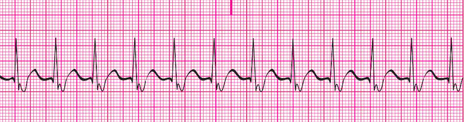
Identify the following rhythm.
Accelerated junctional rhythm
Premature junctional contractions
Junctional escape rhythm
Junctional tachycardia
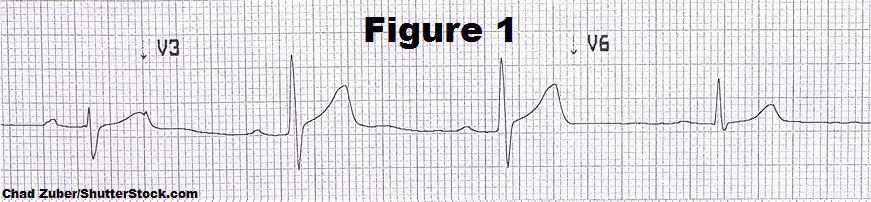
Identify the following rhythm.
Third-degree AV block
Second-degree Type I AV block
Second-degree Type II AV block
First-degree AV block

Identify the following rhythm.
Accelerated junctional rhythm
Junctional bradycardia
Premature junctional contractions
Junctional escapte rhythm
Junctional tachycardia
Which rhythm results in abnormally wide QRS (>0.12s) with a slur caused by pre-excitation?
SVT
WPW
A-Fib
A-Flutter
Increased automaticity and reentry circuits are two examples of mechanisms by which arrhythmias are generated.
True
False
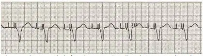
Which of the following rhythms is indicated by the EKG tracing provided?
atrial pacing
atrioventricular pacing
biventricular pacing
atriobiventricular pacing
A dual chamber pacemaker has
the ability to defibrillate the heart
electrodes in the right atrium and right ventricle
a pacing wire in the RV only
leads in the RA and both ventricles

Which of the following correctly describes this EKG rhythm?
atrial flutter
atrial fibrillation
ventricular tachycardia
ventricular fibrillation

Which of the following correctly describes this EKG rhythm?
atrial flutter
atrial fibrillation
ventricular tachycardia
ventricular fibrillation
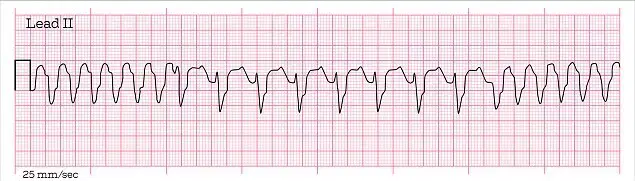
Which of the following correctly describes this EKG?
monomorphic (unifocal) ventricular tachycardia
polymorphic (multifocal) ventricular tachycardia
The electrocardiogram rhythm strip shows the following:
The P waves and QRS complexes are regular. The PR interval is 0.16s and the QRS complex measures 0.06s. The overall heart rate is 64 bpm. This cardiac rhythm is which of the following?
normal sinus rhythm
sinus bradycardia
sick sinus syndrome
first-degree heart block
The electrocardiogram rhythm strip shows the following:
There are no P waves, the QRS complexes are wide, and the ventricular rate is regular but does not exceed 100 bpm. Which of the following cardiac rhythms is this?
PVCs
VFib
VTach
Sinus Tach
A sonographer is viewing the cardiac monitor in a patient's room and notes that the patient has just gone into ventricular tachycardia. The patient is awake, alert, and has good skin color. The tech would prepare to do which of the following?
immediately defibrillate
start chest compressions
prepare for cardioversion
assess the patient's pulse and blood pressure
A patient is having frequent premature ventricular contractions. Which of the following assessments are priority to evaluate if the patient is experiencing changes in cardiac output?
blood pressure & peripheral perfusion
sensation of palpitations
causative factors such as caffeine
precipitating factors such as infection
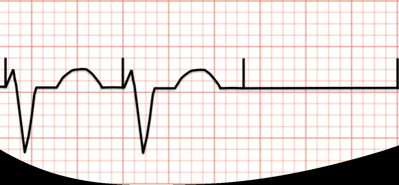
This EKG tracing shows which of the following rhythms?
atrial pacing
ventricular pacing with failure to capture
atrioventricular pacing with failure to sense
atriobiventricular pacing with failure to pace
A healthcare worker notes that a patient with sinus rhythm has a premature ventricular contraction that falls on the T wave of the preceding beat. The client's rhythm suddenly changes to one with no P waves or definable QRS complexes. Instead, there are coarse wavy lines of varying amplitude. This rhythm is?
VTach
VFib
AFib
Asystole
A sonographer is performing a complete echocardiogram on a patient who has sustained an MI. During the scan, the sonographer notes eight PVCs in one minute on the cardiac monitor. The patient is receiving oxygen. The sonographer's first course of action should be to:
notify the nurse caring for the patient
continue the scan until completed
increase the patient's oxygen therapy
request the nurse bring the patient pain medication
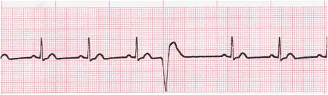
Which of the following correctly describes the EKG?
PVCs
PJCs
PACs
None of the above
Match the PVC types with the corresponding pattern/explanation.
six or more PVCs per minute
frequent PVCs
all complexes have a similar shape, originating from only one irritable focus
unifocal PVC
every other complex is a PVC
bigeminy
every third complex is a PVC
trigeminy
PVCs have varied shapes and forms, originating from more than one irritable foci
multifocal PVC
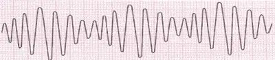
Identify the rhythm.
Torsades de Pointes
VTach
VFib
None of the above
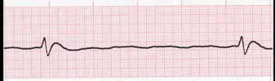
Identify the rhythm.
agonal rhythm
atrial escape rhythm
asystole
sinus exit block
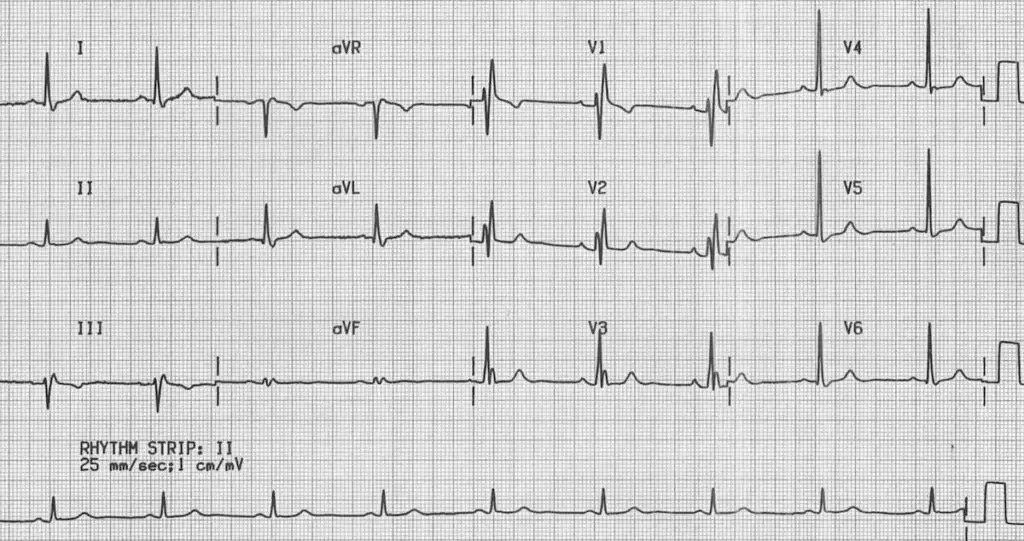
Identify the rhythm.
Normal Sinus Rhythm
Right Bundle Branch Block
Left Bundle Branch Block
Complete Heart Block
Which of the following does NOT cause PVCs?
stress
caffeine/alcohol
ischemia
pacemaker insertion
hypoxia
If ventricular fibrillation is not corrected, the patient will decline into (a) .
(a) refers to the changes that occur on a 12-lead ECG as a result of the position of the heart within the patient's chest?
What is the most common axis deviation?
left
right
extreme right
extreme left
Match the leads with the corresponding portion of the heart viewed.
septal wall
Leads V1 and V2
anterior wall
Leads V3 and V4
lateral wall
Leads I, aVL, V5, and V6
inferior wall
Leads II, III, and aVF
Match the coronary artery & branch with the corresponding portion of the heart viewed.
anterior wall of LV
LCA - left anterior descending
septal wall of LV
LCA - septal branch
inferior wall of LV
RCA - posterior descending
lateral wall of LV
LCA - left circumflex
Which two leads do we refer to when determining axis deviation? Select all that apply.
Lead I
Lead II
Lead III
aVF
aVL
Which is NOT a cause of left axis deviation?
LVH
pregnancy
obesity
emphysema
tall, thin adults & children
Which part of the heart does the standard 12-lead ECG focus on directly?
RA
RV
LV
LA
Which of these are factors that determine the seriousness of a Myocardial Infarction? Select all that apply.
coronary arteries involved
location & extent of infarction
vital heart structures involved
time
age
Which is the most helpful indicator for differentiating between a myocardial injury and myocardial infarction?
pathologic Q wave
physiologic Q wave
T wave inversion
ST elevation
An extreme right axis deviation can be caused by situs inversus or dextrocardia.
True
False
Infarction must be seen in at least ___ anatomically contiguous leads.
2
3
4
only required to see it in 1 lead
When totaled, how many millimeters is the minimum necessary to clinical suspect hypertrophy?
3 mm
5 mm
3.5mm
35mm
Where does the initial electrical impulse originate from in a junctional rhythm?
SA node
AV node
Bundle of HIS
Purkinje fibers
The sonographer notices the rhythm on the echo machine changes from normal sinus to ventricular fibrillation. Which of the following interventions should they do next?
assess if the patient is awake and/or has a pulse
request the nurse assigned to the patient call a rapid response
run to get the crash cart and oxygen tank
immediately call a code blue and start CPR
In LVH, more electricity flows through the heart. In practical terms, how will the QRS complexes appear?
wider than normal and bizarre
narrower than normal and bizarre
taller and deeper than normal
shorter and wider than normal
How many electrodes are used in a 12-lead EKG?
6
10
12
13
Put the steps to 12-lead ECG interpretation in the correct order.
Determine the rhythm & regularity, then calculate the atrial & ventricular rate
Identify the P wave morphology, then measure the PR interval
Measure the QRS duration & analyze the configuration
Evaluate the leads in a group
Identify morphological changes

