
NEW
Font size
 Worksheets
type shi
Total questions: 66
Worksheet time: 33mins
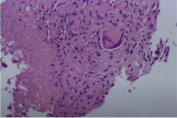
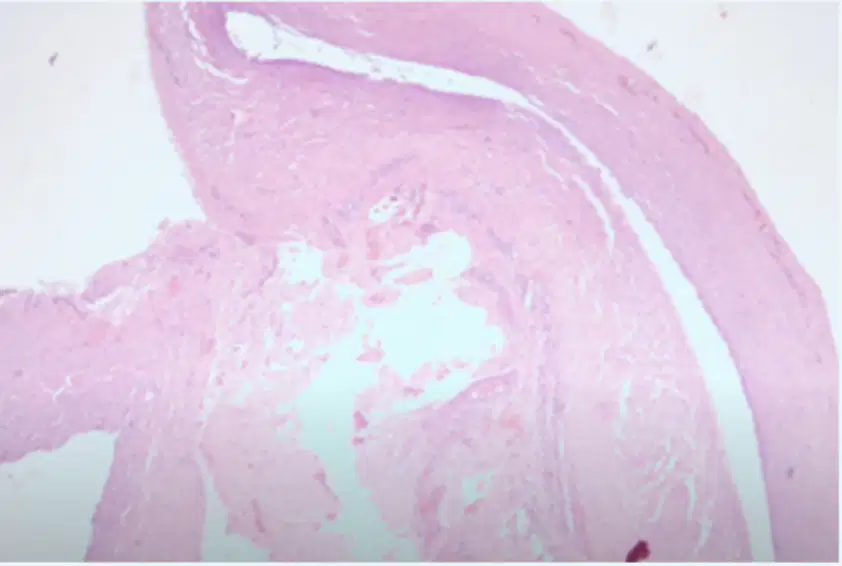
A 20-year age girl had her right humerus fractured after a car accident happened in last three weeks. As the fracture did not properly cured, a yellowish fluid was then developing on the skin around the spot of fracture. The X-ray showed a focus bone loss near the lower end of the humerus. A curettage biopsy was performed and its microscopic appearance is as shown here. What is the microscopic characteristic of the diagnosis?
Lymphocytes cells, plasma cells, fibrotic tissue, necrosis
Necrosis caseous, lymphocytes cells, fibrotic tissue
Lymphocytes cells, plasma cells, fibrotic tissue
Necrosis caseous, datia Langhans, fibrotic tissue
Necrosis caseous, datia Langhans cells, epitheloid cells
The most sensitive and specific for supporting diagnosis Rhematoid arthritis:
Uric acid serum
Anti Cyclic Citrillinated Peptide (anti CCP) serum
Hydroxyproline Urine
Osteocalcin serum
Acid Phosphatase serum
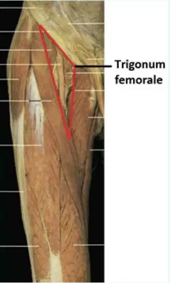
The anatomical structures present in the trigonum femorale from medial to lateral sequentially are:
N. femoralis, a. femoralis, v. femoralis
V. femoralis, n. femoralis, a. femoralis
V. femoralis, a. femoralis, n. femoralis
A. femoralis, v. femoralis, n. femoralis
N. femoralis, v. femoralis, a. femoralis
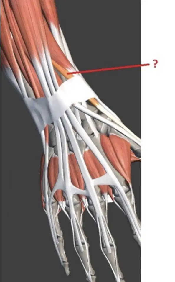
The muscle that marked by this red line is:
M. adductor pollicis brevis
M. adductor pollicis longus
M. extensor pollicis brevis
M. extensor pollicis longus
M. extensor indicis
SEORANG PENDERITA DATANG DENGAN KELUHAN NYERI LUTUT KANAN. PADA PEMERIKSAAN CAIRAN SENDI LUTUT DIDAPAT HASIL: Volume > 3,5 ml, Clarity: Opaque, Color: Mixed, WBC >100.00/mm3, PMN > 75 %, Gram stain: often positive. Kesimpulan dari hasil cairan sendi diatas adalah:
Hemorrhagic
Normal
Non inflammatory
Septic
Inflammatory
Result synovial fluid examination from laboratory: Volume: 3,7 ml, Viscosity: high, Clarity: clear, WBC: 400/ml, PMN: 20% According data what is your conclusion about this synovial fluid:
Septic
Inflammatory
Non inflammatory
Normali
Hemorrhagic
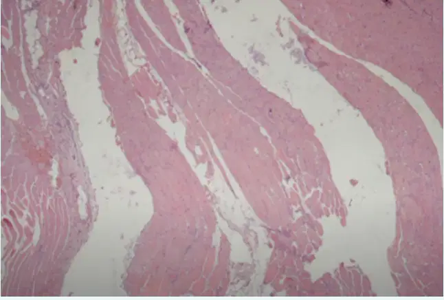
A 10-years old boy appears with muscular weakness. He is unable to play with other children, unable to keep up and quickly becoming tired. A muscle biopsy is performed and its microscopic appearance at low magnification is as shown here. The condition of his muscle cells are:
Atrophy
Dystrophy
Hypertrophy
Malignant tumor
Neoplasma
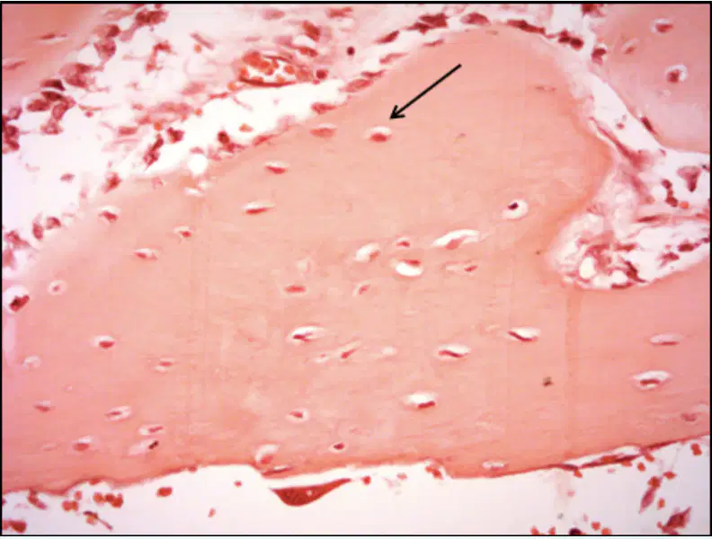
What is the function of the pointed cell?
Bone resorption
Maintain mineral concentration of matrix
Bone formation

Sulcus nervi ulnaris is located at:
Bone D
Bone A
Bone B
Bone C
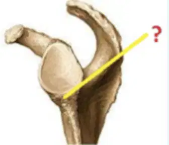
What is the name of the muscle that originates from the area marked with this yellow line?
M. biceps brachii caput breve
M. biceps brachii caput longum
M. triceps brachii caput longum
M. triceps brachii caput laterale
M. deltoideus
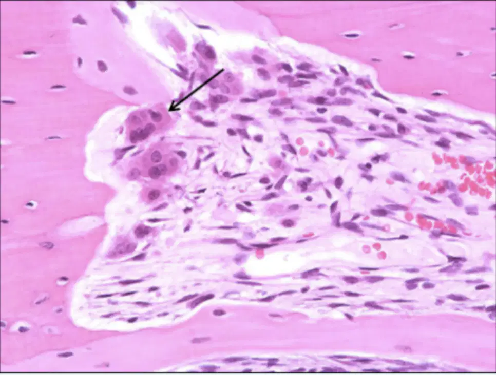
What is the pointed cell name?
Osteoblast
Osteocyte
Osteoclast
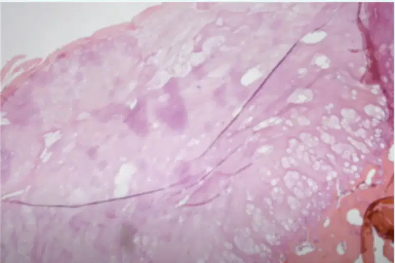
A -40-years old lady consulted for the exophityc mass in the interphalanx joint digiti V. The mass was excised and its microscopic appearance is as shown here. The best diagnosis is:
Chondrosarcoma
Osteoma
Osteochondroma
Chondroma

What is the characteristic of this tissue?
Murky/dirty matrix
Chondrocytes located between collagenous fibers
Many isogenic cells in clear matrix
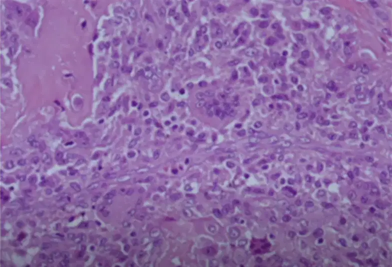
A 63-year-old man go to the clinic with pain in his right distal femur. He says that it has been last 3 months. The mass was excised and its microscopic appearance is as shown here. The best diagnosis is
Bursitis Benign
Granuloma
Ganglion cyst Berian
Fibroma
Giant cell tumor

A 33-year-old woman presented with an 8-month history of progressive right-sided headache, ipsilateral facial pain, and hearing impairment. CT shows a large, right middle cranial fossa mass that measured 6.7 × 5.6 × 4.2 cm. A curettage biopsy was performed and its microscopic appearance is as shown here. Based upon the appearance of that microscopic-characteristics to support your diagnosis are:
Lobular capillary hemangioma.
Osteosarcoma
Osteochondroma
Ganglion cyst
Bursitis

What is the function of this calcification?
To form bones at the base of the skull and long bones
To repair damage cartilage
To form the flat bones of the face, most of the cranial bones, & the clavicles
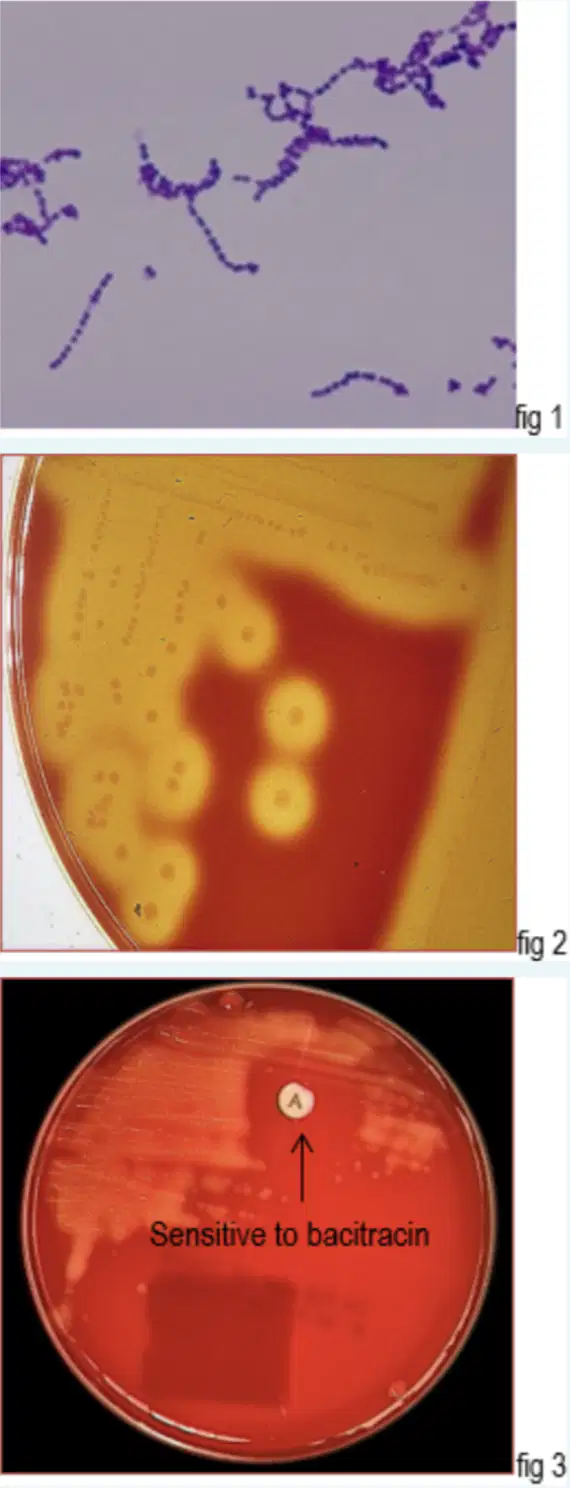
A woman complaint of pain in his knee. A synovial fluid was taken for Gram staining (fig. 1) and culture on blood agar (fig. 2 and 3). What is likely identity of the organism? State the name of these organism!
Streptococcus pneumoniae
Streptococcus agalactiae
Streptococcus pyogenes
Staphylococcus epidermidis
Staphylococcus aureus
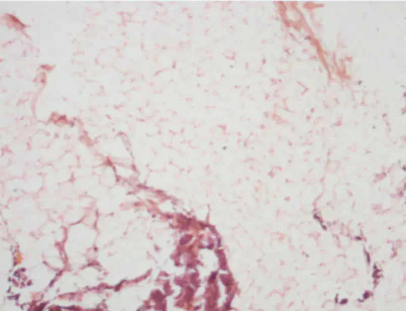
A 3 cm soft mass/tumor is palpable in the right shoulder of a 42-years old male. The excised mass is performed and it showed a yellow cut surface. Based on the appearance of that microscopic, your best diagnosis is
Schwanoma
Fibroma
Lipoma
Hemangioma
Neurinoma

A purulent fluid was taken for Gram staining (fig. 1) and culture on MacConkey Agar medium revealed round, small, smooth, rough, and mucoid colonies (fig. 2). What is likely identity of the organism? State the name of these organism!
Candida albicans
Klebsiella pneumoniae
Escherichia coli
Pseudomonas
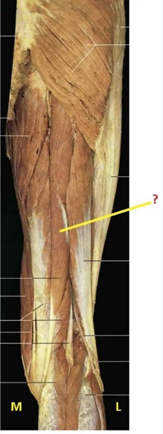
The muscle that marked by this yellow line is:
M. biceps femoris caput longum
M. semimembranosus
M. semitendinosus
M. adductor magnus
M. rectus femoris
The inferior margin of the Trigonum auscultationis is:
M. rhomboideus minor
M. levator scapulae
M. trapezius
M. teres minor
M. latissimus dorsi
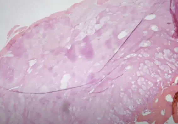
A 3 cm soft mass/tumor is palpable in the right shoulder of a 42-years old male. T performed and it showed a dark brown cut surface. The most likely condition of his mass /tumor is:
Malignant neoplasm of epithelial tissue
Malignant neoplasm
Benign neoplasma of epithelial tissue
Benign neoplasm of soft tissue
Myoglobin is exclusive to striated and non striated skeletal or cardiac muscle. After a myocardial infark or damage to any muscle tissue, myoglobin will start elevated in serum in
1 hours after damage muscle tissue/ myocardial infark
3 hours after damage muscle tissue/ myocardial infark
4 hours after damage muscle tissue/ myocardial infark
2 hours after damage muscle tissue/ myocardial infark
Less than 1 hours after damage muscle tissue/myocardial infark
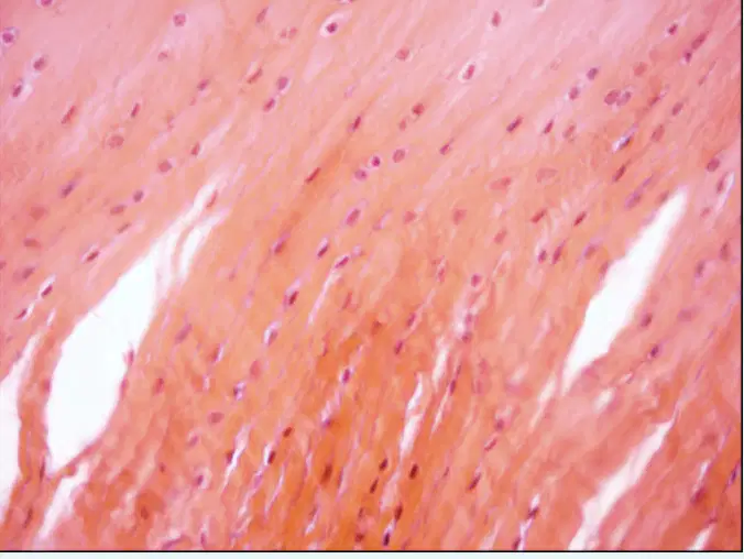
Where is this tissue located in the body?
Auricle
Intervertebral disc
Trachea
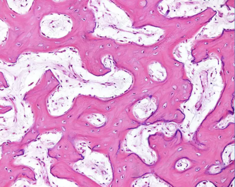
What is the function of this calcification?
To repair damage cartilage
To form bones at the base of the skull and long bones endochandral
To form the flat bones of the face, most of the cranial bones, & the clavicles
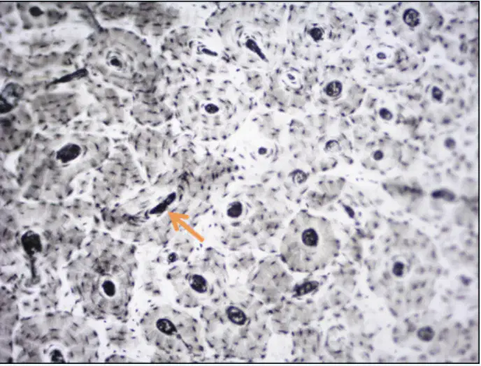
What is the canal name?
Interstitial canal
Haversiann canal
Volkmann canal.
Facies poplite is located at:
Anterior part of os. femur
Posterior part of os. humerus
Posterior part of os. femur
Anterior part of os. tibia
Anterior part of os. Fibula
A-40-years old lady consulted for the exophityc mass in the interphalanx joint digiti V. The mass was excised and its microscopic appearance is as shown here. The most likely causative of her exophytic mass is:
Ganglion cyst
Infectious disease
Osteomielitis
Malignant neoplasm

A young man is diagnosed with osteomyelitis. The result of gram-stained is GRAM POSITIVE COCCI IN CLUSTER The result of culture is performed in fig. 1 What is the most possible causative organism? State the name of this organism!
Methicillin-resistant Staphylococcus aureus
Oxacillin-sensitive Staphylococcus aureus
Carbapenemase resistance Klebsiella pneumoniae
Extended-spectrum beta-lactamases
Vancomycin-resistant Enterococci
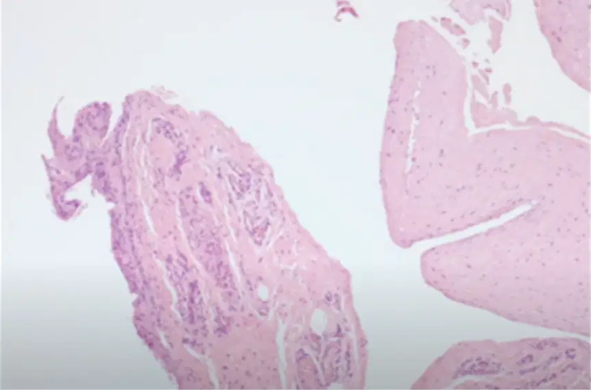
A 40 year age lady consulted for a 1 cm cystic mass in her right elbow. The X-ray showed a cystic mass in outer articular joint of the right elbow but no destruction on the bone. The enucleation of cystic mass was performed. Your best diagnosis based on the microscopic appearance is
Bursitis
Simple cyst of the bone
Synovitis
Ganglion cyst of the bone

Tuberositas pronatoria is located at:
Bone C
Bone A
Bone B
Bone D
Result macroscopic examination synovial fluid analysis to supporting diagnosis inflammation is:
Green
Clear
Light yellow
Cloudy
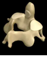
This is:
Os. axis
Os. atlas
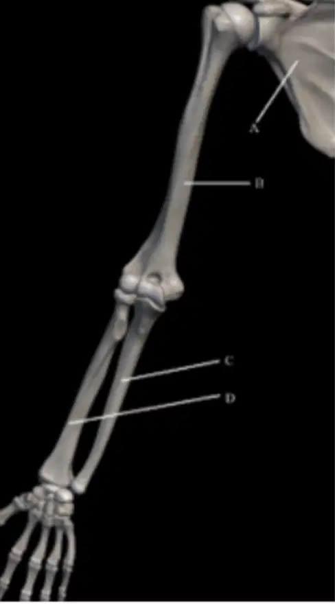
Tuberositas pronatoria can be found in:
Bone A
Bone B
Bone C
Bone D
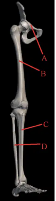
Linea aspera can be found in:
Bone A
Bone B
Bone C
Bone D
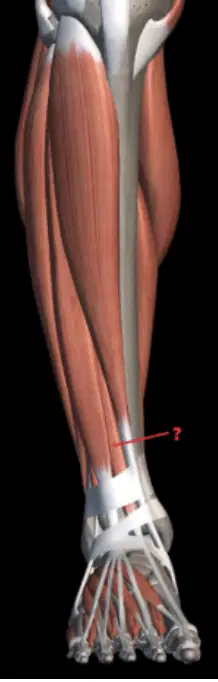
What muscle is this (Red line)?
M. tibialis anterior
M. extensor digitorum longus
M. extensor hallucis longus
M. flexor hallucis brevis
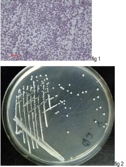
A purulent fluid was taken for Gram staining (fig. 1) and culture on nutrient media revealed round, raise shiny, and white-gray colonies. These colonies are sensitive to novobiocin (fig. 2) What is likely identity of the organism? State the name of these organism!
Escherichia coli
Pseudomonas aeruginosa
Staphylocococcus epidermidis
Candida albicans
Klebsiella pneumoniae
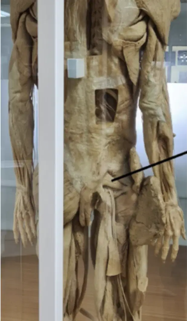
What muscle is this (Red line)?
M. quadratus femoris
M. gemellus superior
M. gemellus inferior
M. obturator internus
M. piriformis
Pertanda adanya infeksi / inflamasi di cairan sendi bila pada pemeriksaan cairan sendi didapat hasil
Glucose - less than 40 mg/dl; Protein - greater than or equal to 3 g/dl; LDH - less than 333 IU/L
Glucose - less than 40 mg/dI; Protein - greater than or equal to 3 g/di; L DH - greater than 333 IU/L
Glucose - greater than 40 mg/dl; P Protein - greater than or equal to 3 g/dl; DH - greater than 333 IU/L
Glucose - less than 40 mg/dI; Protein - less than 3 g/dl; LDH - less than 333 IU/L
Glucose - less than 40 mg/dl; Protein - less than 3 g/dl; LDH - greater than 333 IU/L
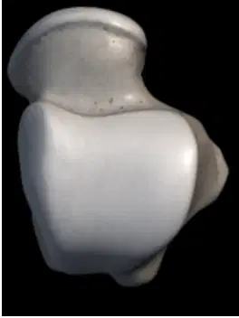
What bone is this?
Os. talus sinsitra
Os. calcaneus sisnistra
Os.calcaneus dextra
Os. talus dextra
A. vertebralis runs through foramen transversarium, and runs from ..... to ....
Cl-C7
C5- C2
C7 -C1
C2 - C7
C6 - C1
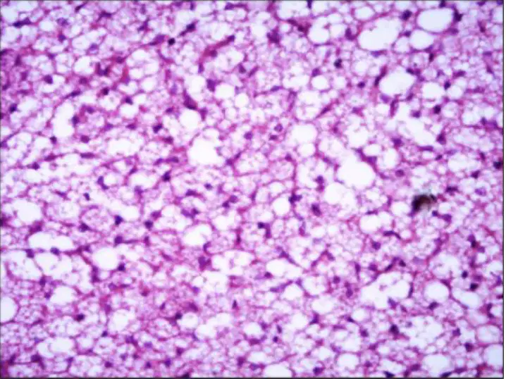
What is this tissue called?
Brown Adipose tissue
Reticular tissue
White adipose tissue
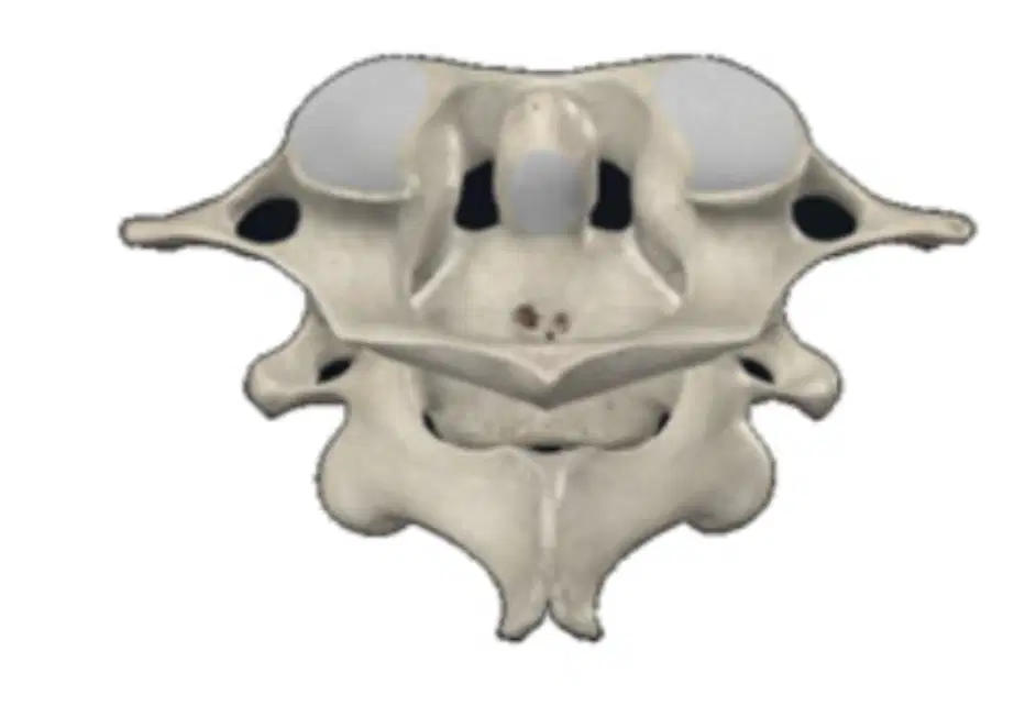
How many articulation locations are there between the two vertebrae in this picture?
1
3
4
5
2
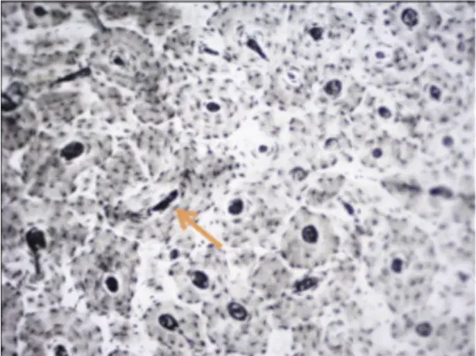
What is the canal name?
Interstitial canal
Haversian canal
Volkmann canal
What kind laboratory examination do you ask for supporting diagnosis if you think she suffer rheumatoid arthritis:
Uric Acid concentration
Erythrocyte Sedimentation Rate
Anti Cyclic Citrillinated Peptide
C Reactive Protein
WBC Count

Woman 25 year old , in the ICU with a ventilator for 1 month. The doctor suggested culturing the ventilator tube. The results of the bacteria growing look like the following:
Candida albicans
Pseudomonas aeruginosa
Escherichia coli
Klebsiella pneumoniae
Serratia marcescens
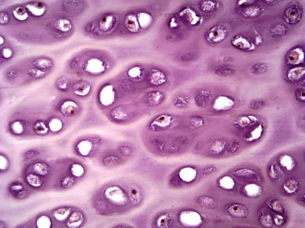
Where is this tissue located in the body?
Auricle
Trachea
Intervertebral disc
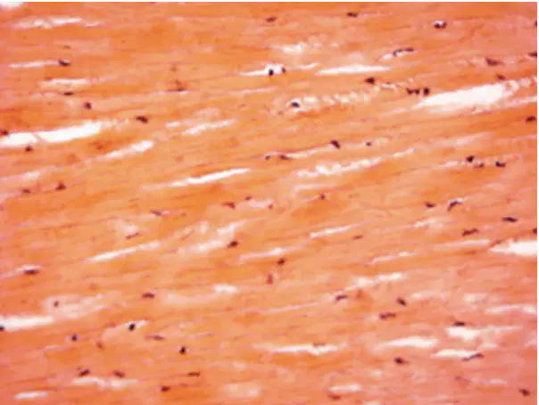
What is this tissue?
ELASTIC FIBRE
COLLAGEN FIBRE
RETICULAR FIBRE
Catalase test POSITIVE have for bacteria :
Staphylococcus aureus
Streptococcus pneumoniae
Streptococcus pyogenes
Streptococcus viridans
Enterococcus faecalis
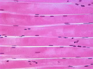
The characteristic of this tissue are...
Striated, the cell nuclei are located at the edge
Wavy, nuclei located between collagenous fiber
Thick & homogenous fiber, elongated shaped
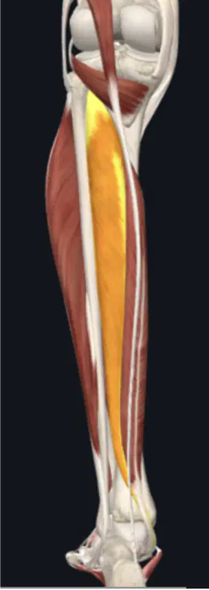
Image of the inferior extremity, seen from the posterior side. The muscle highlighted in yellow, if it becomes paralyzed, the person will have difficulty performing :
Plantarfleksi
Dorsifleksi
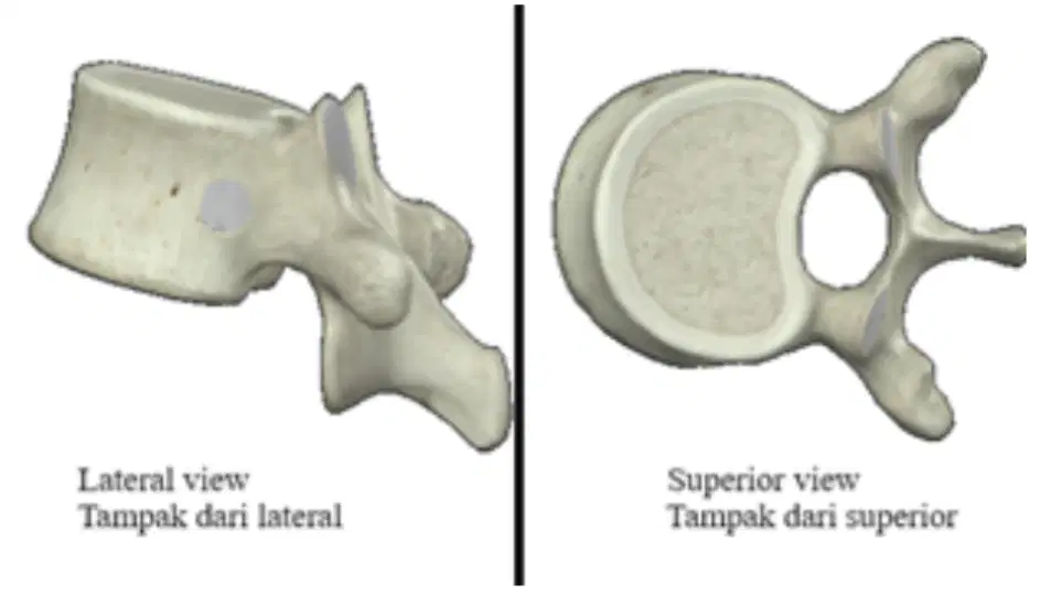
From the following picture, what is the name of this vertebra?
Vertebra cervicale
Vertebra Lumbales
Vertebra thoracicae
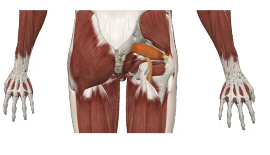
What is the name of the muscle highlighted in yellow?
M.obturator externus
M.quadriceps femoris
M.quadratus femoris
M.obturator internus
M.piriformis
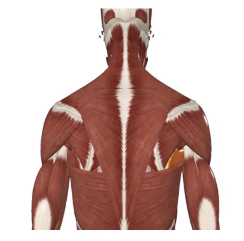
What is the name of the muscle highlighted in yellow?
M.teres mayor
M.latissimus dorsi
M.teres minor
M.trapezius
M.serratus anterior
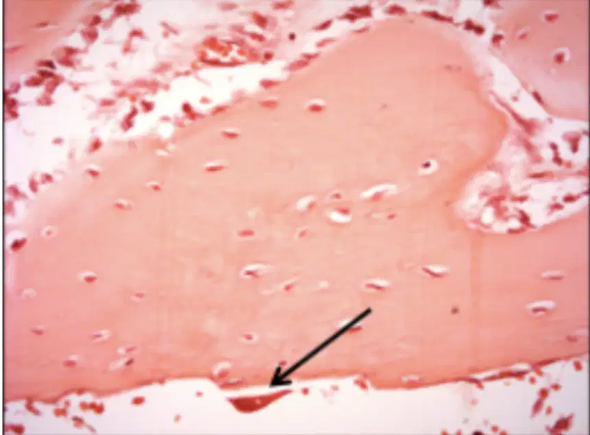
What is the pointed cell name?
Osteocyte
Osteoblast
Osteoclast
Disc antibiotics to detect MRSA and MSSA are:
Cefalosporin
Oxacillin
Floroquinolon
Ampicillin sulbactam
Amoxycillin
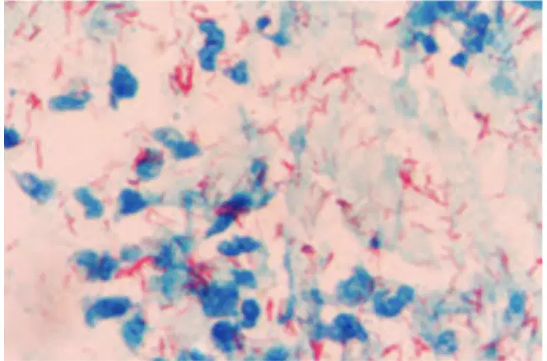
Man, 35 years old , a construction worker, came to the Regional Hospital with a complaint that his leg bones showed a lump that he had been suffering from for a long time. Then the doctor recommended tissue culture, the microscopic results are shown in the image below. What media needed ?
MacConkey
Agar Darah
Lowenstein Jensen
Nutrient Agar
Manitol Salt Agar
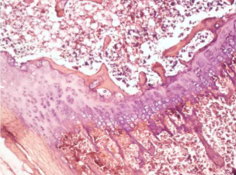
What is the function of this calcification?
To repair damage cartilage
To form bones at the base of the skull and long bones
To form the flat bones of the face, most of the cranial bones, & the clavicles
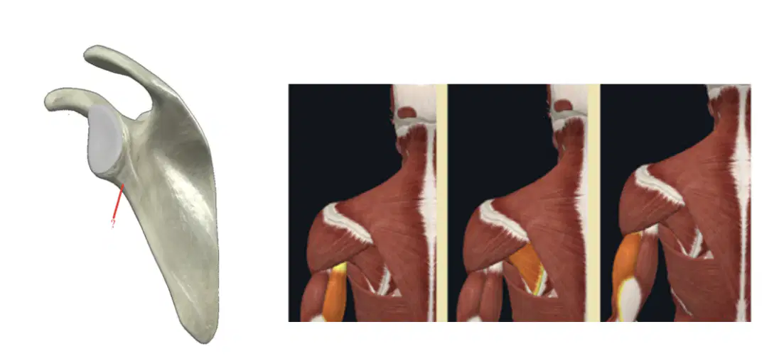
The tendons of what muscles do we find in the area marked with the red line?
Muscle A
Muscle B
Muscle C
We can find the pronatorial tuberosity at os. :
Ulna
Scapula
Radius
Humerus
Clavicula
Laboratory examination for “marker bone formation” :
Osteocalcin
Acid Phosphatase
Creatine kinase
Hydroxyproline Urine
Alkaline Phosphatase
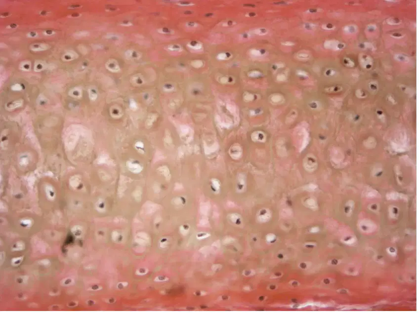
What is the characteristic of this tissue?
Murky / dirty matrix
Many isogenic cells in clear matrix
Chondrocytes located between collagenous fibers
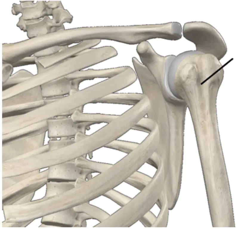
The tendons of what muscle can we find in the area indicated by the black line?
M. biceps branchii caput longum
M. triceps branchii caput laterale
M. triceps branchii caput longum
M. biceps branchii caput brevis
M. brachialis
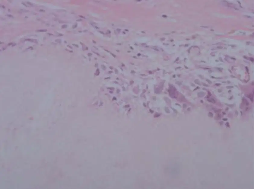
A 63-year-old man go to the clinic with acute pain in his right great toe. He says that it has been excruciatingly tender to touch for the last 2 days. The mass was excised and its microscopic appearance is as shown here.
The best diagnosis is?
gout
bursitis
ganglion cyst
fibroma
granuloma
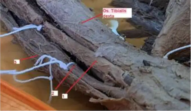
Muscle that function to extend the hallux is:
2
3
1
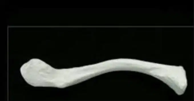
What bone is this?
The surface you see is the rough surface?
Os clavicula dextra
Os costae 1 dextra
Os costae 1 sinistra
Os clavicula sinistra

