
 Worksheets
The Heart and EKG Review
Total questions: 72
Worksheet time: 44mins
Which statement best explains why the muscle wall of the left ventricle is about twice as thick as the wall of the right ventricle?
It pumps blood only a short distance to the lungs.
It must generate higher pressure to pump blood through the entire body.
It contains fewer valves than the right ventricle.
It receives blood with more oxygen and therefore expands.
Pulmonary circulation primarily moves blood between which structures?
Heart and lungs
Heart and brain
Heart and skeletal muscles
Heart and kidneys
Which diagnostic tool records the electrical activity of the heart?
MRI
ECG (Electrocardiogram/EKG)
CT scan
Echocardiogram
In cardiac circulation, blood collected from the heart muscle gathers in the coronary sinus and enters the heart:
Through the superior vena cava
Directly through separate openings in the right atrium
Through the pulmonary veins
Through the left atrium
Systemic circulation begins when oxygenated blood leaves the heart through the:
Pulmonary artery
Pulmonary veins
Aorta
Coronary sinus
According to the lesson, the heart’s task is to pump oxygen-poor blood to the lungs and then pump oxygen-rich blood:
Back to the right atrium
To the rest of the body
Only to the brain
Into the coronary sinus
Which pair correctly identifies the protective structures around the heart that it is "protected by" in the lesson?
Sternum and ribs
Pericardium and rib cage
Pericardium and sternum
Diaphragm and pleura
Blood from the body’s systemic circulation returns to the heart through which vessels to reach the right atrium?
Pulmonary arteries
Superior vena cava and inferior vena cava
Aorta and coronary sinus
Pulmonary veins
From the right atrium, blood passes through which valve into the right ventricle?
Mitral (bicuspid) valve
Aortic valve
Pulmonary valve
Tricuspid valve
After entering the right ventricle, blood goes through the pulmonary valve and travels to the lungs via the:
Aorta
Pulmonary artery
Pulmonary vein
Coronary artery
Heart valve "leaflets" are called what?
Cusps
Chordae
Chordae tendineae
Papillary muscles
The mitral valve’s other common name is the bicuspid valve because it has:
Two cusps
Three cusps
Four cusps
One cusp
Which valves are found between the atria and the ventricles?
Pulmonary and aortic valves
Mitral and tricuspid valves
Aortic and mitral valves
Tricuspid and pulmonary valves
The fibrous cords that hold the atrioventricular valve leaflets in place are called:
Papillary muscles
Chordae tendineae
Coronary ligaments
Trabeculae carneae
These cords are anchored in the ventricles by which muscles?
Intercostal muscles
Papillary muscles
Cardiac skeletal muscles
Smooth muscles
The heart muscle is supplied by which artery, located at the aortic valve and forming the first branches off the aorta?
Pulmonary artery
Carotid artery
Coronary artery
Subclavian artery
Which circulation delivers nutrients and oxygen to all other parts of the human body besides the lungs?
Pulmonary circulation
Cardiac circulation
Systemic circulation
Portal circulation
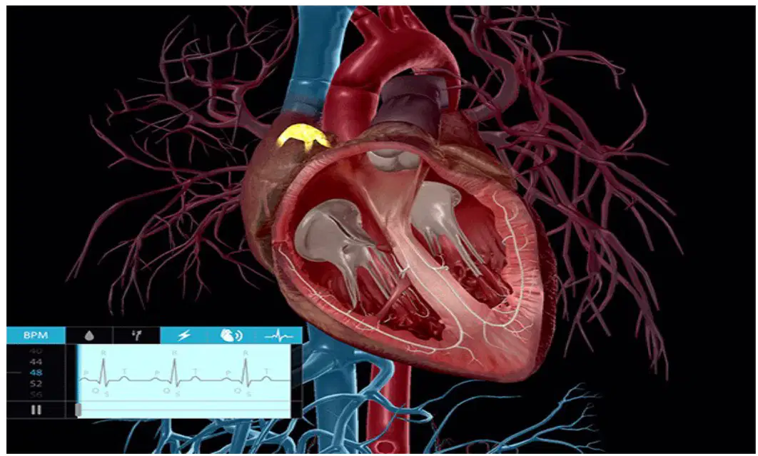
Based on the ECG image of a heart cycle shown, which tool would a healthcare provider use to interpret the heart’s electrical conduction patterns?
Stethoscope
Electrocardiogram (ECG/EKG)
Blood pressure cuff
Pulse oximeter
Which statement best describes an "end artery" in the coronary circulation?
An artery with many alternate pathways supplying the same area
An artery that has no significant collateral routes to the tissue it supplies
An artery that drains deoxygenated blood into the coronary sinus
An artery that only functions during diastole
If any branch of a coronary artery becomes blocked, what is the most likely effect on the heart muscle supplied by that branch?
The muscle is still fine because collateral vessels immediately take over
The muscle receives reduced blood flow but continues contracting normally
The muscle can become damaged due to lack of oxygen and nutrients
The muscle switches to receiving blood from the pulmonary veins
A right-dominant heart primarily receives posterior interventricular (descending) blood supply from which coronary artery?
Left coronary artery
Right coronary artery
Circumflex artery
Pulmonary artery
In a left-dominant heart, the posterior interventricular (descending) artery most commonly arises from which vessel?
Right coronary artery
Left anterior descending artery
Circumflex branch of the left coronary artery
Coronary sinus
Which node is considered the natural pacemaker that initiates the heartbeat?
Atrioventricular (AV) node
Sinoatrial (SA) node
Bundle of His
Purkinje fibers
A special branch of the right coronary artery that supplies the sinoatrial node is called the SA nodal artery. What is its primary role?
To carry deoxygenated blood from the SA node
To deliver oxygenated blood to the pacemaker region
To open the atrioventricular valves during systole
To drain into the coronary sinus directly
What is the main function of the coronary sinus?
Carry oxygenated blood to the left ventricle
Collect venous blood from the heart and return it to the right atrium
Initiate electrical impulses that trigger ventricular contraction
Regulate blood pressure during diastole
During systole and diastole, how do pressures typically compare in a normal blood pressure reading (e.g., 120/80 mmHg)?
Systolic pressure is lower than diastolic pressure
Systolic pressure is higher because the ventricles are contracting and ejecting blood
Both pressures are equal because the heart maintains constant force
Diastolic pressure is higher due to valve closure
Why is systolic pressure commonly around 120 mmHg while diastolic pressure is around 80 mmHg?
Arterial walls are more relaxed during systole
Ventricular contraction during systole generates greater force against arterial walls
Blood volume decreases during diastole
Venous return peaks in diastole
Which component conducts electrical impulses from the AV node into the ventricles?
Sinoatrial node
Bundle of His
Coronary sinus
Pulmonary trunk
Purkinje fibers primarily function to:
Initiate the heartbeat in the right atrium
Depolarize the ventricles rapidly to coordinate contraction
Return deoxygenated blood to the right atrium
Control blood pressure during diastole
What is the main function of the atrioventricular (AV) node in the heart’s conduction system?
Acts as the heart’s primary pacemaker
Delays the electrical impulse before it passes to the ventricles
Collects venous blood from the myocardium
Opens the semilunar valves during systole
Which statement about the pericardium is most accurate?
The heart is covered by a visceral pericardium and the cardiac cavity by a parietal pericardium, with a small amount of fluid between them to reduce friction
The heart is covered only by the parietal pericardium without any fluid
The pericardium’s fluid increases friction to strengthen contractions
The pericardium is a venous vessel that drains into the right atrium
Which vessels receive excess tissue fluid returned to general circulation by the lymphatic system?
Carotid arteries
Subclavian veins
Jugular veins
Aorta
Two main lymphatic ducts carry lymph back to the bloodstream. Which duct drains lymph from most of the body and empties into the left subclavian vein?
Right lymphatic duct
Thoracic duct
Coronary duct
Pulmonary duct
Which duct drains lymph from the right side of the head, neck, thorax, and right arm, emptying into the right subclavian vein?
Thoracic duct
Left lymphatic duct
Right lymphatic duct
Coronary sinus
Which heart layer is the tough, protective sac that surrounds the heart?
Pericardium
Epicardium
Myocardium
Endocardium
Which heart layer is the outer surface of the heart wall and is closely associated with the pericardium?
Endocardium
Epicardium
Myocardium
Septum
Which heart layer is composed primarily of cardiac muscle responsible for pumping action?
Pericardium
Epicardium
Myocardium
Endocardium
Which heart layer lines the interior chambers and helps provide a smooth surface for blood flow?
Pericardium
Epicardium
Myocardium
Endocardium
What structure divides the heart into right and left sides, forming internal walls between chambers?
Chordae tendineae
Interatrial and interventricular septum
Pericardial cavity
Coronary sulcus
Select the best description of the thoracic duct's drainage territory compared to the right lymphatic duct.
Thoracic duct drains only the right upper limb
Thoracic duct drains the majority of the body; right lymphatic duct drains the right head, neck, thorax, and right arm
Both ducts drain identical regions on opposite sides
Right lymphatic duct drains the majority of the body; thoracic duct drains only the left arm
A student traces lymph from the right arm back to the bloodstream. Which vessel does it enter last before becoming part of the blood?
Left subclavian vein
Right subclavian vein
Superior vena cava
Pulmonary vein
Which side of the heart primarily pumps oxygenated blood to the body?
Right heart
Left heart
Both sides equally
Neither side
Which side of the heart sends deoxygenated blood to the lungs?
Left heart
Right heart
Coronary circulation
Atria only
What is the main role of the coronary arteries?
Carry oxygen-poor blood to the lungs
Supply the heart muscle with oxygen and nutrients
Drain blood from the heart muscle into the right atrium
Open and close to prevent backflow in the ventricles
Which vessel collects most venous blood from the heart muscle and empties into the right atrium?
Aorta
Pulmonary trunk
Coronary sinus
Superior vena cava
Where does blood go after leaving the coronary sinus?
Left atrium
Right atrium
Left ventricle
Pulmonary veins
Which combination lists the two atria of the heart?
Left atrium and left ventricle
Right atrium and right ventricle
Left atrium and right atrium
Left ventricle and right ventricle
Blood entering the right atrium most directly arrives from which structures?
Pulmonary veins carrying oxygenated blood
Aorta carrying systemic blood
Superior and inferior vena cavae carrying systemic venous blood
Coronary arteries carrying nutrient-rich blood
Blood entering the left atrium most directly arrives from which structures?
Pulmonary veins carrying oxygenated blood
Pulmonary arteries carrying deoxygenated blood
Superior vena cava carrying venous blood
Coronary sinus collecting cardiac venous blood
Which combination lists the two ventricles of the heart?
Left ventricle and right ventricle
Left atrium and right atrium
Aorta and pulmonary trunk
Right atrium and left ventricle
To which circulation does the left ventricle pump blood?
Pulmonary circulation via the pulmonary trunk
Systemic circulation via the aorta
Coronary sinus to the right atrium
Pulmonary veins to the left atrium
Which valve is located between the right atrium and right ventricle?
Mitral (bicuspid) valve
Tricuspid valve
Aortic semilunar valve
Pulmonary semilunar valve
Which valve is located between the left atrium and left ventricle?
Tricuspid valve
Pulmonary semilunar valve
Mitral (bicuspid) valve
Aortic semilunar valve
Which structure prevents atrioventricular valve leaflets from inverting during ventricular contraction by anchoring to papillary muscles?
Chordae tendineae
Interventricular septum
Coronary sinus
Pulmonary veins
Where are papillary muscles found, and what is their function?
In the atria; they initiate electrical impulses
In the ventricles; they contract to tension the chordae tendineae
In the coronary arteries; they regulate blood flow
In the aorta; they open semilunar valves
Which valve opens to allow blood to exit the right ventricle into the pulmonary trunk?
Aortic semilunar valve
Pulmonary semilunar valve
Tricuspid valve
Mitral valve
Which valve opens to allow blood to exit the left ventricle into the aorta?
Pulmonary semilunar valve
Aortic semilunar valve
Tricuspid valve
Mitral valve

Identify this rhythm
Sinus tachycardia
Normal Sinus Rhythm
Supraventricular Tachycardia
Atrial fibrillation

Identify this rhythm
Sinus Tachycardia
Atrial fibrillation
Atrial flutter
Normal Sinus Rhythm

Identify this rhythm
Ventricular Fibrillation
Ventricular Tachycardia
Pulseless Electrical Activity
Atrial fibrillation


A patient is experiencing electrocardiogram changes. Which wave should the nurse recognize as representing ventricular repolarization?
Q wave
R wave
S wave
T wave
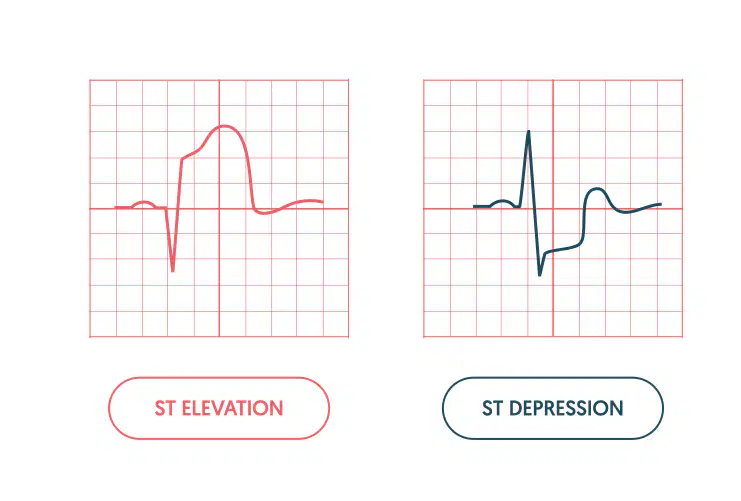
What does the depressed ST wave in the image possibly indicate
A heart block leading to ischemia
Rapid hear beat
low blood pressure
heart attack (myocardial infarction)
What does an elevated ST wave in the image possibly indicate
A heart block leading to ischemia
Rapid hear beat
low blood pressure
heart attack (myocardial infarction)
There are (a) pulmonary veins that return to the left atrium of the heart.
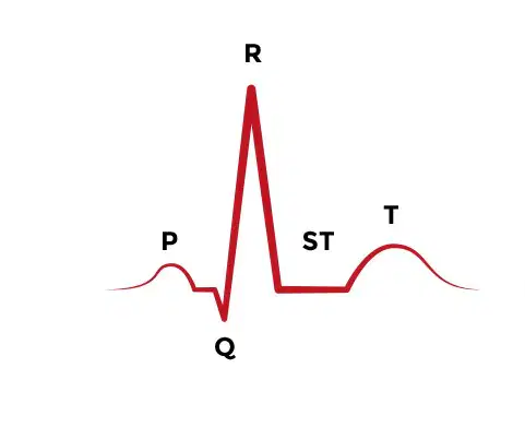
Look at the image. Why is the line labeled ST flat?
The heart is resting
The atria are repolarizing
The person has damage from a heart attack
They have a heart block
Label the heart
right atrium
left atrium
left ventricle
right ventricle
Correctly identify the following areas of the heart.

Match the following terms to their description
When blood leaves this chamber of the heart, it heads to the lungs.
Right ventricle
The main artery which distributes oxygenated blood to the entire body
Aorta
This blood vessel splits in two (L/R) and sends deoxygenated blood to the lungs to be refreshed.
Pulmonary artery
The chamber in the heart that receives oxygenated blood from the lungs.
Left atrium

