
 Worksheets
Neuron structures identification and slides
Total questions: 132
Worksheet time: 1hrs 6mins

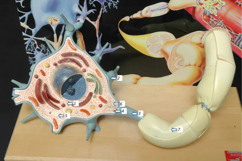
In the neuron model image with numbered labels, which structure is indicated by label 1 (major part of a neuron with nucleus and other organelles)?
Soma (cell body)
Dendrite
Axon
Schwann cell
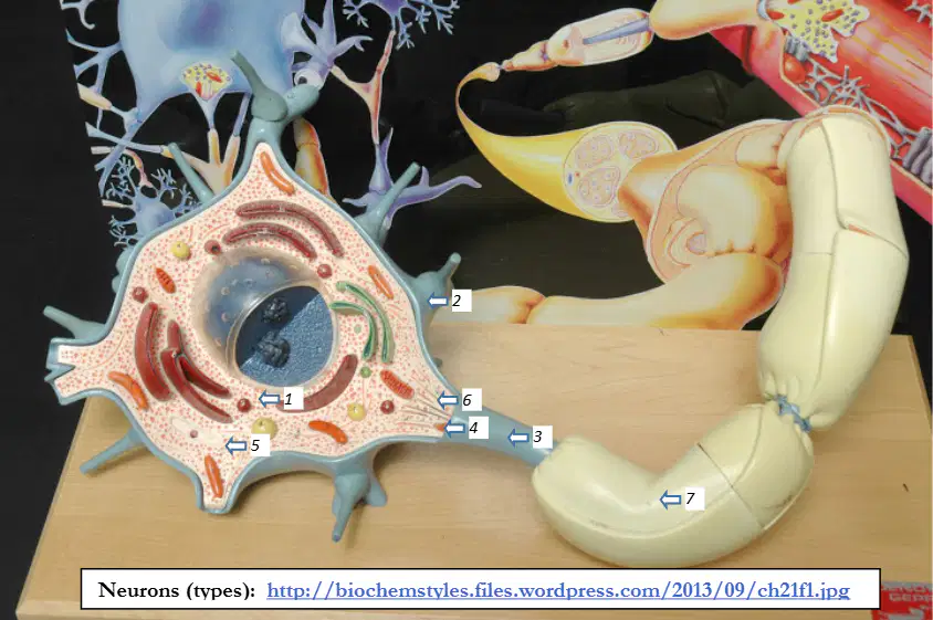
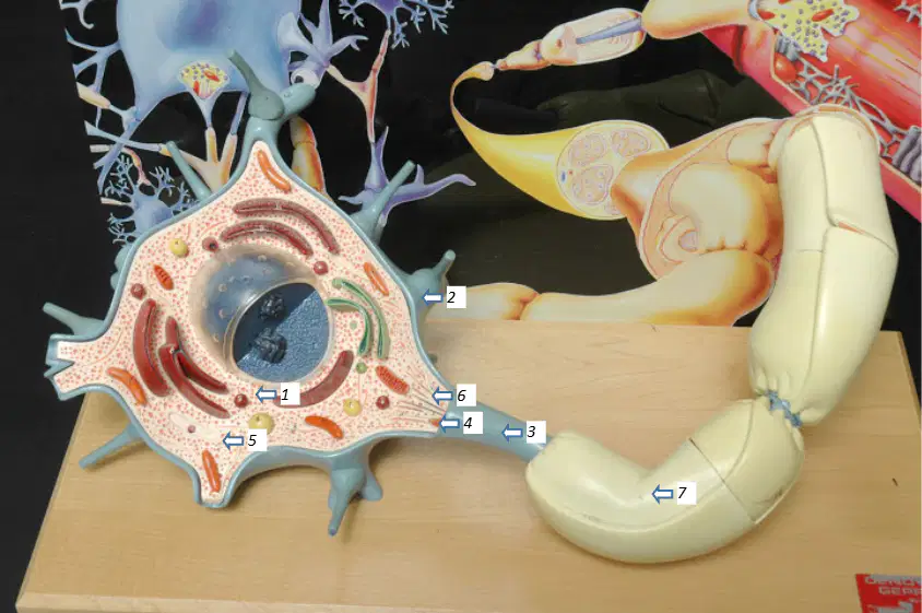
In the neuron model image with numbered labels, which structure is indicated by label 2 (thick, short projections from the soma that transmit signals to the soma)?
Dendrites
Axon
Node of Ranvier
Neurofibrils
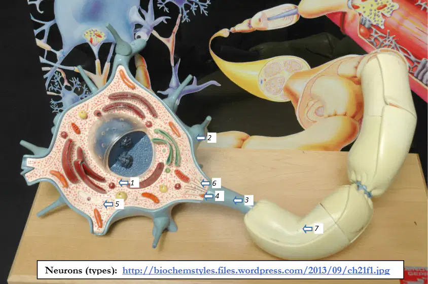
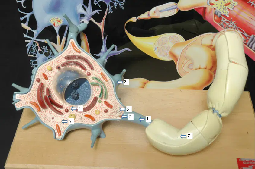
In the neuron model image with numbered labels, which structure is indicated by label 3 (long projection going away from the soma that transmits signals away)?
Axon
Dendrites
Axolemma
Neurilemma
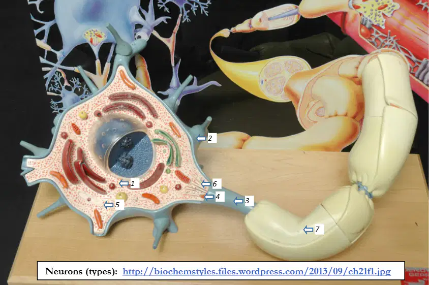
In the neuron model image with numbered labels, which structure is indicated by label 4 (part of the soma that connects to the axon)?
Axon hillock
Node of Ranvier
Myelin sheath
Nissl bodies
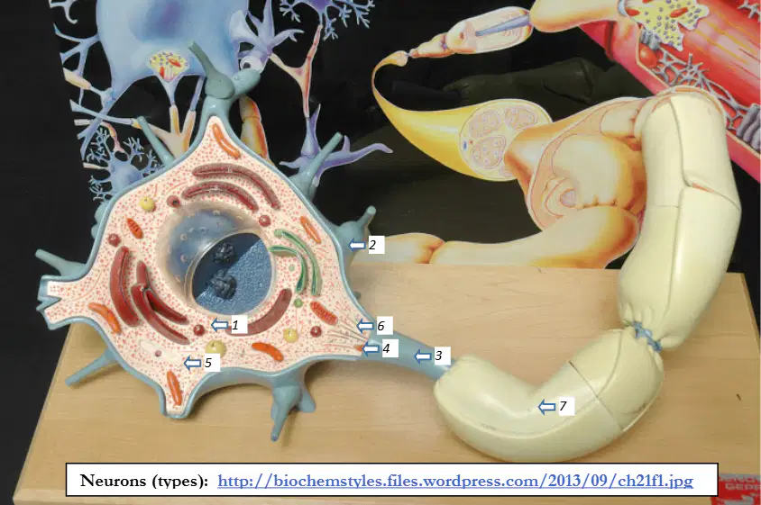
In the neuron model image with numbered labels, which structure is indicated by label 5 (red dots in the soma that represent sites of protein synthesis)?
Nissl bodies
Neurofibrils
Axoplasm
Schwann cell nucleus
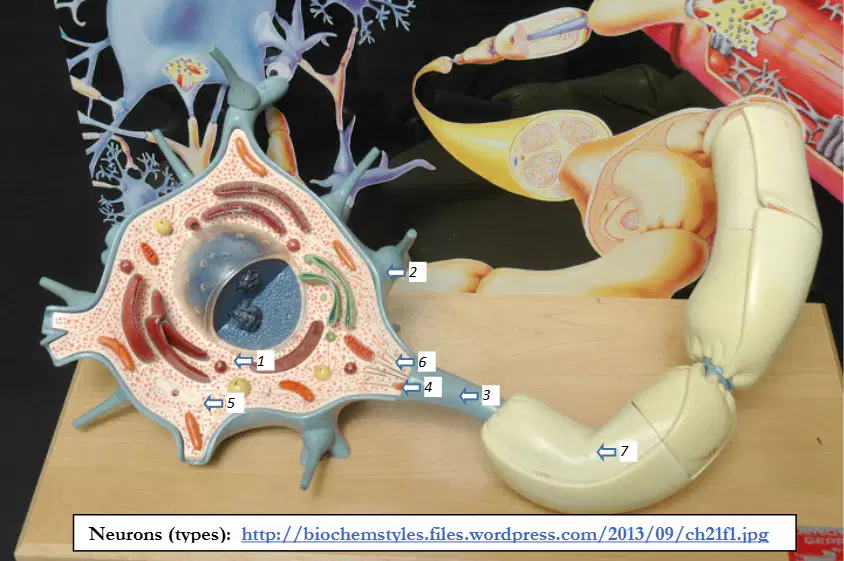
In the neuron model image with numbered labels, which structure is indicated by label 6 (lines inside the axon hillock that form part of the cytoskeleton)?
Neurofibrils
Axolemma
Schwann cell
Node of Ranvier
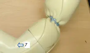
In the neuron model image with numbered labels, which structure is indicated by label 7 (fatty covering that wraps around an axon)?
Myelin sheath
Neurilemma
Axoplasm
Dendrite
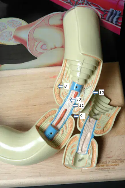
In the neuron model image with numbered labels, which structure is indicated by label 8 (a myelin sheath segment with its own tiny nucleus)?
Schwann cell
Node of Ranvier
Nissl bodies
Soma
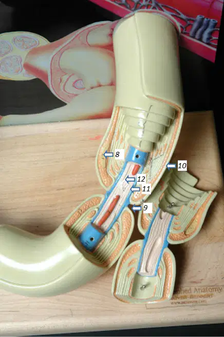
In the neuron model image with numbered labels, which structure is indicated by label 9 (gap between adjacent myelin sheaths)?
Node of Ranvier
Axon hillock
Axolemma
Neurofibrils
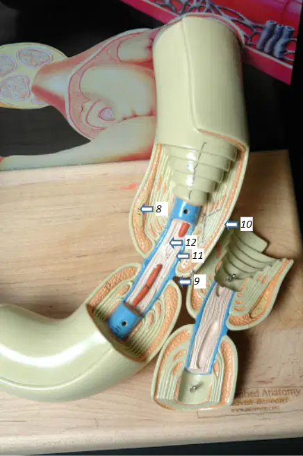
In the neuron model image with numbered labels, which structure is indicated by label 10 (cell membrane of the neuron seen outside)?
Neurilemma
Axolemma
Axoplasm
Myelin sheath
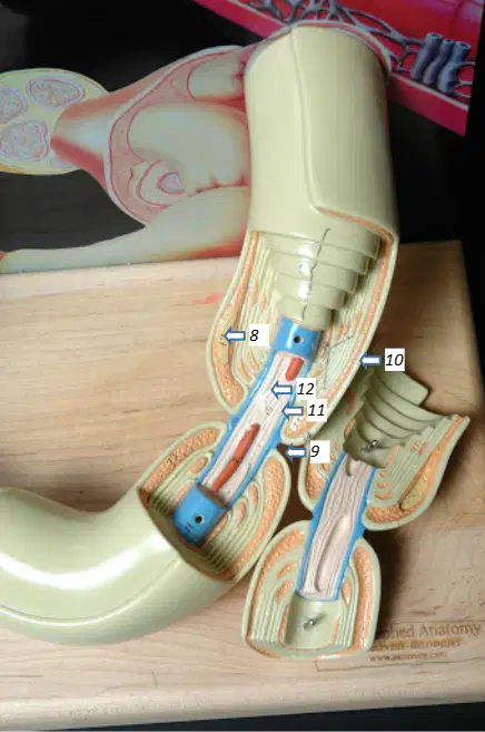
In the neuron model image with numbered labels, which structure is indicated by label 11 (cell membrane of the axon)?
Axolemma
Neurilemma
Myelin sheath
Schwann cell
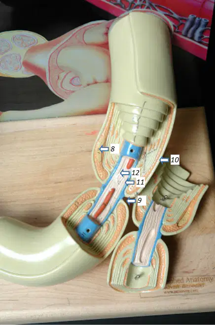
In the neuron model image with numbered labels, which structure is indicated by label 12 (cytoplasm of the axon)?
Axoplasm
Axolemma
Neurofibrils
Nissl bodies
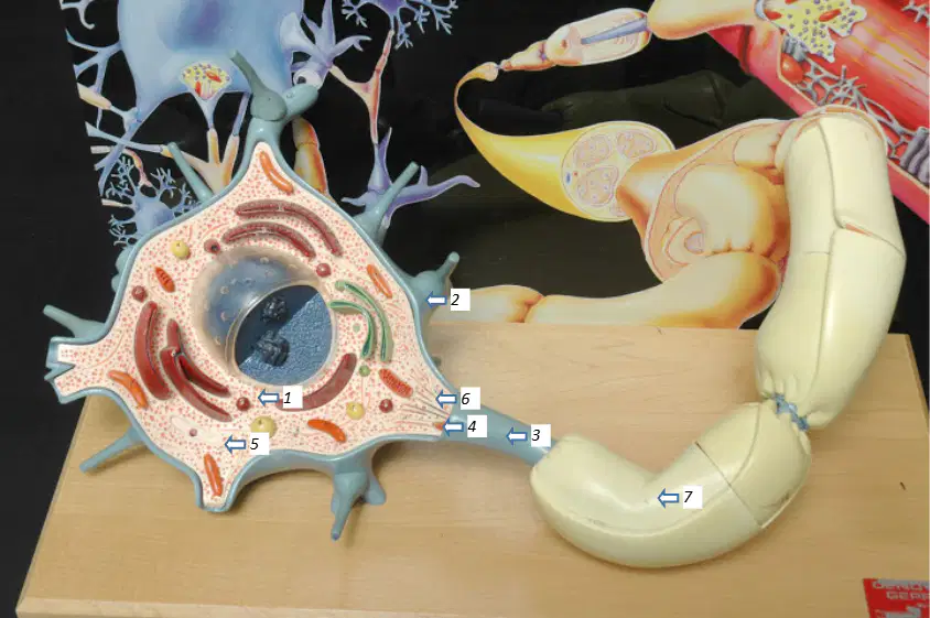
In the neuron model image labeled 1–12, which number corresponds to the soma (cell body)?
1
4
7
10
In the neuron model image labeled 1–12, which number corresponds to the dendrites?
2
5
8
11
In the neuron model image labeled 1–12, which number corresponds to the axon?
3
6
9
12
In the neuron model image labeled 1–12, which number corresponds to the myelin sheath (fatty covering)?
7
4
2
10
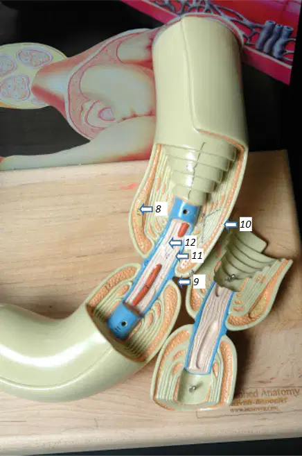
In the neuron model image labeled 1–12, which number corresponds to a Schwann cell (myelin sheath segment with a tiny nucleus)?
8
1
5
11
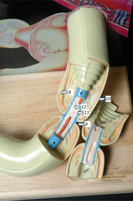
In the neuron model image labeled 1–12, which number corresponds to a node of Ranvier (gap between adjacent myelin sheaths)?
9
6
3
12
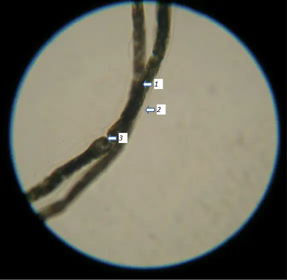
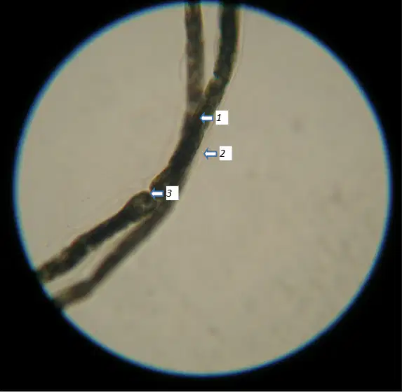
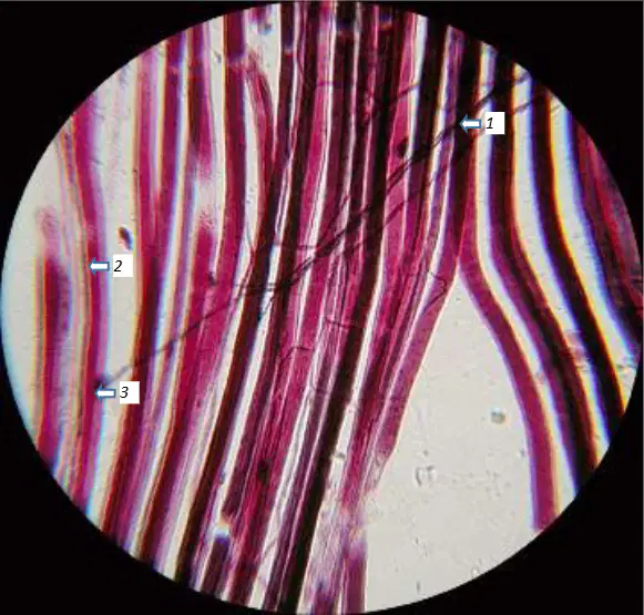
In the axon-slide micrograph with labels 1–3, which structure is indicated by label 1 (dark central fiber)?
Axon
Myelin sheath
Node of Ranvier
Schwann cell
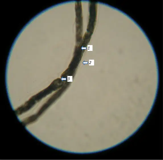
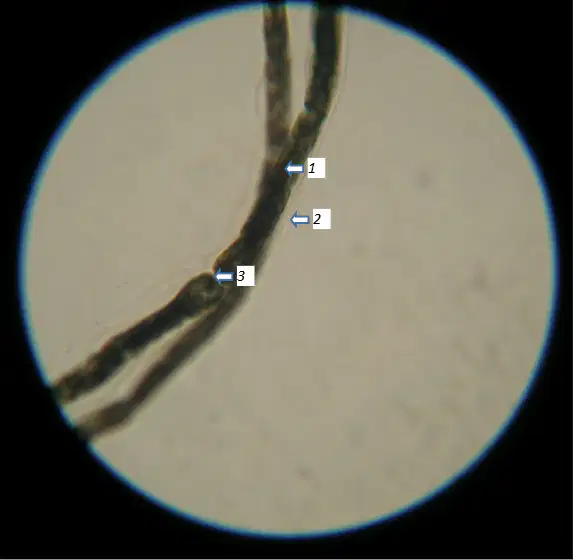
In the axon-slide micrograph with labels 1–3, which structure is indicated by label 2 (transparent layer seen outside, around the dark-colored axon)?
Myelin sheath
Axon
Node of Ranvier
Neurilemma
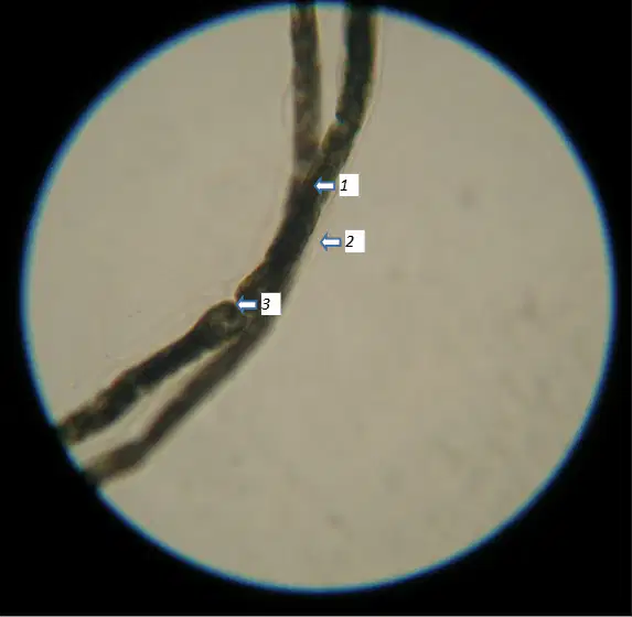
In the axon-slide micrograph with labels 1–3, which structure is indicated by label 3 (gap between adjacent myelin sheaths)?
Node of Ranvier
Schwann cell nucleus
Axon hillock
Axoplasm
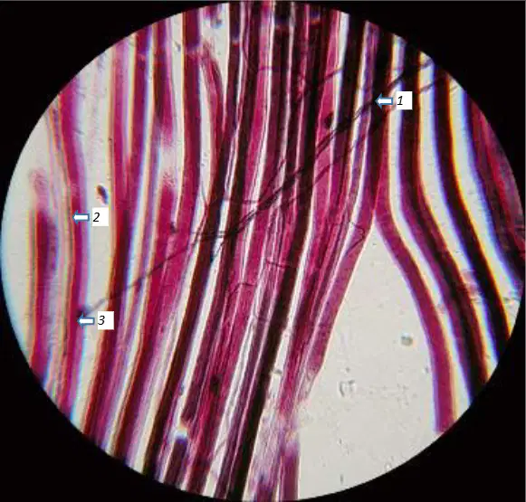
In the myoneural junction slide with labels 1–3, which structure is indicated by label 1 (axon and its synaptic terminals, dark with tiny branches)?
Axon with terminals
Muscle fibers
Synapse/motor end plate
Myelin sheath
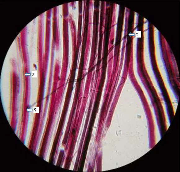
In the myoneural junction slide with labels 1–3, which structure is indicated by label 2 (long, cylindrical, thread-like pink fibers)?
Muscle fibers
Axon with terminals
Synapse/motor end plate
Neurofibrils
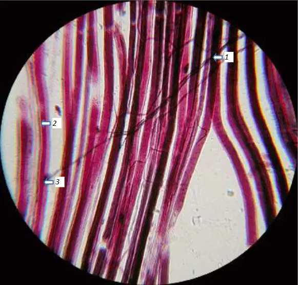
In the myoneural junction slide with labels 1–3, which structure is indicated by label 3 (dark spots at the junction where the axon meets the muscle fiber)?
Synapse/motor end plate
Axon with terminals
Muscle fibers
Node of Ranvier
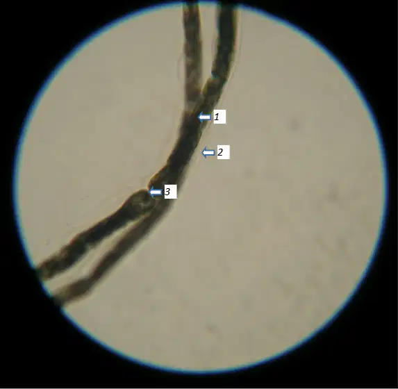
On the myelinated axon slide image, identify the structure indicated by label 1.
Myelin sheath
Node of Ranvier
Schwann cell nucleus
Axon hillock
On the myelinated axon slide image, identify the structure indicated by label 2.
Node of Ranvier
Myelin sheath
Axon terminal
Dendrite
On the myelinated axon slide image, identify the structure indicated by label 3.
Schwann cell nucleus
Myelin sheath
Node of Ranvier
Synaptic bouton
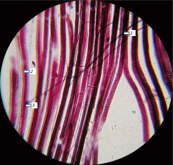
On the myoneural junction slide image, identify the feature indicated by label 1.
Motor end plate (neuromuscular junction)
Nucleus of a muscle fiber
Capillary
Satellite cell
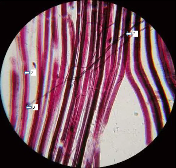
On the myoneural junction slide image, identify the structure indicated by label 2.
Striations of a skeletal muscle fiber
Elastic connective tissue
Smooth muscle layer
Myelin sheath
On the myoneural junction slide image, identify the structure indicated by label 3.
Motor axon branch
Sarcomere Z-line
Mitochondrion
Blood vessel lumen
In the sheep brain images, label 5 indicates which lobe of the cerebrum.
Frontal lobe
Temporal lobe
Occipital lobe
Parietal lobe
In the sheep brain images, label 6 indicates which lobe of the cerebrum.
Parietal lobe
Frontal lobe
Temporal lobe
Insula
In the sheep brain images, label 7 indicates which visible lobe on the lateral surface.
Temporal lobe
Occipital lobe
Frontal lobe
Parietal lobe
In the sheep brain images, label 8 indicates which lobe at the posterior end of the cerebrum.
Occipital lobe
Temporal lobe
Parietal lobe
Frontal lobe
In the sheep brain images, the raised ridges of the cerebral cortex indicated by label 9 are called what.
Gyri
Sulci
Meninges
Fissures
In the sheep brain images, the shallow grooves of the cerebral cortex indicated by label 10 are called what.
Sulci
Gyri
Arachnoid villi
Ventricles
In the sheep brain images, label 11 indicates which part located inferior to the cerebrum and anterior to the cerebellum.
Brain stem
Corpus callosum
Thalamus
Pineal gland
In the sheep brain images, label 12 indicates which structure at the rear of the brain.
Cerebellum
Midbrain
Pons
Medulla oblongata
In the sheep brain images, label 13 indicates which midline feature between the two cerebellar hemispheres.
Vermis
Falx cerebri
Longitudinal fissure
Fourth ventricle
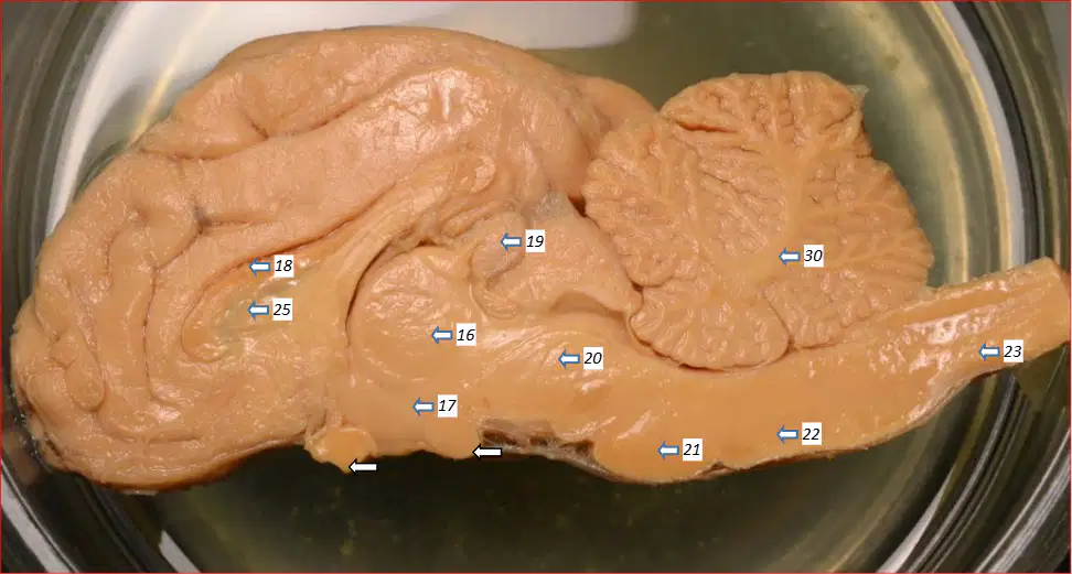
In the mid-sagittal sheep brain, identify the structure labeled 16 described as a round or oval-shaped structure in the middle.
Thalamus
Pineal gland
Hypothalamus
Corpus callosum
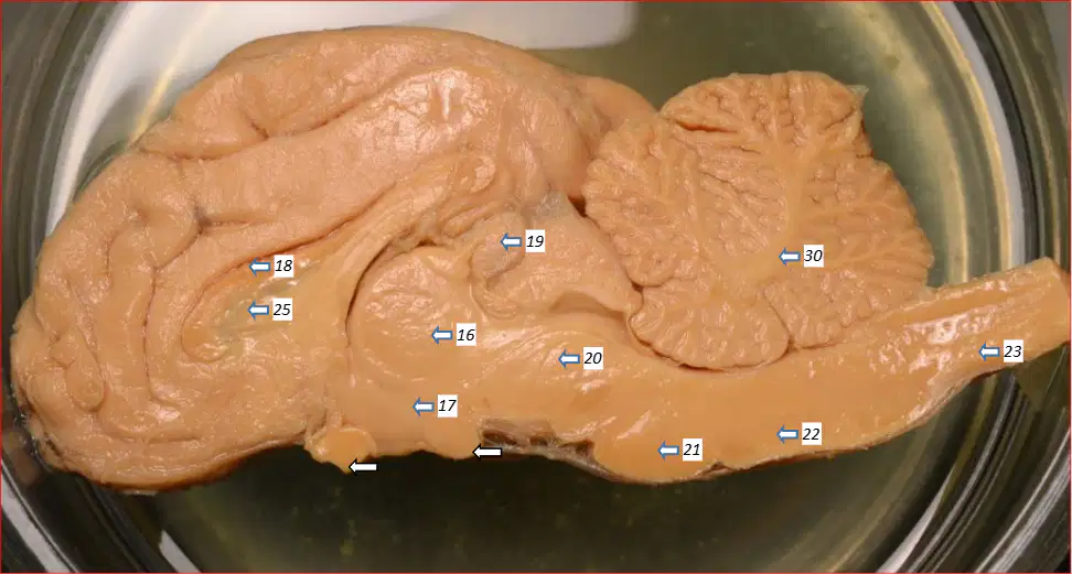
In the mid-sagittal sheep brain, which structure labeled 17 lies inferior to the thalamus?
Hypothalamus
Pons
Medulla oblongata
Cerebral peduncle
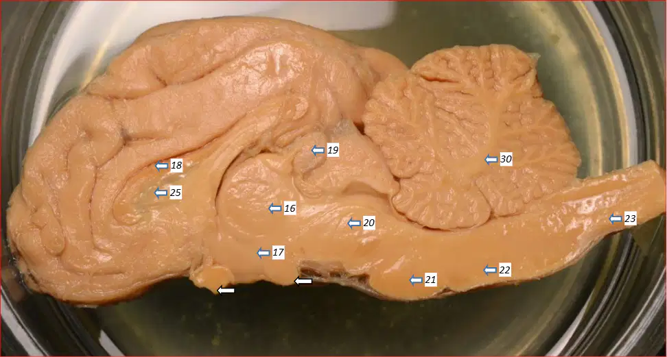
In the mid-sagittal sheep brain, the C‑shaped band above the thalamus labeled 18 is which structure?
Corpus callosum
Septum pellucidum
Lateral ventricle
Arbor vitae
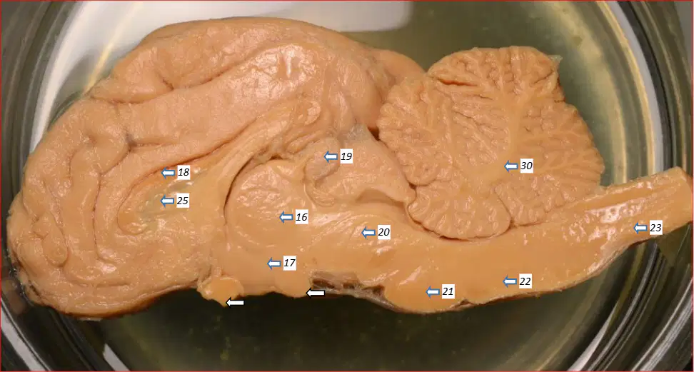
In the mid-sagittal sheep brain, the pine cone–shaped structure superior and posterior to the thalamus labeled 19 is which of the following?
Pineal gland
Pituitary gland
Superior colliculus
Inferior colliculus
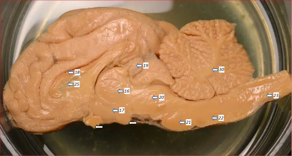
In the mid-sagittal sheep brain, which structure labeled 20 is immediately inferior to the thalamus and superior to the pons?
Cerebral peduncle
Medulla oblongata
Spinal cord
Cerebral aqueduct
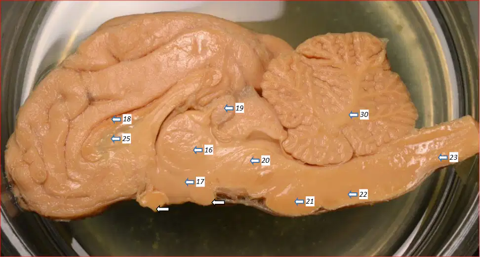
In the mid-sagittal sheep brain, the rounded bulge of the brainstem labeled 21 is which structure?
Pons
Midbrain tectum
Medulla oblongata
Spinal cord
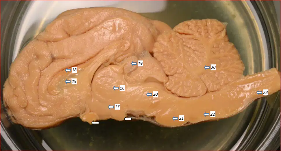
In the mid-sagittal sheep brain, the continuation of the brainstem inferior to the pons labeled 22 is which structure?
Medulla oblongata
Spinal cord
Cerebral peduncle
Cerebellum
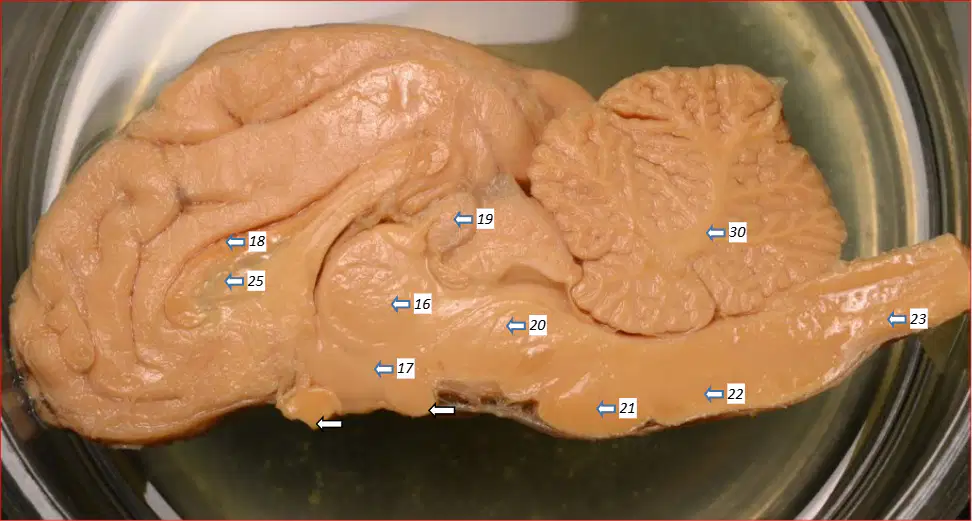
In the mid-sagittal sheep brain, the nervous tissue beginning inferior to the medulla oblongata labeled 23 is which structure?
Spinal cord
Pons
Cerebral peduncle
Cerebellar peduncle
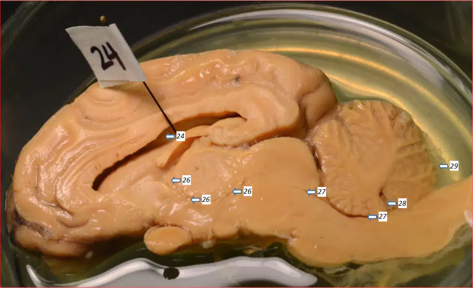
In the mid-sagittal sheep brain, the cerebrospinal fluid–filled cavity labeled 24 (one of a pair covered by septum pellucidum) is which ventricle?
Lateral ventricle
Third ventricle
Fourth ventricle
Cerebral aqueduct
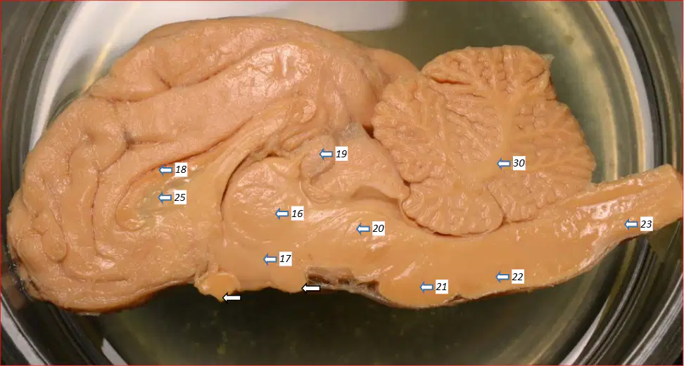
In the mid-sagittal sheep brain, the thin membrane covering the lateral ventricles labeled 25 is which structure?
Septum pellucidum
Arachnoid mater
Dura mater
Choroid plexus
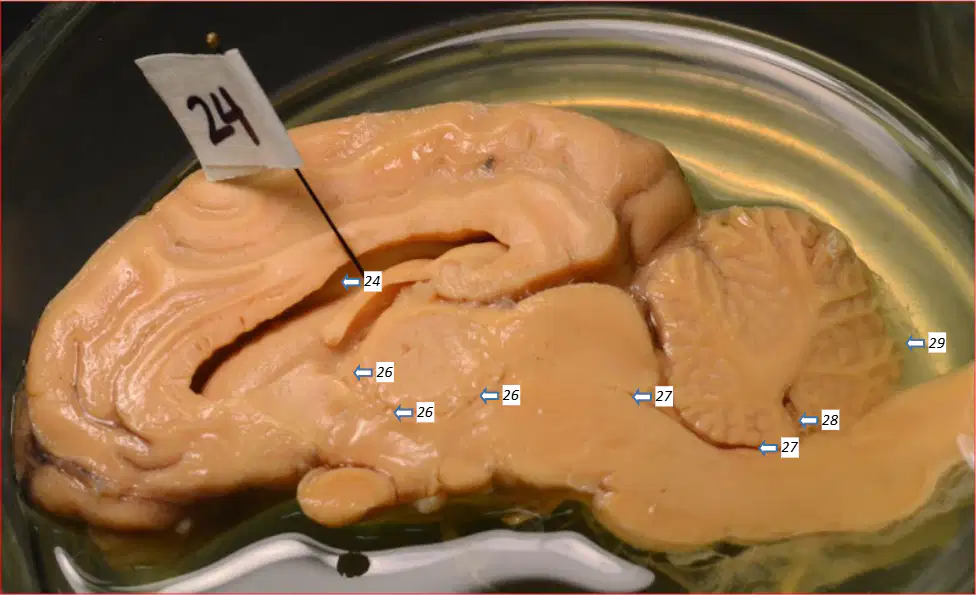
In the mid-sagittal sheep brain, the ventricle labeled 26, located in the diencephalon, is which ventricle?
Third ventricle
Fourth ventricle
Lateral ventricle
Cisterna magna
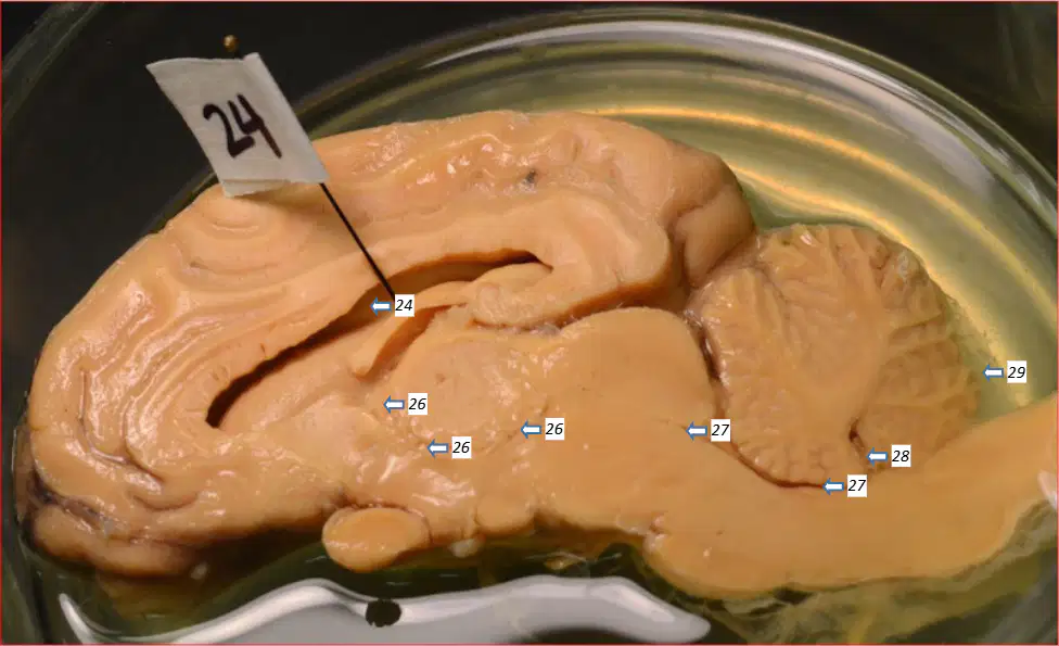
In the mid-sagittal sheep brain, the narrow passage labeled 27 that leads to the fourth ventricle is which structure?
Cerebral aqueduct
Central canal
Interventricular foramen
Longitudinal fissure
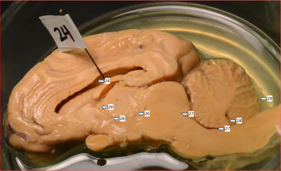
In the mid-sagittal sheep brain, the V‑shaped cavity located between the cerebellum and medulla oblongata labeled 28 is which ventricle?
Fourth ventricle
Third ventricle
Lateral ventricle
Cerebral aqueduct
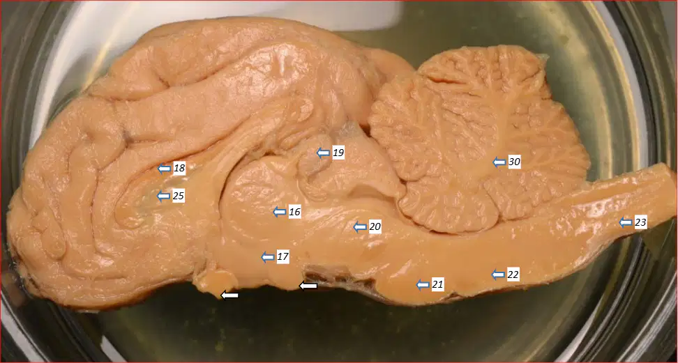
In the mid-sagittal sheep brain, the tree‑like white matter within the cerebellum labeled 30 is which structure?
Arbor vitae
Cerebellar cortex
Corpus callosum
Vermis

On the posterior view of the midbrain, which structure is indicated by the right‑side pin labeled 12 as part of the corpora quadrigemina?
Right superior colliculus
Right inferior colliculus
Pineal gland
Vermis

On the posterior view of the midbrain, which structure is indicated by the right‑side pin labeled 13 as part of the corpora quadrigemina?
Right inferior colliculus
Right superior colliculus
Pineal gland
Vermis

In the posterior midbrain image, a white arrow points to the upper left colliculus without a number. Which structure does this arrow identify?
Left superior colliculus
Left inferior colliculus
Pineal gland
Thalamus

In the posterior midbrain image, a white arrow points to the lower left colliculus without a number. Which structure does this arrow identify?
Left inferior colliculus
Left superior colliculus
Pineal gland
Thalamus
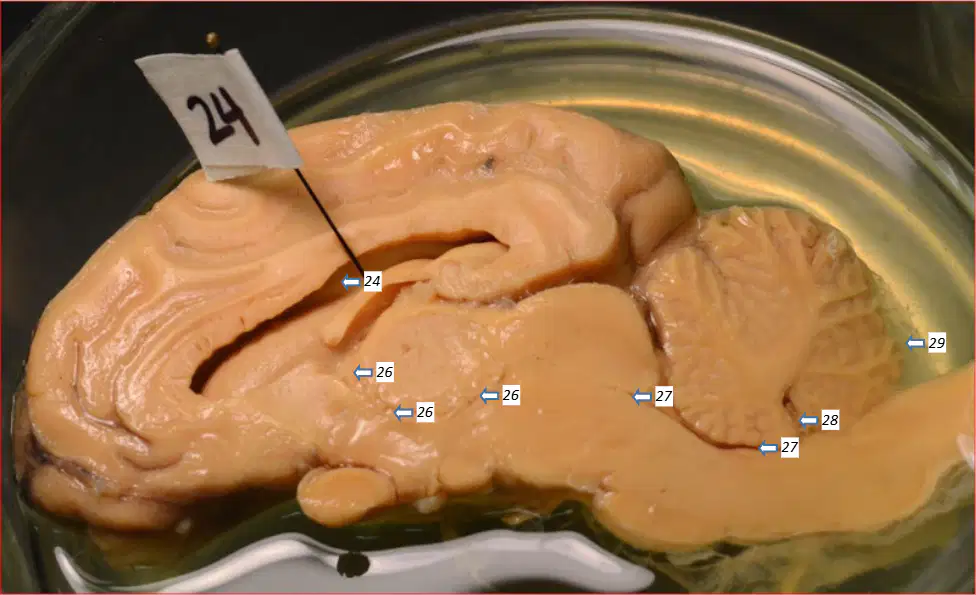
According to the mid-sagittal description, cerebrospinal fluid flows through the cerebral aqueduct to which ventricle located between the cerebellum and medulla oblongata?
Fourth ventricle
Third ventricle
Lateral ventricle
Central canal
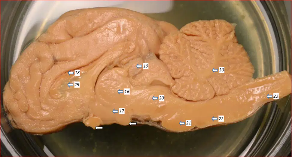
Which statement best describes the arbor vitae as presented in the mid-sagittal sheep brain description?
It is white matter within the cerebellum
It is gray matter of the cerebrum
It is a membrane covering the lateral ventricles
It is the pine cone–shaped endocrine organ
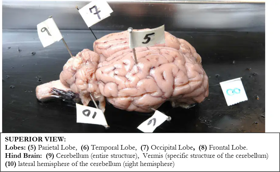
In the superior view of a dissected brain specimen, the flag labeled 5 identifies which cerebral lobe.
Parietal lobe
Temporal lobe
Occipital lobe
Frontal lobe
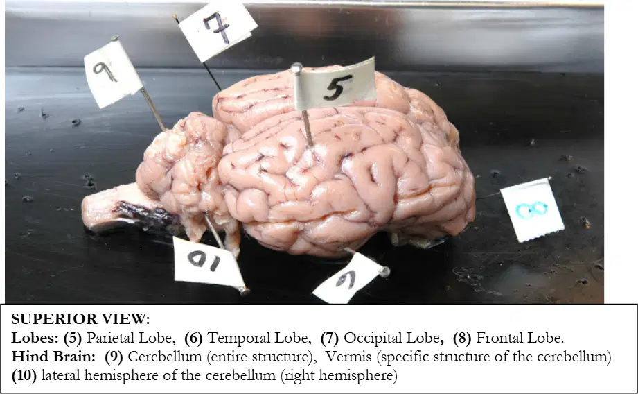
In the same superior view, the flag labeled 6 marks which cerebral lobe.
Frontal lobe
Temporal lobe
Parietal lobe
Occipital lobe
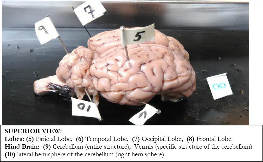
In the superior view, the flag labeled 7 corresponds to which cerebral lobe.
Occipital lobe
Parietal lobe
Frontal lobe
Temporal lobe
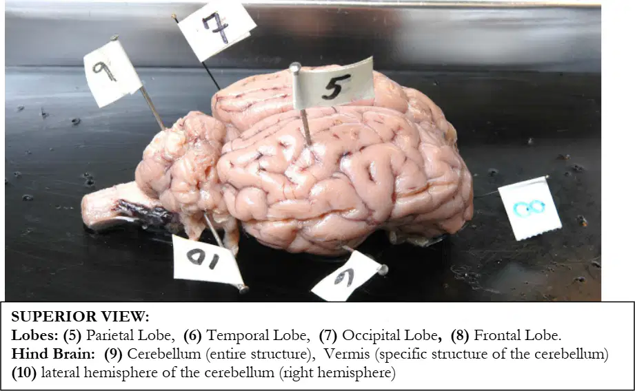
In the superior view, the flag labeled 8 indicates which cerebral lobe.
Frontal lobe
Occipital lobe
Temporal lobe
Parietal lobe
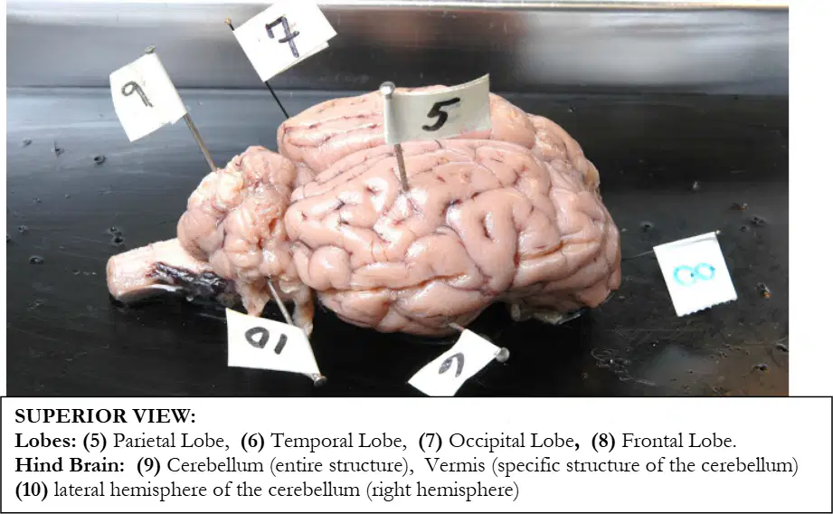
In the superior view focusing on the hindbrain, the flag labeled 9 identifies which structure.
Cerebellum (entire structure)
Vermis of the cerebellum
Medulla oblongata
Pons
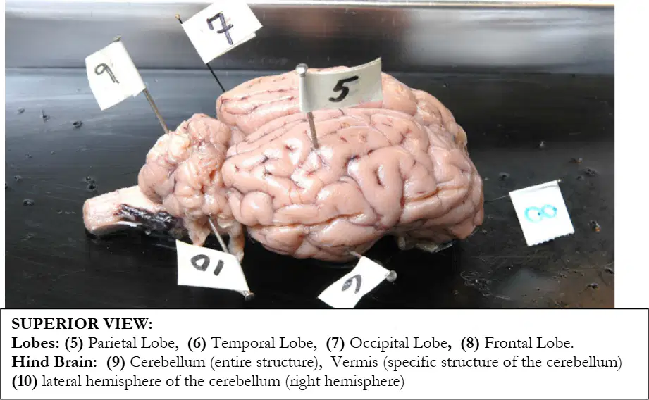
In the superior view of the hindbrain, the flag labeled 10 marks which part of the cerebellum.
Lateral hemisphere of the cerebellum (right hemisphere)
Vermis (midline of the cerebellum)
Flocculus
Arbor vitae
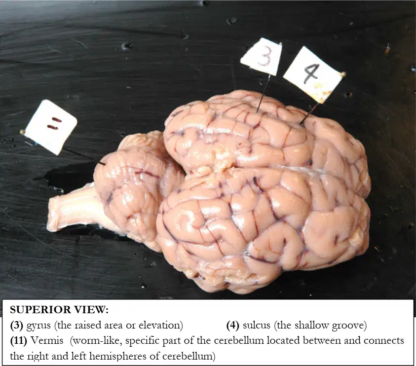
In the superior view highlighting surface features, the flag labeled 3 points to which feature.
Gyrus (raised fold)
Sulcus (shallow groove)
Fissure (deep groove)
Pia mater
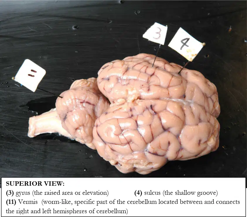
In the same superior view, the flag labeled 4 indicates which feature.
Sulcus (shallow groove)
Gyrus (raised fold)
Central canal
Longitudinal fissure
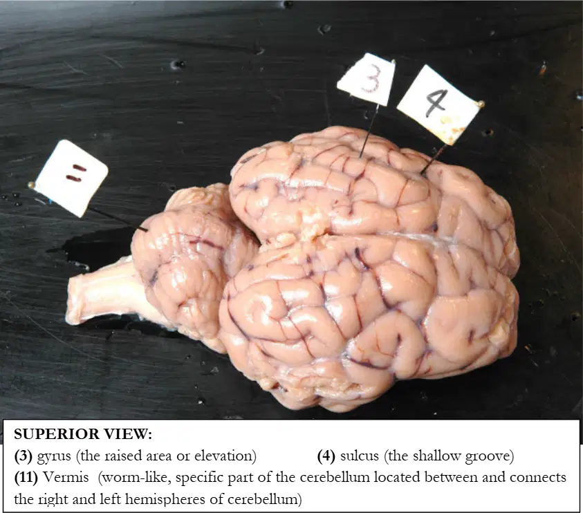
In the superior view of the cerebellum, the flag labeled 11 identifies which midline structure connecting the cerebellar hemispheres.
Vermis
Arbor vitae
Fourth ventricle
Cerebral peduncle
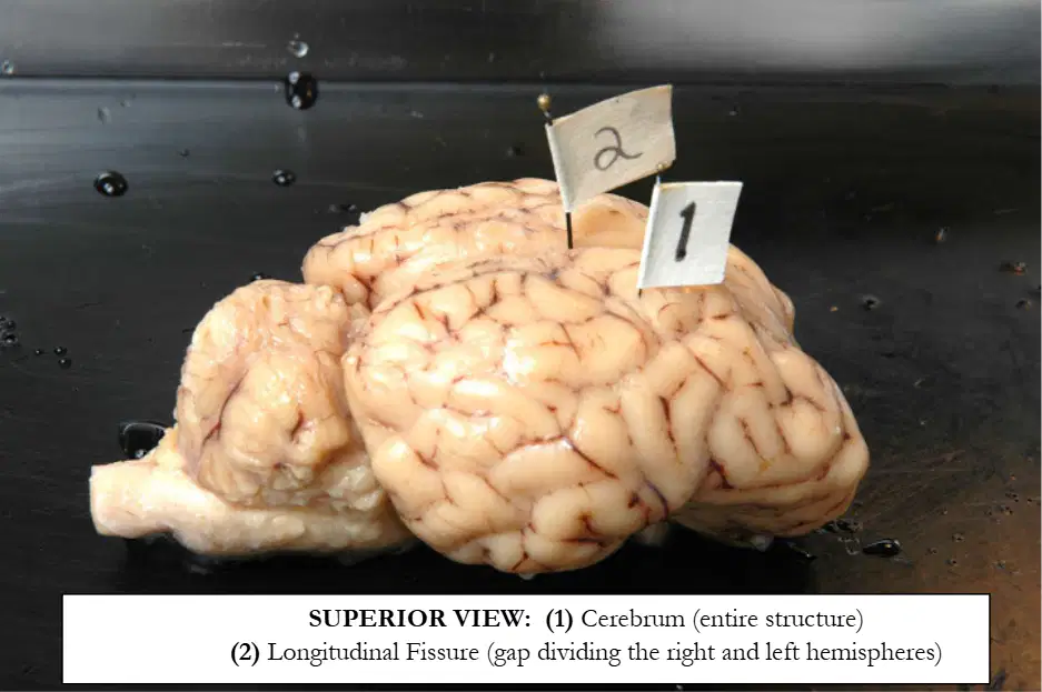
In the superior view of the brain, the flag labeled 1 designates which overall structure.
Cerebrum (entire structure)
Cerebellum
Brainstem
Diencephalon
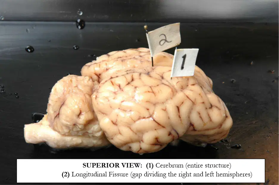
In the same superior view, the flag labeled 2 marks which landmark dividing the hemispheres.
Longitudinal fissure
Central sulcus
Lateral sulcus
Parieto-occipital sulcus
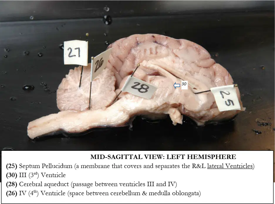
In the mid‑sagittal view of the left hemisphere, the flag labeled 25 indicates which membrane separating the right and left lateral ventricles.
Septum pellucidum
Falx cerebri
Tentorium cerebelli
Arachnoid mater
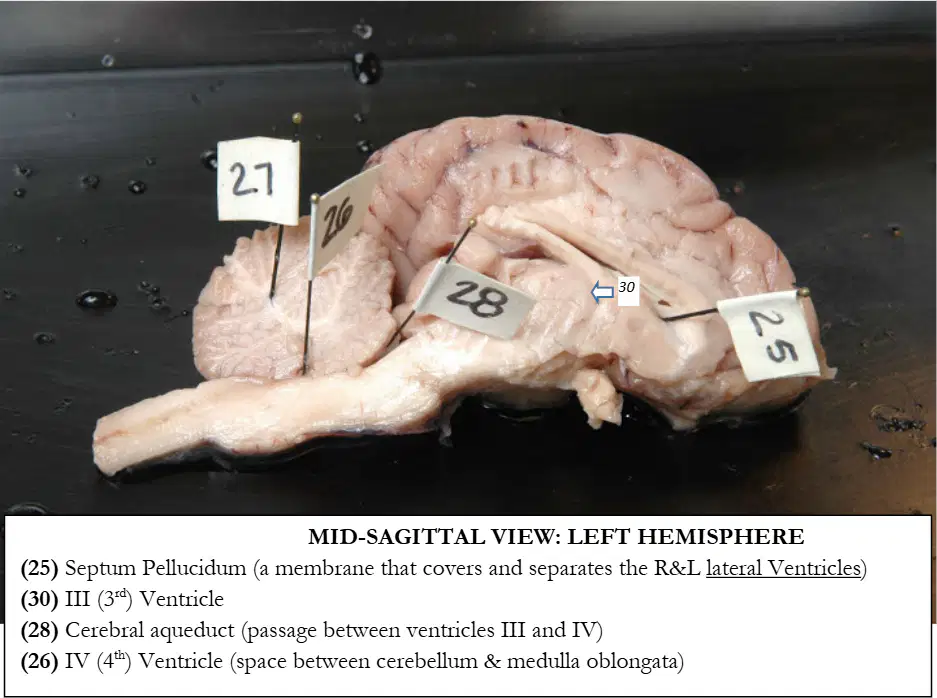
In the same mid‑sagittal view, the flag labeled 30 identifies which ventricle.
Third ventricle
Lateral ventricle
Fourth ventricle
Cerebral aqueduct
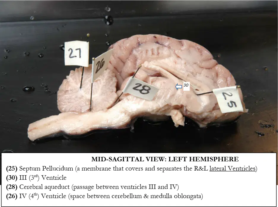
In the same mid‑sagittal view, the flag labeled 28 marks which narrow passage connecting ventricles III and IV.
Cerebral aqueduct
Central canal
Interventricular foramen
Longitudinal fissure
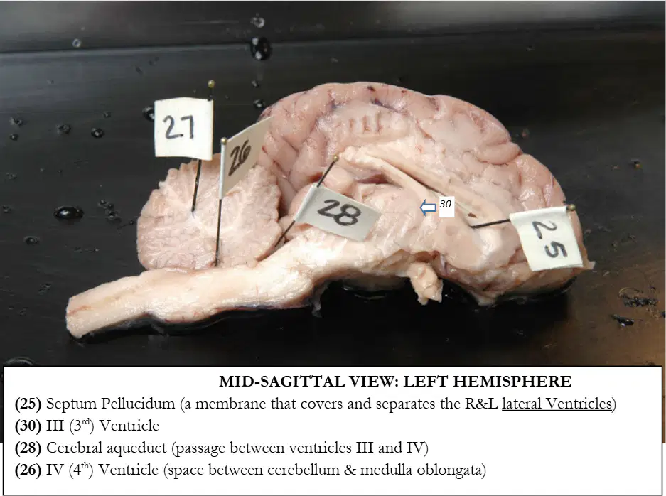
In the same mid‑sagittal view, the flag labeled 26 corresponds to which ventricle located between the cerebellum and medulla oblongata.
Fourth ventricle
Lateral ventricle
Third ventricle
Central canal
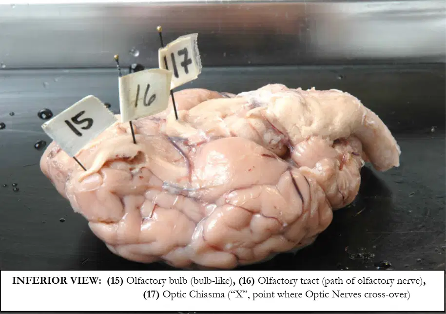
In the inferior view of the brain, the flag labeled 15 identifies which anterior olfactory structure.
Olfactory bulb
Olfactory tract
Optic chiasma
Pituitary stalk
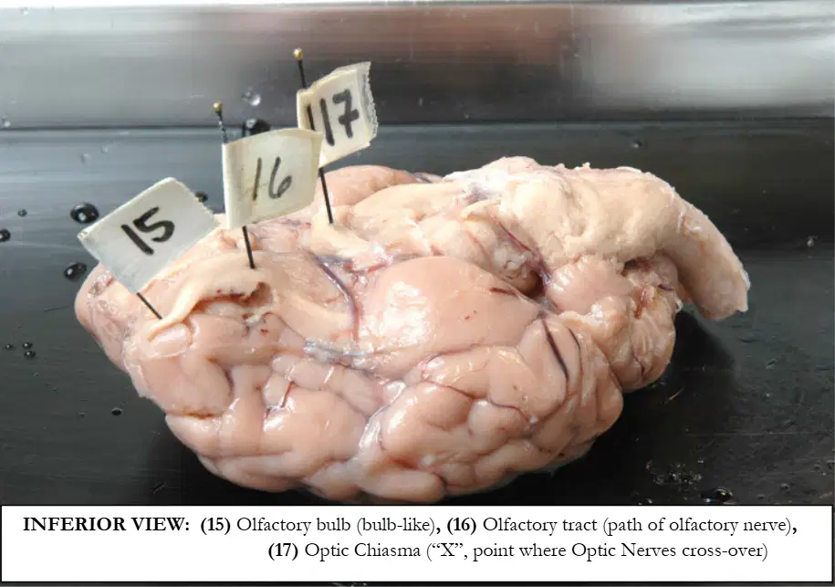
In the same inferior view, the flag labeled 16 indicates which pathway of the olfactory nerve.
Olfactory tract
Olfactory bulb
Optic tract
Trochlear nerve
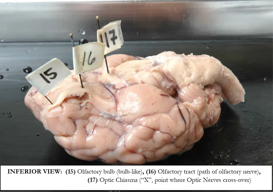
In the same inferior view, the flag labeled 17 marks which X-shaped crossing of the optic nerves.
Optic chiasma
Optic tract
Mammillary body
Infundibulum
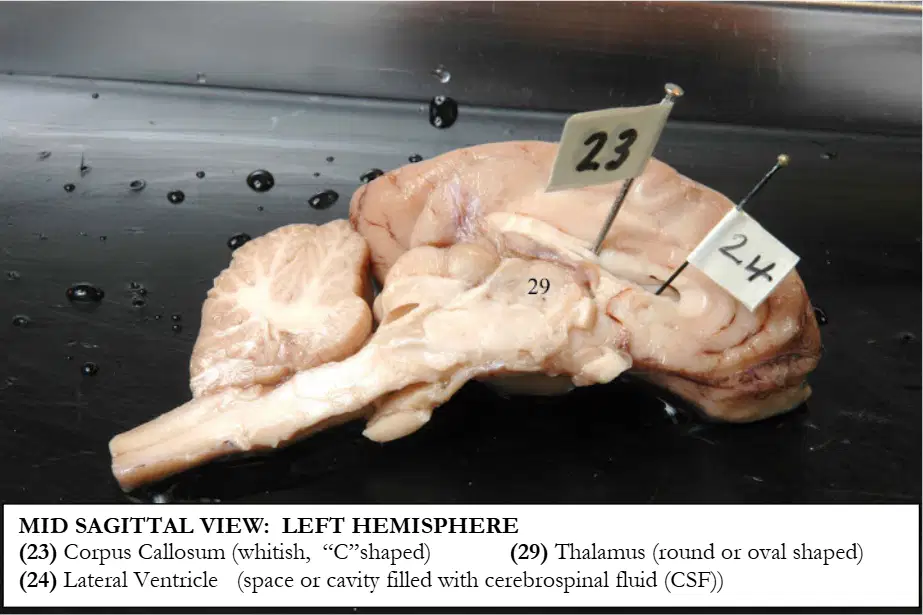
In the mid‑sagittal view of the left hemisphere, the flag labeled 23 designates which C‑shaped white matter structure.
Corpus callosum
Fornix
Anterior commissure
Internal capsule
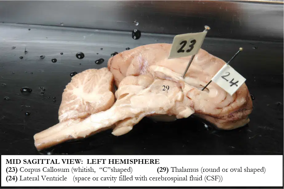
In the same mid‑sagittal view, the flag labeled 24 points to which cerebrospinal‑fluid–filled cavity.
Lateral ventricle
Third ventricle
Fourth ventricle
Cisterna magna
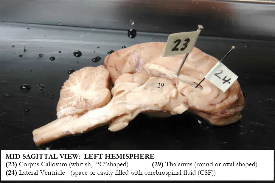
In the same mid‑sagittal view, the flag labeled 29 corresponds to which diencephalic mass.
Thalamus
Hypothalamus
Pineal gland
Midbrain tectum
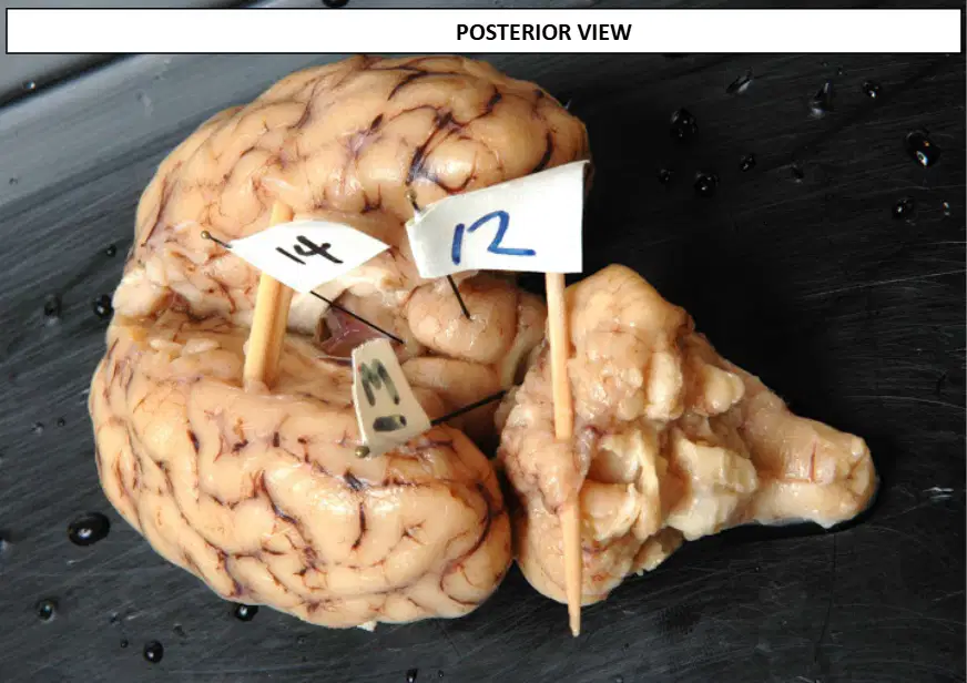
In the posterior view of the midbrain and diencephalon, the flag labeled 12 identifies which colliculus on the right side.
Right superior colliculus
Right inferior colliculus
Left superior colliculus
Pineal gland
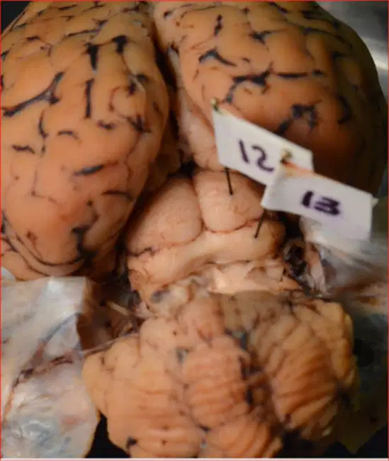
In the same posterior view, the flag labeled 13 indicates which colliculus on the right side.
Right inferior colliculus
Right superior colliculus
Left inferior colliculus
Pineal gland
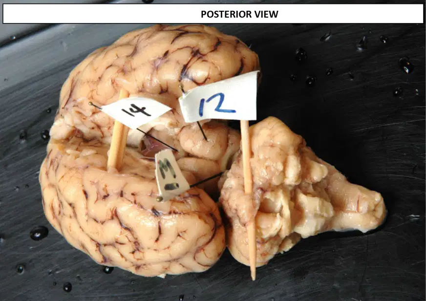
In the same posterior view, the flag labeled 14 marks which endocrine structure projecting posteriorly from the diencephalon.
Pineal gland
Superior colliculus
Inferior colliculus
Mammillary body

In the sagittal view image of the human brain, which endocrine structure is shown hanging from the hypothalamus?
Pituitary gland
Pineal gland
Thyroid gland
Adrenal medulla

In the sagittal view image of the human brain, the gland shown hanging is attached to which specific brain region?
Hypothalamus
Thalamus
Medulla oblongata
Cerebellum
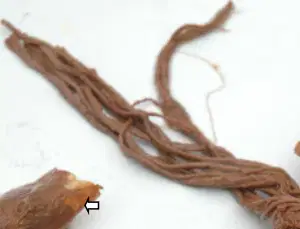
In the specimen shown, which meningeal layer encloses the spinal cord at the superior end until about halfway?
Dura mater
Arachnoid mater
Pia mater
Perineurium
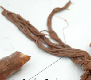
Which named structure in the spinal cord images refers to the bundle of lumbar–sacral–coccygeal nerves that descend inferiorly?
Cauda equina
Conus medullaris
Filum terminale
Spinal nerve
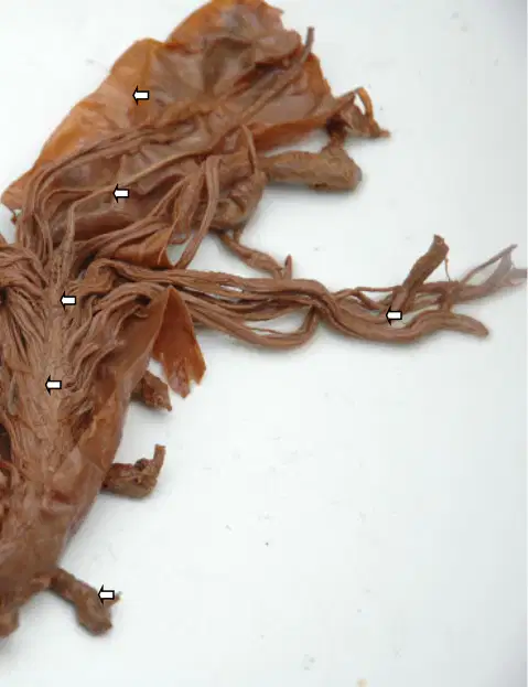
Which term identifies the narrow, conical end of the spinal cord shown in the images?
Conus medullaris
Filum terminale
Cauda equina
Dorsal root ganglion
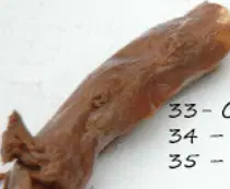
Which structure is the very end of the spinal cord in the specimen?
Filum terminale
Conus medullaris
Cauda equina
Dural sac
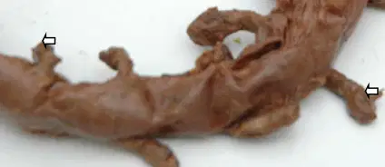
In the spinal cord photographs, which labeled structure carries signals between the spinal cord and the body by exiting between vertebrae?
Spinal nerve
Spinal cord
Dura mater
Filum terminale
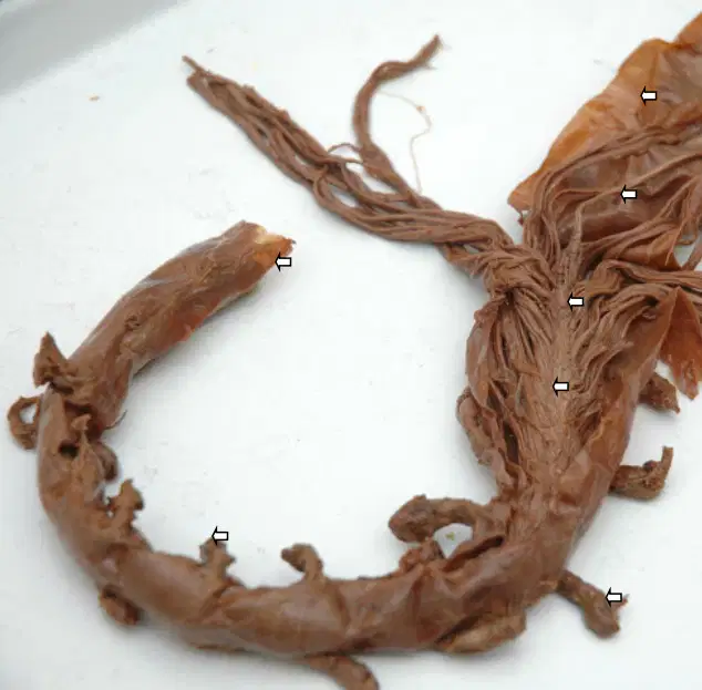
According to the images and description, what cord can be seen inside the dura mater at the superior end of the specimen?
Spinal cord
Notochord
Sciatic nerve
Sympathetic chain

Spinal cord cross section—identify the midline groove that is deeper on the model described: HORS (butterfly‑shaped gray matter areas), COLUMNS (white matter), CENTRAL CANAL (hole in the middle), GRAY COMMISSURE (area around the hole), ANTERIOR MEDIAN FISSURE (deep), POSTERIOR MEDIAN SULCUS (shallow). Which structure is the deeper midline groove?
Anterior median fissure
Posterior median sulcus
Lateral sulcus
Intervertebral foramen

Spinal cord cross section—identify the small circular opening at the center of the gray commissure on the model.
Central canal
Dorsal root ganglion
Anterior gray horn
Posterior gray horn

Spinal cord cross section—name the thin bridge of gray matter surrounding the central canal on the model.
Gray commissure
White commissure
Dorsal column
Lateral funiculus

Spinal cord cross section—what term identifies the butterfly‑shaped gray matter regions in the model?
Horns
Columns
Roots
Ganglia

Spinal cord cross section—what is the name for the white matter areas that surround the gray matter in the model?
Columns
Horns
Tracts only
Pia mater

Spinal nerve with roots—on the model, the swelling on the dorsal root is identified as which structure?
Dorsal root ganglion
Ventral root
Posterior median sulcus
Anterior median fissure

Spinal nerve with roots—on the model, which root is associated with the dorsal root ganglion and carries sensory fibers?
Dorsal root
Ventral root
Lateral root
Anterior horn

Spinal nerve with roots—what is the mixed nerve formed by the union of the dorsal and ventral roots as shown on the model?
Spinal nerve
Accessory nerve
Sympathetic trunk
Intercostal artery
Cranial nerves on sheep brain model—what is the name of Cranial Nerve I in the sequence shown?
Olfactory
Optic
Oculomotor
Trigeminal
Cranial nerves on sheep brain model—what is the name of Cranial Nerve II in the sequence shown?
Optic
Olfactory
Abducens
Facial
Cranial nerves on sheep brain model—what is the name of Cranial Nerve V as shown?
Trigeminal
Trochlear
Vagus
Hypoglossal
Cranial nerves on sheep brain model—what is the name of Cranial Nerve VII as shown?
Facial
Glossopharyngeal
Abducens
Accessory
Cranial nerves on sheep brain model—what is the name of Cranial Nerve VIII as shown on the label list?
Vestibulocochlear (Acoustic)
Vagus
Oculomotor
Spinal accessory
Cranial nerves on sheep brain model—what is the name of Cranial Nerve X in the sequence shown?
Vagus
Glossopharyngeal
Accessory
Hypoglossal
Cranial nerves on sheep brain model—what is the name of Cranial Nerve XII in the sequence shown?
Hypoglossal
Accessory
Trochlear
Oculomotor

Cranial nerves on sheep brain model—according to the note, which cranial nerves are exceptions to the simple I‑to‑XII sequence in this view? Select all that apply.
IV
V
VI
VII
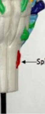
Cranial nerves on sheep brain model—the red nerve labeled near the lower lateral brainstem is identified as which structure?
Spinal nerve
Accessory nerve (XI)
Facial nerve (VII)
Vagus nerve (X)

On the labelled eye model, number 1 indicates which external structure of the anterior eye (dome‑shaped, convex outside)?
Cornea
Iris
Pupil
Lens

On the labelled eye model, number 2 identifies which colored circular structure located behind the cornea that contains wavy muscle lines?
Iris
Pupil
Lens
Ciliary body

On the labelled eye model, number 3 marks which opening that allows light to enter the eye?
Pupil
Fovea centralis
Optic disc
Canal of Schlemm

On the labelled eye model, which extraocular muscle is indicated by number 4?
Superior rectus
Inferior rectus
Medial rectus
Lateral rectus

On the labelled eye model, which extraocular muscle is indicated by number 5?
Inferior rectus
Superior rectus
Lateral rectus
Superior oblique

On the labelled eye model, which extraocular muscle is indicated by number 6?
Medial rectus
Lateral rectus
Superior oblique
Inferior oblique

On the labelled eye model, which extraocular muscle is indicated by number 7?
Lateral rectus
Medial rectus
Superior oblique
Inferior oblique

On the labelled eye model, which extraocular muscle is indicated by number 8?
Superior oblique
Inferior oblique
Lateral rectus
Medial rectus

On the labelled eye model, which extraocular muscle is indicated by number 9?
Inferior oblique
Superior oblique
Superior rectus
Inferior rectus
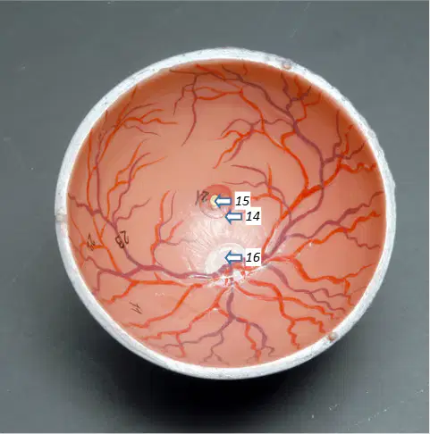
Inside the retinal bowl model, number 14 identifies which circular reddish area near the optic disc with high cone density for central vision?
Macula lutea
Fovea centralis
Optic disc (blind spot)
Ora serrata

Inside the retinal bowl model, number 15 indicates which small yellowish depression within the macula that provides the sharpest vision?
Fovea centralis
Macula lutea
Optic disc (blind spot)
Ciliary processes
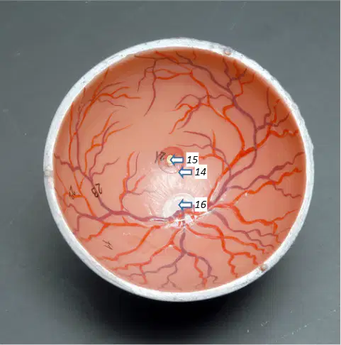
Inside the retinal bowl model, number 16 marks which site where the optic nerve exits and there are no rods or cones?
Optic disc (blind spot)
Fovea centralis
Macula lutea
Central retinal artery
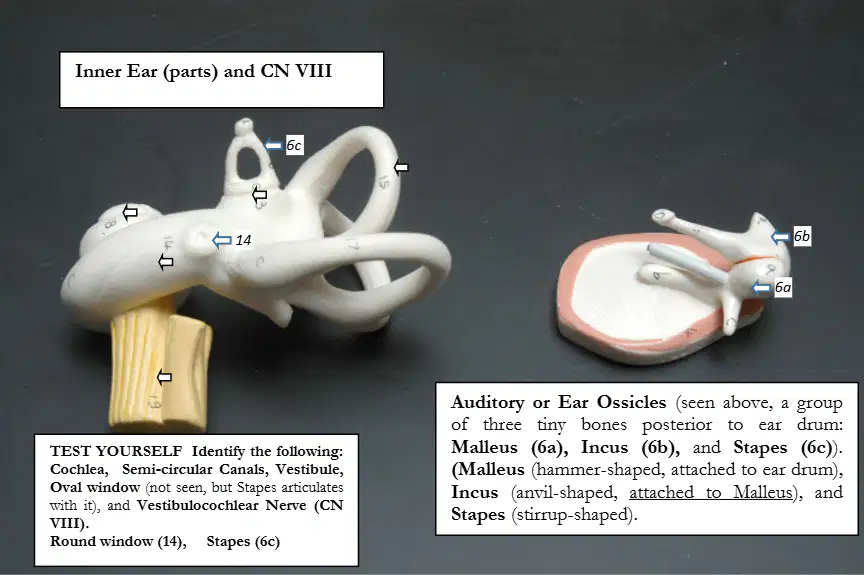
In the ear ossicles inset, which bone is described as hammer‑shaped and attached to the eardrum?
Malleus
Incus
Stapes
Cochlea
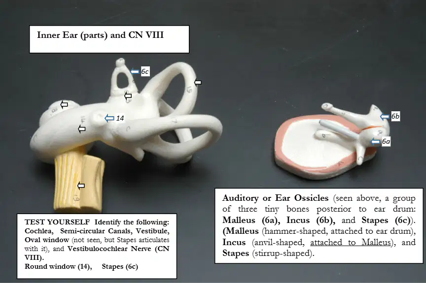
In the ear ossicles inset, which bone is anvil‑shaped and articulates with the malleus?
Incus
Malleus
Stapes
Vestibule
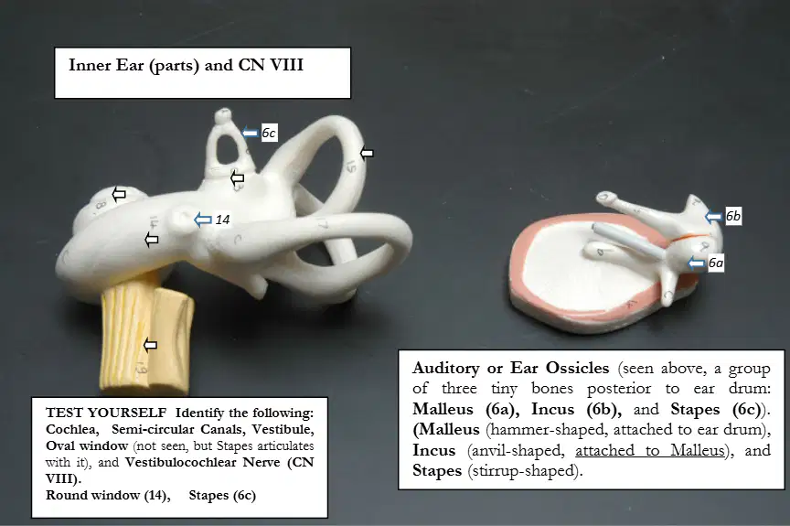
In the ear ossicles inset, which bone is stirrup‑shaped and articulates with the oval window?
Stapes
Malleus
Incus
Round window
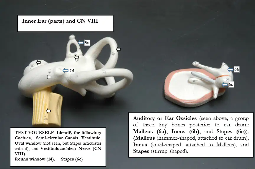
On the inner ear model, number 14 marks which opening in the cochlear wall?
Round window
Oval window
Endolymphatic duct
Auditory tube
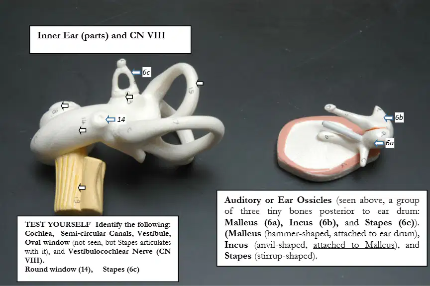
On the inner ear model, which cranial nerve is shown exiting the cochlea and vestibular apparatus as CN VIII?
Vestibulocochlear nerve
Facial nerve
Trigeminal nerve
Glossopharyngeal nerve

According to the model notes, which structure articulates with the oval window (not seen on the casting)?
Stapes
Malleus
Incus
Round window
In the right-hand photograph of the sheep brain (dorsal view), which structure is the midline groove dividing the two cerebral hemispheres indicated by the top-center arrow?
Longitudinal fissure
Lateral sulcus
Central sulcus
Transverse fissure
In the right-hand photograph of the sheep brain (dorsal view), which structure is the rounded posterior mass located dorsal to the brainstem and indicated by an arrow near the back of the brain?
Cerebellum
Thalamus
Olfactory bulb
Corpus callosum
In the right-hand photograph of the sheep brain, which brainstem part is the broad bulge inferior to the cerebrum and superior to the medulla that is indicated by a ventral brainstem arrow?
Pons
Midbrain
Medulla oblongata
Spinal cord
In the right-hand photograph of the sheep brain, which structure is the tapered inferior continuation of the brainstem indicated by the lowest arrow?
Medulla oblongata
Pons
Cerebellar peduncle
Corpus callosum
In the right-hand photograph of the sheep brain (dorsal view), the arrow pointing to a shallow groove on the cortical surface indicates which feature?
Sulcus
Gyrus
Fissure
Commissure
In the right-hand photograph of the sheep brain (dorsal view), which structures are the large paired lobes forming the bulk of the anterior brain that are indicated by lateral surface arrows?
Cerebral hemispheres
Cerebellar hemispheres
Temporal poles
Superior colliculi

