
Font size
 Worksheets
Human Eye Structure and Function Quiz
Total questions: 163
Worksheet time: 2hrs 58mins
What is the white part of the external eye that supports the eye called?
Sclera
Cornea
Iris
Pupil
Which part of the eye is the most anterior, clear, and protective?
Cornea
Sclera
Choroid
Pupil
What is the function of the choroid in the eye?
It nourishes the eye and prevents light scattering
It controls the amount of light entering the eye
It supports the eye
It allows light to enter the eye
Which part of the eye is composed of the ora serrata and ciliary process?
Ciliary body
Iris
Cornea
Sclera
What does the ciliary process secrete?
Aqueous humor
Tears
Pigment
Light
How does the iris help control the amount of light entering the eye?
By dilating and constricting
By nourishing the eye
By supporting the eye
By secreting aqueous humor
What is the hole in the iris that allows light to enter the eye called?
Pupil
Cornea
Sclera
Choroid
If a person’s pupil cannot dilate or constrict, what might happen to their vision in bright or dim light? (DoK Level 3)
They may have trouble seeing because too much or too little light enters the eye
Their eye will always be protected
They will see more colors
Their eye will produce more tears
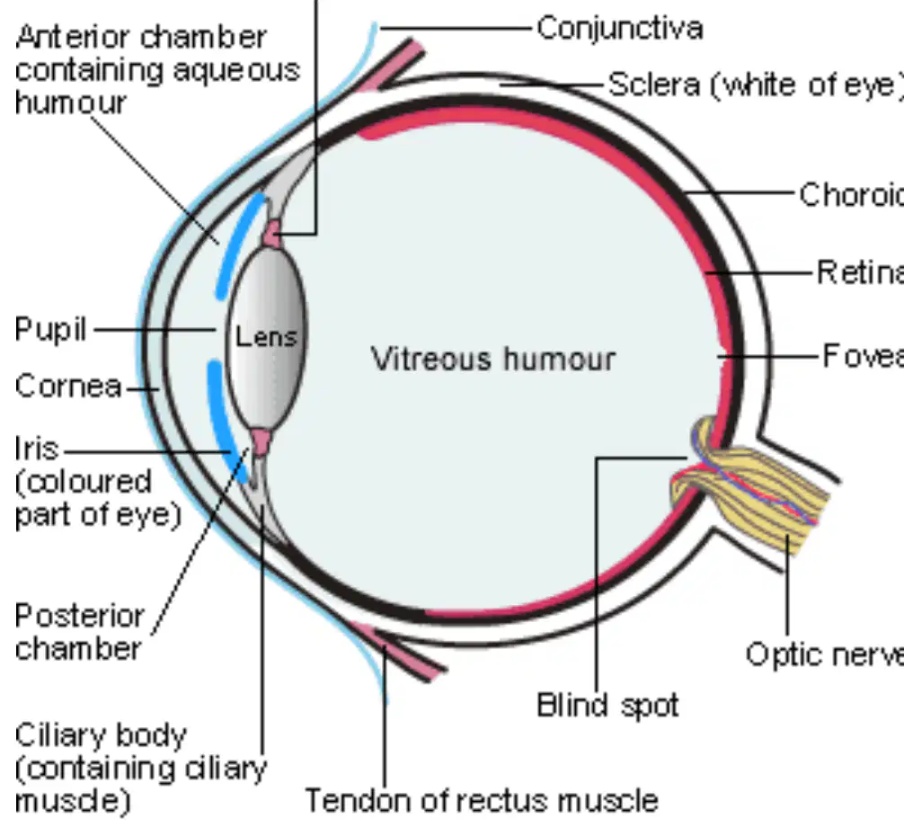
Which part of the eye is known as the "white of the eye"?
Sclera
Retina
Lens
Iris
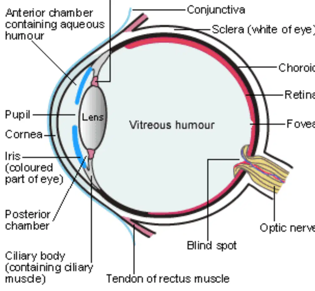
What is the function of the lens in the human eye?
To focus light onto the retina
To produce tears
To control eye movement
To give color to the eye

If a person has trouble seeing because their retina is damaged, which part of the eye is affected?
Retina
Cornea
Lens
Iris
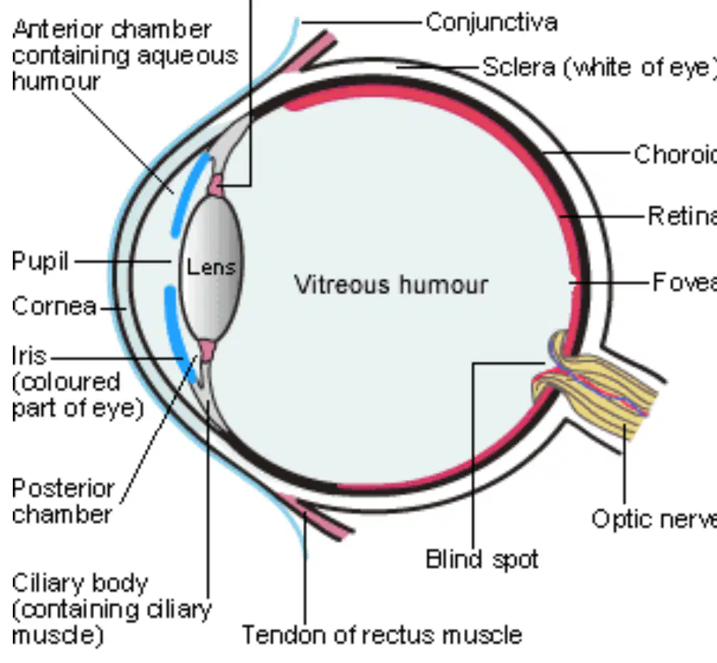
Which part of the eye contains the colored part?
Iris
Pupil
Sclera
Retina

Why is the optic nerve important for vision?
It carries visual information from the eye to the brain
It helps the eye change shape
It produces tears
It gives color to the eye
What is the innermost layer of the eye called?
Retina
Cornea
Lens
Sclera
What is the function of rods and cones in the retina?
They help you blink
They transduce light energy into nerve impulses
They produce tears
They control eye movement
Which part of the eye is known for having a high density of cones and is important for color vision?
Macula lutea
Optic disc
Cornea
Iris
Where is the sharpest image produced in the eye?
Fovea centralis
Optic disc
Retina
Sclera
What is the optic disc also known as?
Blind spot
Color spot
Sharp spot
Light spot
If a person cannot see color well, which part of their eye might not be working properly?
Macula lutea
Optic disc
Cornea
Lens
Why is the optic disc called the "blind spot"?
Because it has no rods or cones and cannot detect light
Because it is the sharpest point of vision
Because it helps you see color
Because it is where tears are produced
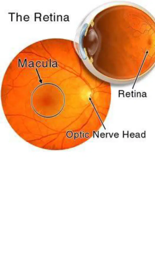
What is the name of the part of the eye labeled as "Macula" in the diagram?
Macula
Cornea
Lens
Iris
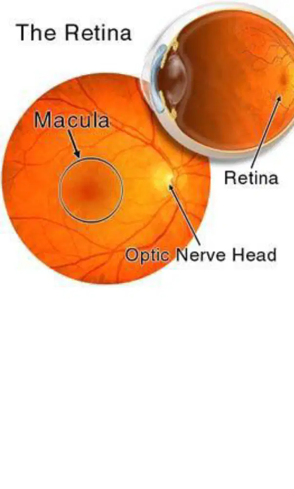
Which part of the eye is responsible for sending visual information to the brain and is labeled as "Optic Nerve Head" in the diagram?
Optic Nerve Head
Retina
Macula
Sclera
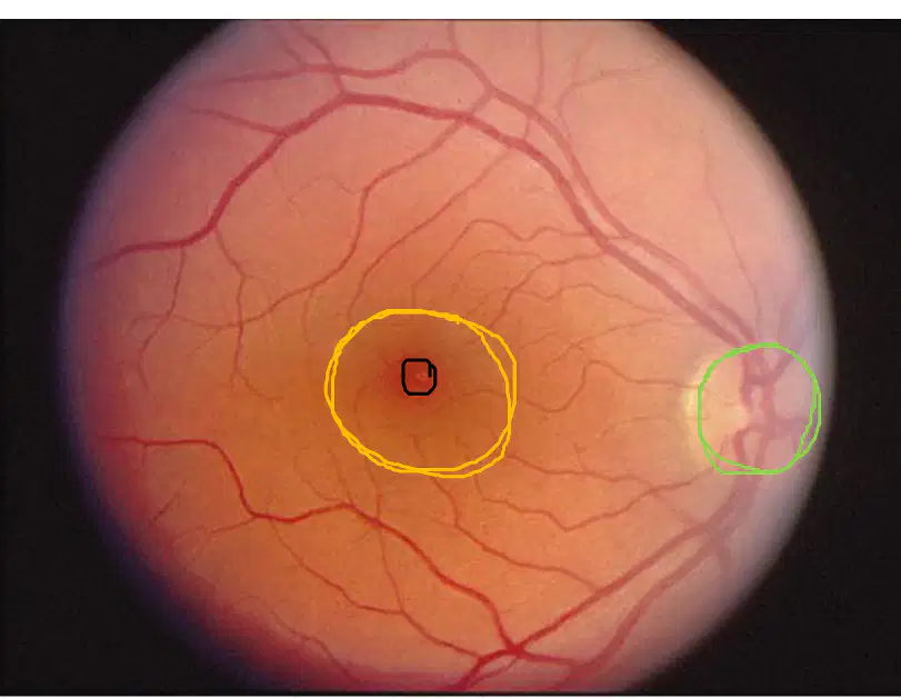
If you were looking at the retina through an ophthalmoscope, which area would you find surrounded by a yellow circle to show the macula lutea?
The area in the center of the retina
The area at the edge of the retina
The area with the green circle
The area with no blood vessels
What is the main function of the lens in the eye?
To focus light on the retina
To produce tears
To change the color of the eye
To protect the eye from dust
Where is the anterior chamber of the eye located?
Between cornea and iris
Between iris and lens
From lens to back of eye
Behind the retina
What keeps the eye lubricated in the anterior segment?
Aqueous humor
Vitreous humor
Tears
Blood
Which part of the eye supports its shape by filling the posterior chamber with a gel-like fluid?
Vitreous humor
Aqueous humor
Lens
Cornea
If the ciliary muscles in the eye did not work properly, what would most likely happen?
The lens would not change shape to refocus light
The eye would produce too many tears
The cornea would become cloudy
The iris would change color
Why is it important for the eye to have both aqueous humor and vitreous humor?
To keep the eye lubricated and to support its shape
To help the eye blink and cry
To change the color of the eye and help it see in the dark
To protect the eye from germs and dust

Which part of the eye is known as the "white of the eye"?
Sclera
Retina
Lens
Iris
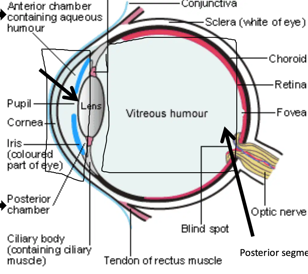
What is the function of the lens in the human eye?
To focus light onto the retina
To produce tears
To control eye movement
To protect the eye from dust

Which part of the eye contains the colored part?
Iris
Retina
Cornea
Optic nerve

If a person cannot see because the optic nerve is damaged, which part of the eye is affected?
Optic nerve
Cornea
Iris
Lens

Why is the retina important for vision?
It receives light and sends signals to the brain
It controls the movement of the eye
It produces tears
It gives color to the eye
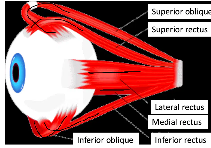
Which of the following is NOT an extrinsic eye muscle?
Superior rectus
Inferior rectus
Medial rectus
Biceps brachii
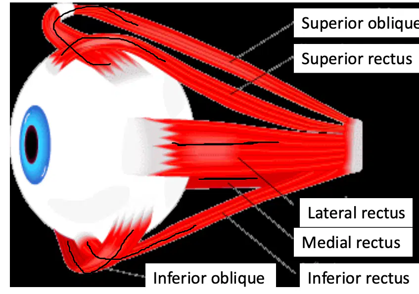
What is the function of the extrinsic eye muscles?
They help move the eyeball.
They help us hear sounds.
They help us taste food.
They help us breathe.
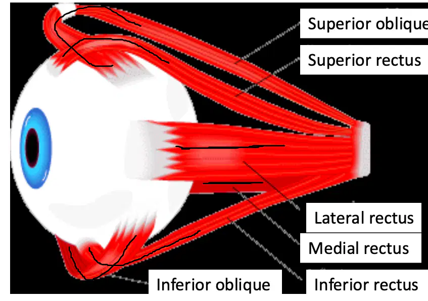
If a person cannot move their eye upward, which muscle might be affected?
Superior rectus
Inferior oblique
Medial rectus
Lateral rectus
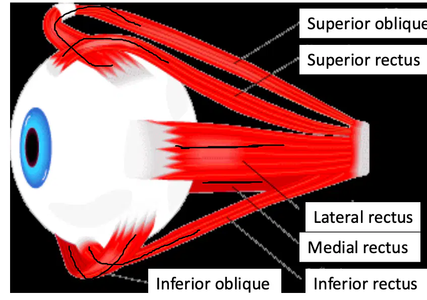
Which two extrinsic eye muscles have "oblique" in their names?
Superior oblique and Inferior oblique
Medial rectus and Lateral rectus
Superior rectus and Inferior rectus
Superior oblique and Medial rectus
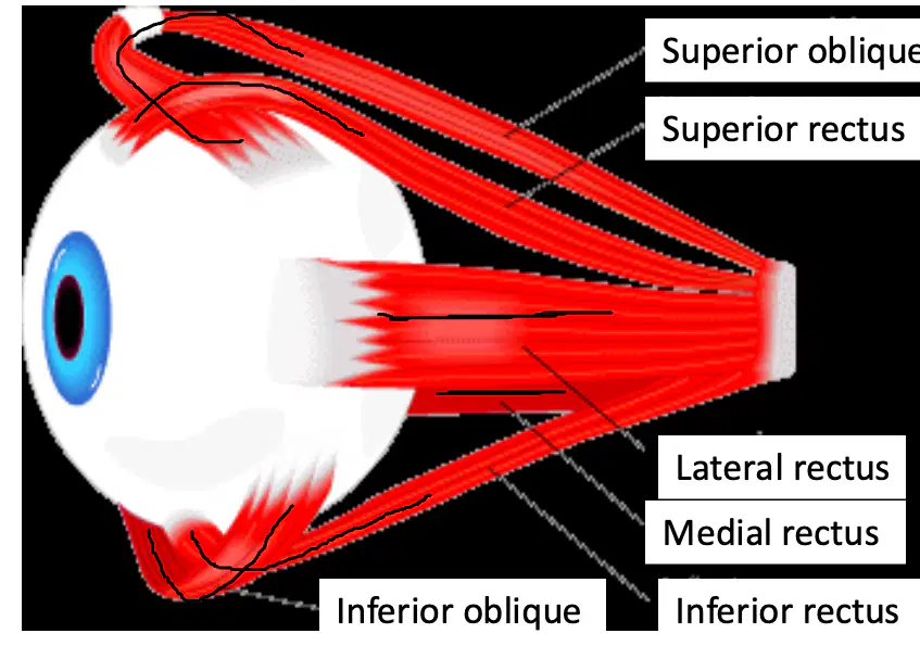
If you want to look to the side, which extrinsic eye muscle helps you do that?
Lateral rectus
Superior rectus
Inferior oblique
Medial rectus

What is the name of the clear covering on the front of the eye?
Cornea
Iris
Sclera
Retina

Which part of the eye is the colored part that surrounds the pupil?
Iris
Sclera
Cornea
Lens

What is the function of the pupil in the eye?
It is the opening in the iris that lets light into the eye
It is the colored part of the eye
It is the white part of the eye
It covers the eye to protect it

If the cornea is damaged, which part of the eye's function would be most affected?
The eye's ability to let light in
The eye's color
The eye's white outer layer
The eye's ability to move

Which part of the eye is labeled as "Sclera" in the diagram?
The white outer layer of the eye
The yellow part at the bottom
The blue area with blood vessels
The part labeled "Optic nerve"
What is the function of the optic nerve in the eye?
It sends visual information from the eye to the brain
It protects the eye from dust
It helps the eye change shape
It produces tears

Which part of the eye is shown as a blue area with blood vessels in the diagram?
Choroid
Sclera
Optic nerve
Retina

What is the clear, dome-shaped part at the front of the eye called?
Cornea
Retina
Sclera
Choroid

Which part of the eye is responsible for focusing light onto the retina?
Lens
Sclera
Ora Serrata
Canal of Schlemm

What is the name of the white outer layer of the eye?
Sclera
Retina
Choroid
Ciliary body

If a person has trouble focusing on objects, which part of the eye might not be working properly?
Lens
Sclera
Choroid
Ora Serrata

Which part of the eye contains the blood vessels and provides nutrients to the eye?
Choroid
Cornea
Lens
Canal of Schlemm

What is the function of the ciliary muscle in the eye?
Helps the lens change shape to focus
Protects the eye from dust
Produces tears
Sends signals to the brain

Which part of the eye is known as the area around the central fovea?
Macula Lutea
Retina
Sclera
Choroid

What is the name of the part of the eye labeled as the "Blind spot"?
Blind spot
Central Fovea
Retina
Sclera

Which layer of the eye is located between the sclera and the retina?
Choroid
Sclera
Macula Lutea
Anterior segment

If you were to trace the path of light entering the eye, which segment would it pass through first: the anterior segment or the posterior segment?
Anterior segment
Posterior segment
Retina
Choroid

Imagine you are a scientist studying the eye. Which part would you examine to learn about the area responsible for sharp, central vision?
Central Fovea
Sclera
Posterior segment
Choroid
Which part of the ear is the outermost part?
Pinna/Auricle
Tympanic membrane
Eustachian tube
Ceruminous glands
What is the function of ceruminous glands in the ear?
They secrete cerumen (ear wax)
They help you hear sounds
They separate the middle ear from the inner ear
They connect the ear to the throat
Which structure separates the external ear from the middle ear?
Tympanic membrane
Pinna/Auricle
Eustachian tube
Ceruminous glands
The Eustachian tube connects the middle ear to which part of the body?
Nasopharynx
Outer ear
Ear drum
Ceruminous glands
If the tympanic membrane is damaged, which two parts of the ear would no longer be separated?
External ear and middle ear
Middle ear and inner ear
Outer ear and Eustachian tube
Ceruminous glands and pinna
Why is the middle ear important for hearing?
It contains ossicles that help transmit sound
It produces ear wax
It connects to the nose
It is the outermost part of the ear

Which part of the ear is responsible for collecting sound from the environment?
Cochlea
Pinna
Eardrum
Auditory nerve

Which of the following is NOT a part of the external ear?
Ear lobe
Cartilage
Cochlea
Pinna

If the eardrum (tympanic membrane) is damaged, which process would be most affected?
Collecting sound
Transmitting sound vibrations
Balancing the body
Smelling odors

How do the semicircular canals help us in our daily life?
They help us hear sounds.
They help us balance and keep our body steady.
They help us taste food.
They help us see things.
Which of the following is NOT one of the ossicles found in the ear?
Malleus
Incus
Stapes
Cochlea
What is the main function of the stapes in the ear?
To send vibrations into the inner ear
To produce earwax
To help with balance
To detect smells
Where is the round window located in relation to the oval window in the ear?
Just inferior and medial to the oval window
Just superior and lateral to the oval window
Directly above the oval window
Far away from the oval window
Imagine you are explaining to a friend how sound travels through the ear. Using what you know about the ossicles, describe their role in hearing.
The ossicles help pass vibrations from the eardrum to the inner ear.
The ossicles help produce earwax.
The ossicles help us smell things.
The ossicles help us see better.
What is the inner ear composed of?
A membrane-filled bony labyrinth
A group of muscles
A layer of skin
A collection of blood vessels
Which part of the inner ear is associated with balance?
Vestibular apparatus
Eardrum
Cochlea
Auditory nerve
Which of the following is NOT a part of the vestibular apparatus?
Cochlea
Utricle
Saccule
Semicircular canals
Where is the ampula receptor located?
At the base of each semicircular canal
Inside the cochlea
On the eardrum
In the outer ear
Imagine you are spinning around quickly and then stop. Which part of your inner ear helps you regain your balance?
Semicircular canals
Eardrum
Auditory nerve
Outer ear

What is the name of the part labeled as "Cochlea" in the diagram of the vestibular system?
Cochlea
Utricle
Saccule
Ampullae

Which part of the vestibular system contains endolymph?
Tubular ducts
Cochlea
Ampullae
Saccule
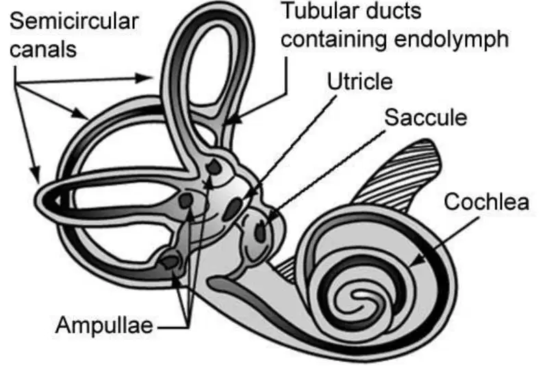
If a person has trouble with balance, which part of the vestibular system might be affected?
Semicircular canals
Cochlea
Saccule
Tubular ducts
What is the cochlea?
A snail-shaped bony cavity that contains the mechanism of hearing
A tube that connects the ear to the throat
A muscle that helps you chew
A bone in your arm
Which part of the ear is a spiral membrane that fills the cochlea?
Cochlear duct
Eardrum
Auditory nerve
Ear canal
What are the two levels of the cochlear duct?
Scala vestibuli and Scala tympani
Outer ear and inner ear
Eardrum and ear canal
Hammer and anvil
Where is the scala vestibuli located in the cochlear duct?
Upper part
Lower part
Middle part
Outside part
What does the Spiral Organ of Corti contain?
Receptors for hearing
Blood vessels
Muscles for chewing
Bones for movement
The tectorial membrane is part of which structure?
Spiral Organ of Corti (SOC)
Eardrum
Ear canal
Auditory nerve
If a scientist wanted to study the part of the ear that contains receptors for hearing, which structure should they focus on?
Spiral Organ of Corti
Scala vestibuli
Tectorial membrane
Cochlear duct
Imagine you are explaining to a friend how the cochlear duct is organized. How would you describe its two levels?
The upper part is called scala vestibuli and the lower part is called scala tympani.
The upper part is called ear canal and the lower part is called eardrum.
The upper part is called hammer and the lower part is called anvil.
The upper part is called outer ear and the lower part is called inner ear.

Which part of the ear is responsible for collecting sound from the environment?
Pinna
Cochlea
Eustachian tube
Auditory nerve

What is the function of the cochlea in the ear?
It helps collect sound from the environment.
It sends sound signals to the brain.
It balances the body.
It protects the ear from dust.

Which part of the ear connects to the throat and helps balance air pressure?
Eustachian tube
Ear lobe
Incus
Stapes

If a person has trouble hearing, which part of the ear might be damaged if the problem is with sending signals to the brain?
Cochlea
Pinna
Cartilage
Ear lobe

Why is the ear drum important for hearing?
It collects sound from the environment.
It vibrates when sound waves hit it, helping us hear.
It sends signals to the brain.
It balances the body.
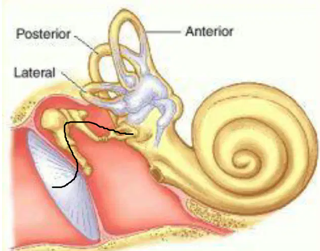
Which part of the ear is responsible for hearing and looks like a spiral or snail shell?
Cochlea
Eardrum
Ear canal
Outer ear
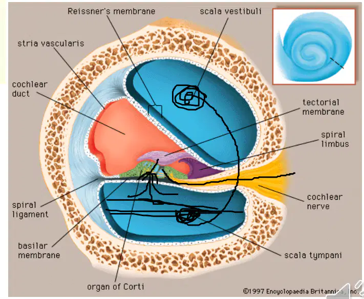
What is the name of the membrane inside the cochlea that helps detect sound vibrations?
Basilar membrane
Tectorial membrane
Reissner's membrane
Spiral ligament
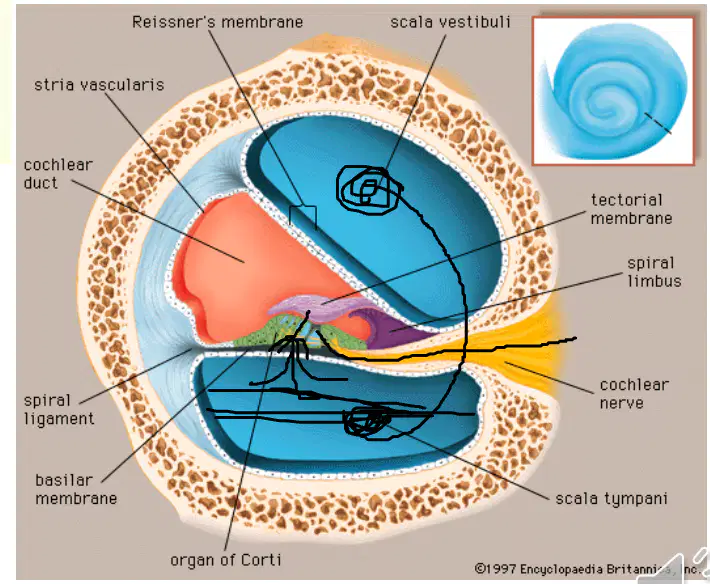
If the cochlear nerve is damaged, what might happen to a person?
They may have trouble hearing sounds.
They may have trouble seeing.
They may have trouble tasting food.
They may have trouble smelling.


Which part is #1?
sclera
cornea
retina
lens
Which part is #2?
iris
cornea
retina
lens
Which part is #3?
sclera
cornea
retina
lens
Which part is #4?
pupil
iris
cornea
lens
Which part is #5?
pupil
iris
cornea
lens
Which part is #6?
sclera
cornea
retina
lens
Which part is #7?
sclera
optic nerve
cornea
retina

#3 Refers to the circular structure that surrounds the pupil and regulates the amount of light entering the eye.
Cornea
Pupil
Retina
Iris

#1 Refers to the clear tissue that covers the front of the eye
Retina
Cornea
Pupil
Iris

#2 refers to
optic nerve
cornea
iris
pupil

#7 Refers to the part of the eye that carried visual information from the eyeball to the brain
lens
optic nerve
retina
sclera

# 5 refers to the layer of receptor cells at the back of the eye in which the image is focused
Retina
Lens
Optic Nerve
Lens

Identify # 1
cornea
iris
lens
retina
Identify #4
cornea
iris
lens
pupil
Identify #5
cornea
iris
lens
retina
Identify #7
cornea
optic nerve
lens
retina
Which part is #2?
iris
cornea
retina
lens
Which part is #3?
sclera
cornea
retina
lens
Which part is #4?
pupil
iris
cornea
lens
Which part is #5?
pupil
iris
cornea
lens
Which part is #6?
sclera
cornea
retina
lens
Which part is #7?
sclera
optic nerve
cornea
retina
#3 Refers to the circular structure that surrounds the pupil and regulates the amount of light entering the eye.
Cornea
Pupil
Retina
Iris
#1 Refers to the clear tissue that covers the front of the eye
Retina
Cornea
Pupil
Iris
#1 Refers to the clear tissue that covers the front of the eye
Retina
Cornea
Pupil
Iris
#2 refers to
optic nerve
cornea
iris
pupil
#7 Refers to the part of the eye that carried visual information from the eyeball to the brain
lens
optic nerve
retina
sclera
# 5 refers to the layer of receptor cells at the back of the eye in which the image is focused
Retina
Lens
Optic Nerve
Lens
Identify # 1
cornea
iris
lens
retina
Identify #4
cornea
iris
lens
pupil
Identify #5
cornea
iris
lens
retina
Identify #7
cornea
optic nerve
lens
retina


