
 Worksheets
Body Systems Basics: Cardiovascular — Student Notes
Total questions: 77
Worksheet time: 39mins
Fill in the blank: The cardiovascular system circulates blood to transport ________ to all parts of the body (nourishment including amino acids, electrolytes, oxygen, and hormones).
nutrients
wastes
antibodies
carbon dioxide
Fill in the blank: The cardiovascular system includes the heart, ________ and blood.
blood vessels
lungs
nerves
bones
Fill in the blank: The heart is a ________ muscular organ.
hollow
voluntary
skeletal
smooth-only
Fill in the blank: The heart is associated with the root cardi(o), derived from the ________ ‘kardia’ meaning heart.
Greek
Latin
French
Arabic
Fill in the blank: The pericardium is double layered; the outer layer is the ________ pericardium (tough connective tissue attached to the central tendon of the diaphragm and the back of the sternum).
fibrous
visceral
serous
parietal
Fill in the blank: The pericardium is double layered; the inner layer is the ________ pericardium (two layers with a pericardial cavity in between).
serous
fibrous
parietal
synovial
Fill in the blank: The heart wall is comprised of epicardium, ________ (cardiac muscle responsible for heart contractions), and endocardium.
myocardium
pericardium
intercardium
sarcolemma
Fill in the blank: The heart wall is comprised of epicardium, myocardium, and ________ (inner lining of the heart’s chambers and valves that regulates contractions and aids cardiac embryological development).
endocardium
pericardial cavity
parietal layer
epithelium
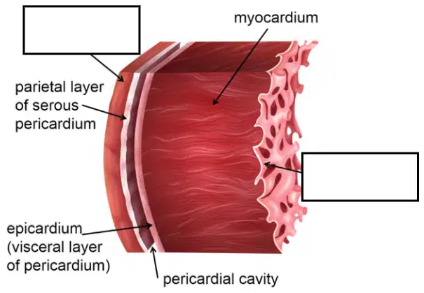
Identify the labeled inner lining in the heart wall cross-section diagram.
endocardium
myocardium
epicardium
pericardial cavity
Fill in the blank: The heart consists of four chambers; the right and left ________ are the upper chambers with thin walls.
atria
ventricles
auricles
valves
Fill in the blank: The heart consists of four chambers; the right and left ________ are the lower chambers with thick walls.
ventricles
atria
sinuses
appendages
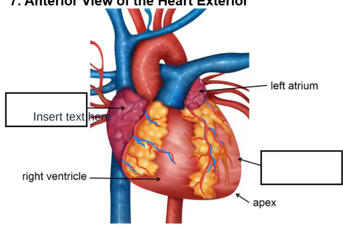
Anterior view of the heart exterior: Identify the structure indicated by the blank label on the left side of the diagram.
right atrium
left atrium
pulmonary trunk
superior vena cava
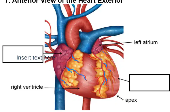
Anterior view of the heart exterior: Identify the structure indicated by the blank label on the right side of the diagram.
left ventricle
right ventricle
apex
left atrium
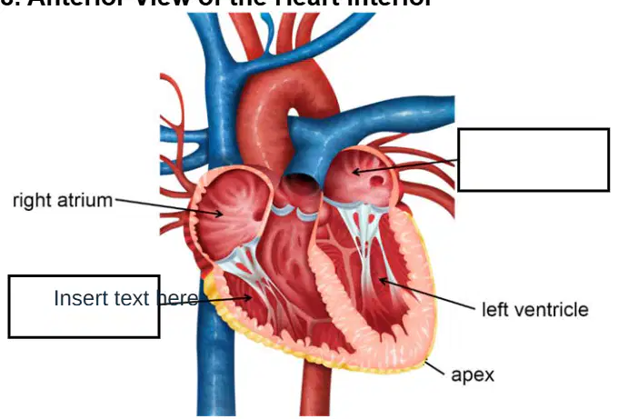
Anterior view of the heart interior: Identify the structure indicated by the blank label near the lower left chamber.
right ventricle
left ventricle
right atrium
left atrium
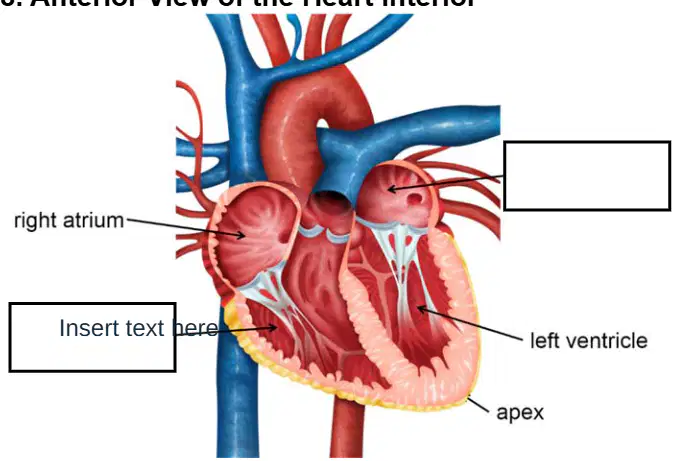
Anterior view of the heart interior: Identify the structure indicated by the blank label adjacent to the valve leaflets on the right side of the diagram.
mitral (bicuspid) valve
tricuspid valve
aortic valve
pulmonary valve
Atria: The right atrium receives _____ blood from the body via the superior and inferior vena cava and from the heart via the coronary veins.
deoxygenated
oxygenated
mixed
carbonated
Atria: The left atrium receives _____ blood from the lungs via the pulmonary veins.
oxygenated
deoxygenated
mixed
venous
Ventricles: Are responsible for pumping blood _____ the heart.
out of
into
around inside
away from the veins
Ventricles: Form the _____ of the heart.
apex
base
septum
atrium
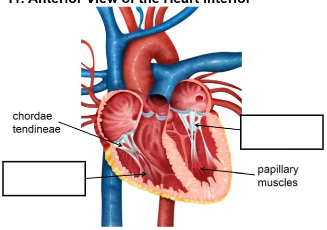
Anterior view of the heart interior: Identify the structure indicated by the blank label near the lower left chamber (separate from the labels for chordae tendineae and papillary muscles).
right ventricle
left atrium
superior vena cava
aorta
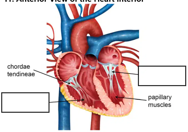
Anterior view of the heart interior: Identify the structure indicated by the blank label near the valve leaflets on the right side (separate from the labels for chordae tendineae and papillary muscles).
mitral (bicuspid) valve
aortic valve
pulmonary valve
tricuspid valve
Great vessels of the heart: The superior and inferior vena cavae are the _____ veins in the body.
largest
smallest
most elastic
most muscular
Great vessels of the heart: Pulmonary arteries carry deoxygenated blood from the right ventricle to the _____.
lungs
left atrium
aorta
systemic capillaries
Great vessels of the heart: Identify the vessel that carries oxygenated blood from the lungs to the left atrium.
pulmonary veins
pulmonary arteries
superior vena cava
coronary sinus
Great vessels of the heart: Identify the vessel that is the largest artery in the body and carries oxygenated blood to be distributed throughout the body.
aorta
pulmonary trunk
coronary artery
inferior vena cava
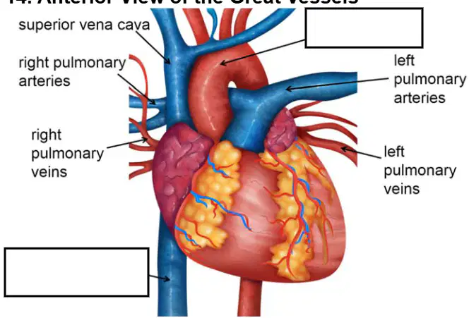
Anterior view of the great vessels: Identify the structure indicated by the blank label on the upper right side of the diagram.
aorta
left pulmonary artery
superior vena cava
pulmonary trunk
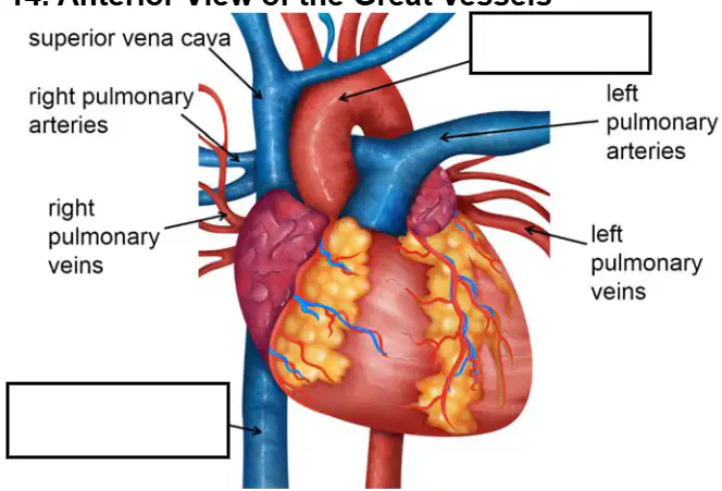
Anterior view of the great vessels: Identify the structure indicated by the blank label on the lower left side of the diagram.
inferior vena cava
right pulmonary vein
pulmonary trunk
left pulmonary artery
Cardiac valves include atrioventricular valves located between the atria and ventricles. Which valve is between the right atrium and right ventricle?
Tricuspid valve
Bicuspid (mitral) valve
Pulmonary valve
Aortic valve
Atrioventricular valves are attached to papillary muscles by chordae tendineae and open during __________ (diastole), allowing the ventricle to fill. Which term correctly completes the statement?
ventricular relaxation
ventricular contraction
atrial systole
isovolumetric contraction
Atrioventricular valves close during ventricular contraction (systole), preventing reflux. Which phase describes when these valves are closed?
systole
diastole
atrial relaxation
isovolumetric relaxation
Cardiac valves include semilunar valves located between ventricles and __________. What structure completes the statement?
arteries
atria
veins
venae cavae
Semilunar valves open when ventricles contract and ventricular pressure exceeds arterial pressure. At the beginning of __________, they close when blood flows toward the heart, pushing the valve cusps together. Which cardiac phase completes the statement?
diastole
systole
atrial systole
ventricular ejection
At the beginning of diastole, semilunar valves close as blood flows toward the heart, pushing __________ the valve cusps together. Which word best completes the statement?
backflow against
forward through
laterally away from
downward past
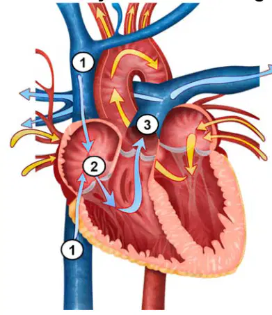
Refer to the labeled heart diagram showing arrows from body vessels into the right atrium. In step 1, __________ from the body enters the right atrium via the vena cavae.
deoxygenated blood
oxygenated blood
lymphatic fluid
plasma only
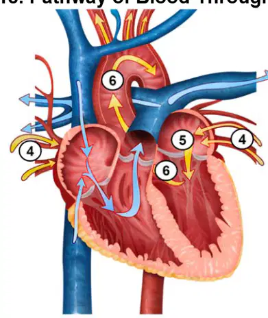
Refer to the labeled heart diagram showing pulmonary veins entering the left atrium. In step 4, oxygenated blood from the lungs enters the __________ via pulmonary veins.
left atrium
right atrium
left ventricle
right ventricle
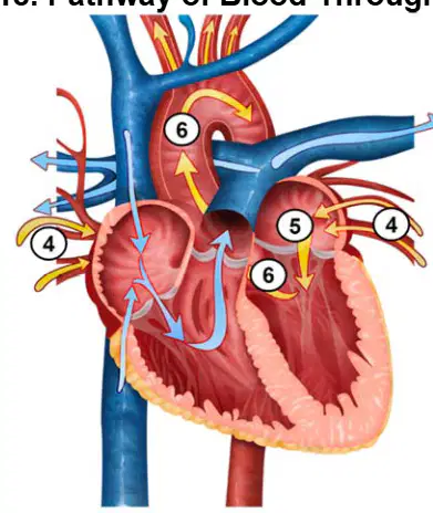
Refer to the labeled heart diagram showing a valve between the left atrium and left ventricle. In step 5, blood travels through the __________ into the left ventricle.
bicuspid (mitral) valve
tricuspid valve
pulmonary valve
aortic valve
Coronary circulation supplies the tissues of the heart with oxygenated blood and drains it of deoxygenated blood. Which components originate at the aortic sinuses within the aorta and provide oxygenated blood?
coronary arteries
cardiac veins
pulmonary arteries
vena cavae
Coronary circulation involves components that drain deoxygenated blood from the heart and open into the coronary sinus in the right atrium. What are these vessels called?
cardiac veins
coronary arteries
pulmonary veins
aortic sinuses
During coronary circulation, oxygenated blood in the __________ is forced into the coronary arteries during diastole due to increased aortic pressure. Which structure completes the statement?
aorta
pulmonary trunk
right ventricle
coronary sinus
In coronary circulation, deoxygenated blood returns to the heart via __________ and drains into the right atrium through the coronary sinus. Which vessels complete the statement?
cardiac veins
coronary arteries
pulmonary arteries
aortic sinuses
The heart conduction system initiates and coordinates __________ of the heart muscle, using electrical impulses (__________) to make the heart beat. Which pair correctly completes both blanks?
contraction; action potentials
relaxation; pressure waves
diffusion; neurotransmitters
filtration; ion pumps
Heart Conduction occurs when the ______ creates an electrical impulse which spreads over the atria, causing them to contract; forcing blood into ventricles.
sinoatrial (SA) node
atrioventricular (AV) node
Purkinje fibers
bundle branches
Heart Conduction: the impulse is spread to the ____________, causing the ventricles to contract; forcing blood out of the aorta and pulmonary arteries.
Purkinje fibers
sinoatrial (SA) node
atrioventricular (AV) node
bundle of His
Cardiac Cycle involves: __________ — atria and ventricles are relaxed; blood flows into atria and through atrioventricular valves into ventricles.
diastole
systole
depolarization
isovolumetric contraction
Cardiac Cycle involves: __________ — atria contract; blood is pushed into ventricles, building pressure.
atrial systole
ventricular diastole
atrial diastole
isovolumetric relaxation
Cardiac Cycle involves: __________ — ventricles contract and eject; pressure within the ventricles builds until outflow valves are forced open and blood is pumped from the heart.
ventricular systole
atrial systole
ventricular diastole
isovolumetric relaxation
Cardiac Cycle involves: __________ (early diastole) — ventricles relax, reducing pressure and forcing atrioventricular valves open to start the cycle again.
isovolumetric relaxation
ventricular systole
atrial systole
depolarization
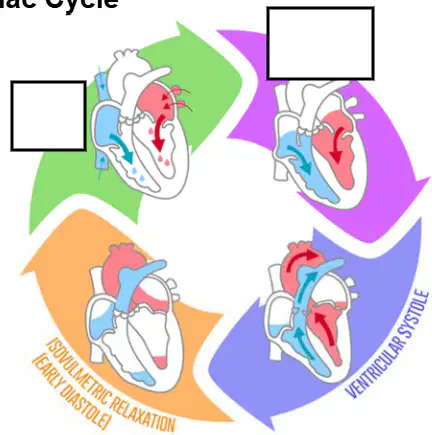
Cardiac Cycle: Based on the diagram of the cardiac cycle showing phases atrial systole, ventricular systole, and isovolumetric relaxation (early diastole), which label correctly names the phase when the ventricles contract to pump blood to the arteries?
ventricular systole
atrial systole
isovolumetric relaxation
atrial diastole
Blood Vessels: Comprise the __________ system.
cardiovascular
respiratory
nervous
lymphatic
Blood Vessels: the hollow passageway through which blood flows is known as the __________.
lumen
atrium
septum
valve
Blood Vessels are composed of three layers: tunica ______ — middle layer of smooth muscle; thickest layer in arteries.
media
intima
adventitia
externa
Blood Vessels are composed of three layers: tunica intima (__________) — inner layer of smooth epithelial and connective tissue; thinnest layer.
interna
media
externa
adventitia
Blood Vessels include: __________, capillaries, and veins.
arteries
arterioles
venules
chambers
Arteries carry oxygenated blood __________ to the capillaries, providing oxygen and nutrients to body tissues (except pulmonary arteries carry deoxygenated blood from the heart to the lungs to be oxygenated).
away from the heart
toward the lungs
into the atria
directly back to the heart
Arteries are thicker and stronger than veins — to withstand the __________.
higher blood pressure
lower blood pressure
valve closure
blood clotting
Arteries include: large __________ conducting arteries. Choose the term that correctly completes the statement.
elastic
muscular
fenestrated
sinusoidal
Arteries include: metarterioles — tiny arteries which supply __________. Select the best completion.
capillary beds
the lungs
cardiac muscle only
large veins
Capillaries are the smallest blood vessels with walls one cell layer thick; walls are __________ so materials can be diffused in and out. Which word best completes the sentence?
permeable
contractile
keratinized
calcified
Capillaries include three types: continuous, fenestrated, and sinusoidal — each of which allow varying amounts and types of __________ to pass through. What completes the statement?
substances
electrical signals
hormone secretion only
bone tissue
Veins carry deoxygenated blood from capillaries toward the heart, relieving body tissues of __________ (except the pulmonary veins, which carry oxygenated blood from the lungs to the heart). Choose the best completion.
carbon dioxide
oxygen
nutrients
antibodies
Veins have a series of __________ that work against gravity to prevent backflow. Select the correct term.
valves
sphincters
cilia
papillae
Veins include: __________ — small veins (with a diameter less than 1 mm). Which term correctly fills the blank?
venules
arterioles
capillaries
large veins
Veins include: __________ — have diameters greater than 10 mm and have a thicker tunica intima than other veins. Pick the correct term.
large veins
medium arteries
venules
capillaries
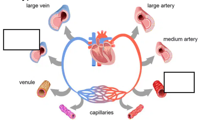
Types of Blood Vessels diagram: Identify the missing vessel on the venous side between capillaries and large veins that completes the set of labels.
small vein
arteriole
medium artery
large artery
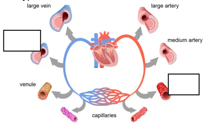
Types of Blood Vessels diagram: Identify the missing vessel on the arterial side between arteries and capillaries that completes the set of labels.
arteriole
small vein
large vein
venule
Blood transports the following to tissues throughout the body: nutrients, electrolytes, __________, vitamins, antibodies, heat, oxygen, immune cells. Choose the correct completion.
hormones
bone marrow
synovial fluid
bile acids
Blood transports __________ and waste from tissues throughout the body to the kidneys and liver for filtering. Select the best completion.
carbon dioxide
oxygen
glycogen
albumin
Blood consists of the following: erythrocytes (__________ blood cells). Which word correctly completes the phrase?
red
white
platelet
stem
Blood consists of the following: leukocytes (__________ blood cells). Which word correctly completes the phrase?
white
red
plasma
scar
Heart rate is the __________ at which the heart beats — the number of beats per minute. Choose the best completion.
rate
pressure
volume
stroke
output
Heart rate can be measured by checking the pulse — __________ of an artery due to blood pumping within it as the heart beats. Select the correct completion.
rhythmic expansion
valve closure
nerve impulse
muscle contraction
Blood pressure is the force exerted by blood on the ____________ of arteries as it pumps through the body.
walls
valves
chambers
veins
Peripheral resistance is created by the arteries as they ____________.
constrict
dilate
thicken
lengthen
Systolic pressure is the maximum pressure during ventricular systole (__________).
contraction
relaxation
filling
repolarization
Diastolic pressure is the minimum pressure during diastole (__________).
relaxation
contraction
ejection
depolarization

Using the Blood Pressure Categories table, the systolic pressure range for Stage 1 (Mild) Hypertension is __________ mmHg.
140–159
160–180
121–139
>180

Using the Blood Pressure Categories table, the diastolic pressure range for Stage 2 (Moderate) Hypertension is __________ mmHg.
100–110
90–99
81–89
>110

