
 Worksheets
Axial & Appendicular Skeleton: Learning Targets 1 & 2 (Part 1)
Total questions: 91
Worksheet time: 46mins
Which set lists ONLY axial skeleton components?
Coxa, femur, patella, tibia
Clavicle, humerus, radius, ulna
Skull, vertebrae, ribs, sternum
Scapula, carpals, metacarpals, phalanges
Fibula, tarsals, metatarsals, phalanges
Which bone group belongs to the appendicular skeleton of the upper limb?
Scapula, clavicle, humerus, radius, ulna
Skull, vertebrae, sacrum, coccyx, ribs
Sternum, ribs, vertebrae, sacrum
Fibula, tarsals, metatarsals, phalanges
Coxa, femur, tibia, patella
Select the correct classification for the lower limb bones listed: fibula, patella, tarsals.
Cranial skeleton components
Axial skeleton components
Vertebral column components
Thoracic cage components
Appendicular skeleton components

On a scapula diagram, which anatomical term names a shallow depression for muscle attachment?
Condyle
Epicondyle
Tubercle
Fossa
Foramen
Which term best describes a rounded bump on a bone, smaller than a tuberosity?
Tubercle
Angle
Process
Ramus
Facet
A student must color-code a skeleton to separate axial and appendicular regions. Which planning step will most reduce mistakes?
Ignore the rib cage until the end
Color all long bones first
Use the brightest colors available
Start coloring from the feet upward
List axial bones before coloring
You are labeling a vertebra. Which feature is an opening that allows passage of nerves or vessels?
Foramen
Facet
Condyle
Ramus
Process
Which term describes a shallow depression on a bone surface commonly found near articular areas?
Fossa shallow depression
Notch deep indentation
Spine ridge on bone
Tuberosity rough bump
On long bones, what is the narrowed region between the head and the body called?
Ramus projecting arm
Foramen round opening
Neck constricted region
Process small extension
Which bones are classified as cranial bones? Select the option that lists only cranial bones.
Frontal, parietal, temporal
Lacrimal, ethmoid, inferior concha
Mandible, vomer, palatine
Maxilla, zygomatic, nasal
Which structure forms the small, flat articular surface on a vertebra that helps joint movement between adjacent vertebrae?
Tubercle rounded bump
Spinous posterior ridge
Foramen nerve opening
Facet articular surface
Which sutures of the skull connect the parietal bones to neighboring bones?
Zygomatic and maxillary
Metopic and palatine
Sagittal and lambdoid
Coronal and squamous
What is the only movable joint of the skull?
Occipital condyles
Nasal cartilages
Suture lines
TMJ at mandible
Which sequence correctly lists vertebral column regions from superior to inferior?
Cervical thoracic lumbar
Lumbar thoracic cervical
Thoracic cervical lumbar
Sacral lumbar cervical
You are comparing cervical and thoracic vertebrae. Which feature best distinguishes a typical cervical vertebra from a typical thoracic vertebra?
Large heart-shaped body
Transverse foramina present
Long inferior spinous
Costal facets for ribs
Which vertebral region has short, bifid spinous processes and transverse foramina?
Thoracic vertebrae T1–T12
Lumbar vertebrae L1–L5
Cervical vertebrae C1–C7
Sacral vertebrae S1–S5
Which vertebra is called the atlas and primarily supports the skull?
L1 with largest vertebral body
T1 with rib facets
C2 with dens odontoid process
C1 with superior articular facets
Which feature distinguishes the axis from other cervical vertebrae?
Long inferior spinous process
Costal facets for ribs
Presence of the dens process
Large kidney-shaped body
Which vertebral region has costal facets on the body for rib articulation?
Lumbar vertebrae L1–L5
Cervical vertebrae C1–C7
Coccygeal vertebrae Co1–Co4
Thoracic vertebrae T1–T12
Which vertebral region typically has the largest vertebral bodies to bear weight?
Lumbar vertebrae L1–L5
Cervical vertebrae C1–C7
Thoracic vertebrae T1–T12
Sacral vertebrae S1–S5
What is the primary function of intervertebral disks?
Anchor ribs to the sternum
Shock absorption and load distribution
Protect cranial nerves in foramina
Enable rotation of the atlas on axis
Which two main components make up an intervertebral disk?
Nucleus pulposus and annulus fibrosus
Trabecular bone and cortical shell
Ligamentum flavum and interspinous ligament
Synovial capsule and articular cartilage
Which set of ribs is classified as true ribs in the thoracic cage?
Rib pairs 3–10 articulate with xiphoid
Rib pairs 1–7 attach directly to sternum
Rib pairs 8–12 attach via cartilage
Rib pairs 11–12 have no anterior attachment
Which ribs are considered floating ribs?
Rib pairs 3–7 connect by costal cartilage
Rib pairs 11–12 lack anterior attachment
Rib pairs 8–10 attach to xiphoid
Rib pairs 1–2 fuse with manubrium
What is the primary function of costal cartilage in the thoracic cage?
Protect spinal cord from compression
Increase rib thickness for force absorption
Anchor clavicle to manubrium firmly
Connect ribs to sternum and add flexibility
Which part of the sternum articulates with the clavicles?
Manubrium at clavicular notches
Sternal angle at rib tubercle
Body at intercostal facets
Xiphoid at costal margin
Which bones form the pectoral girdle?
Scapula and radius
Sternum and rib one
Humerus and ulna
Clavicle and scapula pair
Which upper limb bone contains the trochlea that articulates with the ulna?
Distal humerus at trochlea
Clavicle at acromial end
Proximal radius at head
Scapula at coracoid
Which landmark is found on the ulna and forms the elbow tip?
Greater tubercle superiorly
Coracoid process anteriorly
Radial styloid laterally
Olecranon process posteriorly
Which statement best compares true, false, and floating ribs?
True attach to sternum; false via shared cartilage; floating no anterior
True attach by ligaments; false by tendons; floating by muscles
True are upper; false are lower; floating are middle
True attach to xiphoid; false to manubrium; floating to clavicle
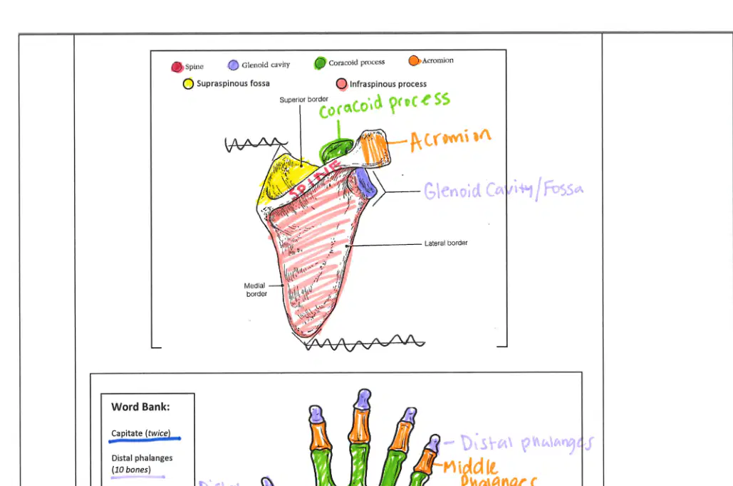
In the labeled scapula diagram, which structure forms the superior-lateral projection that articulates with the clavicle?
Scapular spine on posterior surface
Glenoid cavity on lateral border
Acromion projecting laterally
Coracoid process at superior border

In the anterior view hand diagram, which carpal lies anterior to (palmar to) the triquetrum and is visible as a pea-shaped bone?
Pisiform on palmar side
Capitate central carpal
Scaphoid near radius
Hamate with hook

Which statement correctly distinguishes thumb phalanges compared with other digits in the diagrams?
Thumb lacks a middle phalanx
Thumb has fused proximal and distal
Thumb lacks a distal phalanx
Thumb has four phalanges
Thumb has three metacarpals

Identify the carpal that articulates primarily with the radius at the wrist in the diagrams.
Capitate central carpal
Lunate proximal carpal
Pisiform sesamoid carpal
Hamate ulnar carpal

In the posterior view diagram, which pair of carpals is most radial (lateral) in the proximal row?
Scaphoid and trapezium
Scaphoid and lunate
Triquetrum and pisiform
Hamate and capitate

Which metacarpal is aligned with the little finger in both views of the hand diagrams?
1st metacarpal medial
3rd metacarpal central
2nd metacarpal lateral
5th metacarpal ulnar

A fall onto an outstretched hand often injures the scaphoid. Based on the diagrams, where is the scaphoid located relative to the radius?
Central to the radius, distal row
Radial to the radius, proximal row
Radial to the radius, distal row
Ulnar to the radius, proximal row
Which bones fuse to form a single coxa in the pelvic girdle?
Ischium, acetabulum, coccyx
Sacrum, coccyx, ilium
Femur, pubis, sacrum
Ilium, ischium, pubis
What is the cup-shaped socket where the femoral head articulates with the pelvis?
Acetabulum
Obturator foramen
Iliac fossa
Greater sciatic notch
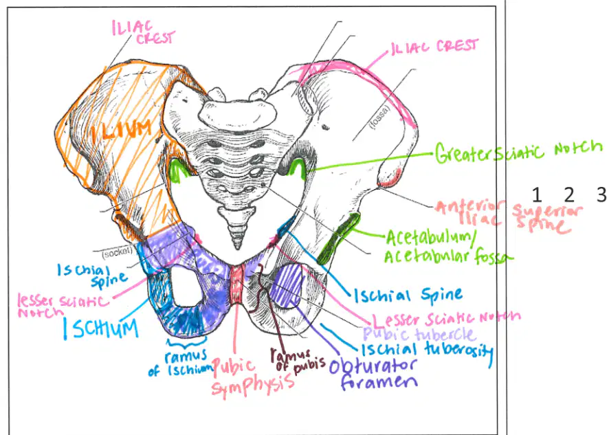
Identify the large opening formed by the ischium and pubis that reduces bone mass while maintaining strength.
Iliac crest
Lesser sciatic notch
Obturator foramen
Acetabular fossa
Which feature is a prominent ridge along the superior border of the ilium used for muscle attachment?
Acetabular rim
Pubic tubercle
Ischial spine
Iliac crest
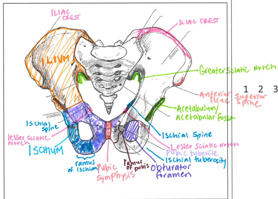
A nerve exits the pelvis through a notch posterior to the acetabulum. Which landmark is this?
Anterior superior iliac spine
Ischial tuberosity
Lesser sciatic notch
Greater sciatic notch
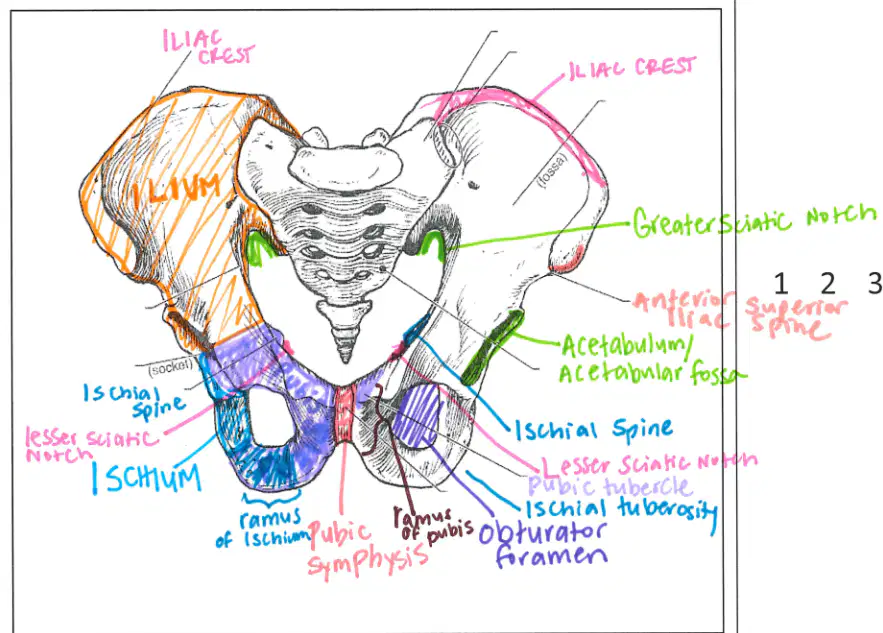
Which feature is located inferior to the ischial spine and transmits structures to the gluteal region?
Acetabular fossa
Pubic symphysis
Iliac crest
Lesser sciatic notch

In the foot diagram, which row correctly orders the phalanges from the toes toward the ankle?
Middle, distal, proximal
Proximal, distal, middle
Proximal, middle, distal
Distal, middle, proximal

Which bone is labeled as the heel bone in the foot diagram?
Talus
Calcaneus
Navicular
Cuboid

Which pairing correctly matches a distal ankle landmark to its bone?
Lateral malleolus—talus
Medial malleolus—tibia
Lateral malleolus—tibia
Medial malleolus—fibula

In the femur diagram, which large projection is lateral and provides muscle attachment?
Intercondylar fossa
Medial condyle
Lesser trochanter
Greater trochanter

Which tarsal bones articulate with metatarsals I–III on the medial side of the foot?
Calcaneus, navicular, cuboid
Medial, intermediate, lateral cuneiforms
Cuboid, navicular, talus
Talus, calcaneus, navicular

A patient has pain along the membrane between tibia and fibula. Which structure is most likely affected?
Head of fibula
Anterior tibial crest
Interosseous membrane
Tibial tuberosity

In a superior view diagram showing two major sutures across the skull vault, which suture runs between the two parietal bones along the midline?
Squamous suture above temporal bone
Sagittal suture between parietal bones
Coronal suture across frontal-parietal
Lambdoid suture across occipital border

In a lateral view highlighting temporal region labels, which opening leads into the ear canal on the temporal bone?
Foramen magnum aperture
Carotid canal entrance
Jugular foramen passageway
External auditory meatus opening

Identify the bone forming the forehead and contributing to the roof of the orbits in an anterior-lateral view.
Frontal bone of the forehead
Maxilla of upper jaw
Ethmoid bone of medial orbit
Zygomatic bone of cheek

Which suture separates the parietal bones from the occipital bone in a posterior-superior view?
Sagittal suture midline vault
Squamous suture lateral margin
Lambdoid suture posterior margin
Coronal suture anterior margin

In an inferior cranial base view, what large opening allows passage of the spinal cord from the cranial cavity to the vertebral canal?
External auditory meatus canal
Foramen magnum central opening
Jugular foramen lateral notch
Carotid canal anterior tunnel

A mandible diagram shows the U-shaped lower jaw with a vertical ramus and an anterior chin region. Which part articulates with the temporal bone at the TMJ?
Mental protuberance chin area
Coronoid process anterior ramus
Mandibular symphysis midline fusion
Condylar process posterior ramus

In the lateral skull diagram, which thin bone contributes to the medial wall of the orbit and is labeled near the nasal cavity?
Sphenoid bone greater wing
Zygomatic bone lateral orbit
Lacrimal bone medial orbit
Nasal bone bridge region

From the superior vault diagram, which suture runs transversely between the frontal bone and parietal bones?
Squamous suture lateral curve
Coronal suture transverse line
Sagittal suture midline seam
Lambdoid suture posterior arc
Which bone is part of the axial skeleton and forms the posterior base of the skull?
Parietal bone of cranial vault
Temporal bone at lateral skull
Occipital bone at skull base
Frontal bone above the orbits
Which paired long bone runs along the lateral side of the forearm in anatomical position?
Humerus of the upper arm
Radius on the lateral forearm
Ulna on the medial forearm
Clavicle across the shoulder
Select the bone that is part of the appendicular skeleton and articulates with the glenoid fossa to form the shoulder joint.
Ribs of the thoracic cage
Humerus of the upper limb
Scapula on the posterior shoulder
Sternum in the thorax
Which axial structure protects the heart by articulating with true ribs via costal cartilages?
Clavicle at the anterior shoulder
Lumbar vertebrae in lower back
Scapula spine posteriorly
Sternum body in the midline
Identify the bone that transmits body weight from femur to the foot and is classified as a tarsal.
Talus at the ankle mortise
Navicular on the medial foot
Cuboid on the lateral foot
Calcaneus forming the heel
Which sequence correctly orders the bones of the hand from proximal to distal?
Phalanges, carpals, metacarpals
Carpals, phalanges, metacarpals
Carpals, metacarpals, phalanges
Metacarpals, carpals, phalanges
Choose the correct classification: Which of the following is axial rather than appendicular?
Fibula along the lateral leg
Sacrum at the pelvic base
Patella at the knee
Femur in the thigh
A student labels the mandible as part of the appendicular skeleton. What correction should be made?
Relabel as pelvic girdle bone
Relabel as vertebral column bone
Relabel as axial skull bone
Relabel as upper limb bone
Which pair correctly matches bone and location for weight-bearing in the leg?
Fibula primarily weight-bearing laterally
Tibia primarily weight-bearing medially
Femur non–weight-bearing proximally
Patella primary shaft for weight-bearing
During labeling, which bone should be identified as part of the pectoral girdle connecting the sternum to the scapula?
Radius of the forearm
Clavicle at the anterior shoulder
Ulna of the forearm
Humerus of the arm
Which pair correctly names the two major parts of the skull?
Cranium and facial bones
Mandible and maxilla
Temporal and parietal bones
Atlas and axis vertebrae
What best describes a suture in the skull?
Movable hinge at the jawline
Flexible cartilage between vertebrae
Immovable joints holding skull bones
Elastic bands connecting facial muscles
Which structure is the only movable joint of the skull?
Mandible (jawbone)
Zygomatic arch
Occipital condyle
Temporal squama
Which feature helps you distinguish cervical vertebrae?
Long spinous processes hooking down
Smallest bodies and transverse foramina
Large articulating surfaces for ribs
Massive bodies and short spinous
Which statement best characterizes thoracic vertebrae?
Smallest and lightest with foramina
No body, supports occipital condyles
Massive bodies and short spinous
Long spinous processes that hook downward
Which statement best characterizes lumbar vertebrae?
Small bodies and bifid spinous processes
Transverse foramina for vertebral arteries
Long spinous processes that hook downward
Massive bodies with short spinous processes
Which pairing of cervical vertebra and property is correct?
Axis: smallest and lightest vertebra
Atlas: no body, nodding “yes” motion
Axis: occipital condyles rest on top
Atlas: dens process, shaking “no” motion
Which term is another name for the coccyx?
Lumbar base
Sacrum
Tailbone
Iliac crest
What is the primary function of intervertebral disks?
Store minerals for bone growth
Control spinal cord reflexes
Anchor ribs to the sternum
Act as cushions between vertebrae
Which statement best describes a herniated disk?
A thickened disk that improves shock absorption
A fused disk that increases flexibility of the spine
A slipped disk that can press on the spinal cord
A dehydrated disk that always heals without pain
How do true ribs differ from false ribs?
True ribs connect only by costal cartilage
True ribs are the last two rib pairs
True ribs attach directly to the sternum
True ribs attach indirectly to the vertebrae
Why are the floating ribs called “floating”?
They are connected solely by costal cartilage
They do not attach to the sternum at all
They are suspended by intervertebral disks
They attach only to the manubrium
Which ribs articulate with the manubrium?
The first pair
Pairs two through seven
Pairs eight through ten
The last two pairs
What is costal cartilage?
Segments of cartilage connecting the sternum to ribs
Elastic tissue cushioning vertebral bodies
Fibrous bands linking vertebrae to pelvis
Cartilage pads protecting the spinal cord
Which bones form the pectoral girdle?
Clavicle and scapula
Humerus and radius
Ulna and carpal bones
Sternum and ribs
Which list correctly names the parts of the upper limb?
Arm, forearm, wrist, hand
Shoulder, elbow, knee, foot
Neck, back, hip, leg
Pelvis, thigh, shin, ankle
To which part of the scapula does the humerus connect?
Glenoid fossa of the scapula
Spine of the scapula
Acromion process of the scapula
Coracoid process of the scapula
What is the primary purpose of the clavicle?
Protects the brachial plexus from injury
Transfers weight from trunk to lower limb
Supports rib movement during breathing
Prevents shoulder dislocation by bracing the arm
Which bones make up the pelvic girdle?
Femur and patella
Sacrum and coccyx
Ilium and sacrum
Two fused coxal bones
Which three bones fuse to form the coxa?
Ilium, ischium, pubis
Sacrum, coccyx, ilium
Femur, tibia, fibula
Patella, talus, calcaneus
Where on the coxa does the head of the femur articulate?
Acetabulum of the coxa
Pubic symphysis
Iliac crest
Greater sciatic notch
What is the function of the greater sciatic notch?
Forms the socket for the femoral head
Serves as attachment for gluteal muscles
Allows vessels and the sciatic nerve to pass to the leg
Anchors ligaments of the pelvic floor
Which statement best distinguishes a typical female pelvis from a male pelvis?
More prominent projections and smaller outlet
Narrower inlet with thicker bones
Heavier with more curved sacrum and coccyx
Smoother and lighter with larger pelvic outlet
Which bones make up the hallux in the human foot?
Proximal phalanx and distal phalanx
Middle phalanx and distal phalanx
Proximal phalanx and middle phalanx
Metatarsal and middle phalanx