
 Worksheets
Abdomen Review
Total questions: 40
Worksheet time: 20mins
The superior curved border of the ilium, which is at the level of the 4th and 5th lumbar, is termed:
Iliac Crest
ASIS
Symphysis Pubis
Sacrum
The prominent bony projection located at the anterior end of the crest is termed:
ASIS
Iliac Crest
Symphysis Pubis
Sacrum
The anterior junction of the pubic bones, where the pubic bones meet medially, is termed:
Symphysis Pubis
Iliac Crest
ASIS
Sacrum
What does KUB stand for?
Kidneys, Ureters, Bones
Kidneys, Urethra, Bladder
Kidneys, Ureters, Bladder
Kidneys, Urethra, Bones
What 3 muscles are of importance in abdominal radiography?
Rt. Psoas Muscle, Lt. Psoas Muscle, Diaphragm
Rt. Psoas Muscle, Lt. Psoas Muscle, Rectus Abdominis
Rt. Psoas Muscle, Diaphragm, Obliques
Lt. Psoas Muscle, Diaphragm, Rectus Abdominis
List the 4 parts of the Urinary System discussed in class and give 1 main function of each part:
Kidneys, Ureters, Bladder, Urethra
Kidneys, Urethra, Bladder, Rectum
Kidneys, Ureters, Bladder, Small Intestine
Kidneys, Urethra, Bladder, Large Intestine
Indicate the abdominal quadrant(s) in which the stomach is located.
RUQ
LUQ
RLQ
LLQ
Which of the following is NOT an accessory organ of digestion?
Liver
Pancreas
Spleen
Gallbladder
What bony landmark is used for the center of the IR when positioning for an AP supine abdomen?
Symphysis Pubis
Iliac Crest
Greater Trochanter
Axilla
What is the purpose of an upright abdomen image?
To check for fractures
To look for free intra-abdominal air
To assess muscle tone
To evaluate spinal alignment
If a patient is too ill or unable to stand, what position may be used instead of the upright abdomen image?
Prone
Left lateral decubitus
Supine
Right lateral decubitus
How many images are typically taken for an upright abdomen?
1 image
2 images
3 images
4 images
When positioning a patient for an upright abdomen image, what anatomical landmark is used for the bottom of the IR?
Iliac Crest
Symphysis Pubis
ASIS
Axilla
When viewing a decubitus abdomen, which side is more important?
Side down
Side up
Both sides are equally important
Neither side is important
What is the routine for an abdomen series?
AP Upright CW, AP Upright LW, and KUB
AP Supine CW, AP Supine LW, and KUB
AP Upright CW, AP Supine LW, and KUB
AP Supine CW, AP Upright LW, and KUB
Is it correct to do a right lateral decubitus abdomen if the patient’s pain is on the right side? Why or why not?
Yes, because it provides a better view of the liver
No, because the liver will trap/block free air
Yes, because it helps identify the source of pain
No, because it increases patient discomfort
Where does the small intestine connect to the large intestine?
Gallbladder
Duodenum
Thoracic spine
Ileocecal valve
Which organ forms bile?
Spleen
Liver
Gallbladder
Duodenum
Which part of the body is made up of 5 vertebrae?
Lumbar spine
Thoracic spine
Greater trochanter
Neck of femur
Which organ is part of the lymphatic system?
Liver
Gallbladder
Spleen
Duodenum
What is the upper flared portion of the ilium called?
Ala/wing
Neck of femur
Greater trochanter
Duodenum
What is the third part of the small intestine?
Duodenum
Ileocecal valve
Gallbladder
Ileum
What stores bile in the body?
Liver
Gallbladder
Spleen
Duodenum
What is the shortest part of the small intestine?
Ileum
Duodenum
Thoracic spine
Lumbar spine
What is the large bump or prominence located at the lateral margin of the upper femur called?
Neck of femur
Greater trochanter
Ala/wing
Lumbar spine
Which part of the body is made up of 12 vertebrae?
Lumbar spine
Thoracic spine
Greater trochanter
Neck of femur
What is the central ray placement for the AP Upright - CW abdomen projection?
Top of IR at axilla, CR to center of IR and down MSP
IR centered at iliac crest, CR to center of IR and down MSP
Top of IR at iliac crest, CR to center of IR and up MSP
IR centered at axilla, CR to center of IR and up MSP
What is the collimation size for the KUB abdomen projection?
14x17-CW
14x17-LW
10x12-CW
10x12-LW
For the Left Lateral Decubitus abdomen projection, where is the top of the IR placed?
At the iliac crest
At the axilla
At the midline of the body
At the pubic symphysis
What image should be taken when the symphysis pubis has been clipped for a KUB?
AP Bladder shot
Lateral Abdomen shot
PA Chest shot
AP Pelvis shot
Where is the IR placed when performing a 2-image crosswise KUB?
Top of IR at axilla for both images
Top of IR at axilla for the first image and bottom of IR at symphysis pubis/greater trochanter for the second image
Bottom of IR at symphysis pubis for both images
Overlap IR at the mid-abdomen for both images
What is one major reason to repeat an AP Upright Abdomen LW image?
Clipped left diaphragm
Clipped symphysis pubis
Incorrect exposure settings
Overexposed image
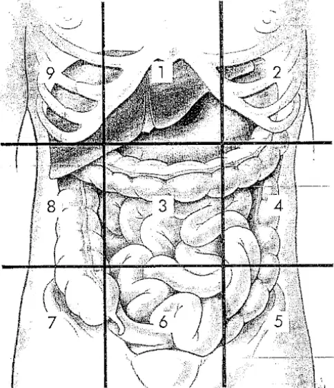
Which abdominal region is located in the center of the diagram, directly above the umbilical region?
Epigastric
Hypogastric
Left Hypochondriac
Right Lumbar
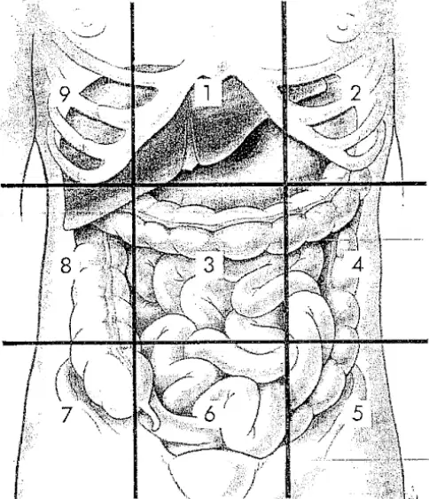
What is the name of the abdominal region located in the lower left corner of the diagram?
Left Lumbar
Left Iliac
Hypogastric
Right Iliac
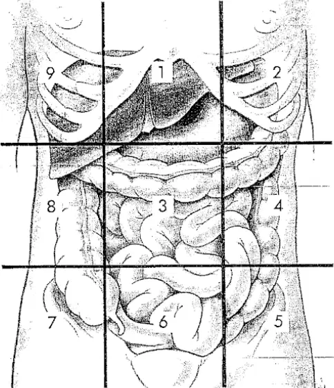
Which abdominal region is located in the upper right corner of the diagram?
Right Hypochondriac
Right Lumbar
Epigastric
Umbilical
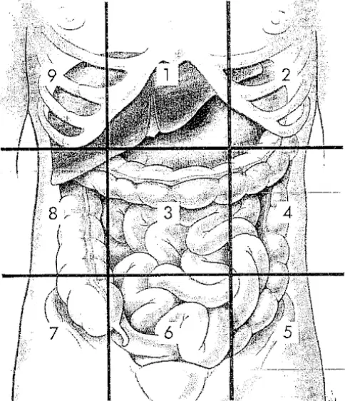
What is the name of the abdominal region located directly below the umbilical region in the diagram?
Hypogastric
Left Lumbar
Right Lumbar
Epigastric
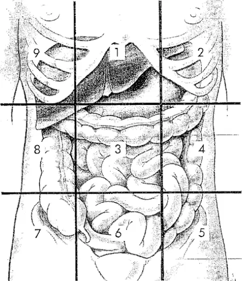
Which abdominal region is located in the middle left side of the diagram?
Left Lumbar
Left Hypochondriac
Left Iliac
Umbilical
Structures located in the right lower quadrant (RLQ) include which of the following?
Spleen
Vermiform Appendix
Sigmoid
In which of the following positions can small amounts of free air in the peritoneal cavity be demonstrated?
Left lateral decubitus
Right lateral decubitus
AP Trendelenburg
AP Supine
Which of the following will best demonstrate the size and shape of the liver and kidneys?
Lateral abdomen
AP abdomen
Dorsal decubitus abdomen
Ventral decubitus abdomen

