
NEW
Font size
 Worksheets
Introduction and Mitral Stenosis Basics
Total questions: 12
Worksheet time: 6mins
Which statement best defines mitral stenosis in clinical terms?
Failure of mitral leaflets causing systolic regurgitant jet
Narrowing of mitral valve orifice impeding diastolic flow
Stiff tricuspid valve reducing venous right atrial inflow
Thickening of aortic cusps restricting systolic ejection
In a patient with mitral stenosis, which hemodynamic change is most expected?
Increase in left atrial pressure during diastole
Decrease in left ventricular systolic pressure
Increase in right ventricular afterload at rest
Reduction in systemic arterial diastolic pressure
What is the most common etiologic cause of mitral stenosis worldwide?
Rheumatic fever causing commissural fusion
Degenerative calcification of the aortic annulus
Infective endocarditis destroying leaflet tissue
Congenital bicuspid mitral valve malformation
Rheumatic fever–related changes in the mitral valve typically include which combination?
Prolapse due to elongated chordae and thin leaflets
Annular dilation with papillary muscle rupture
Leaflet perforation with flail posterior scallop only
Commissural fusion with chordal thickening and shortening
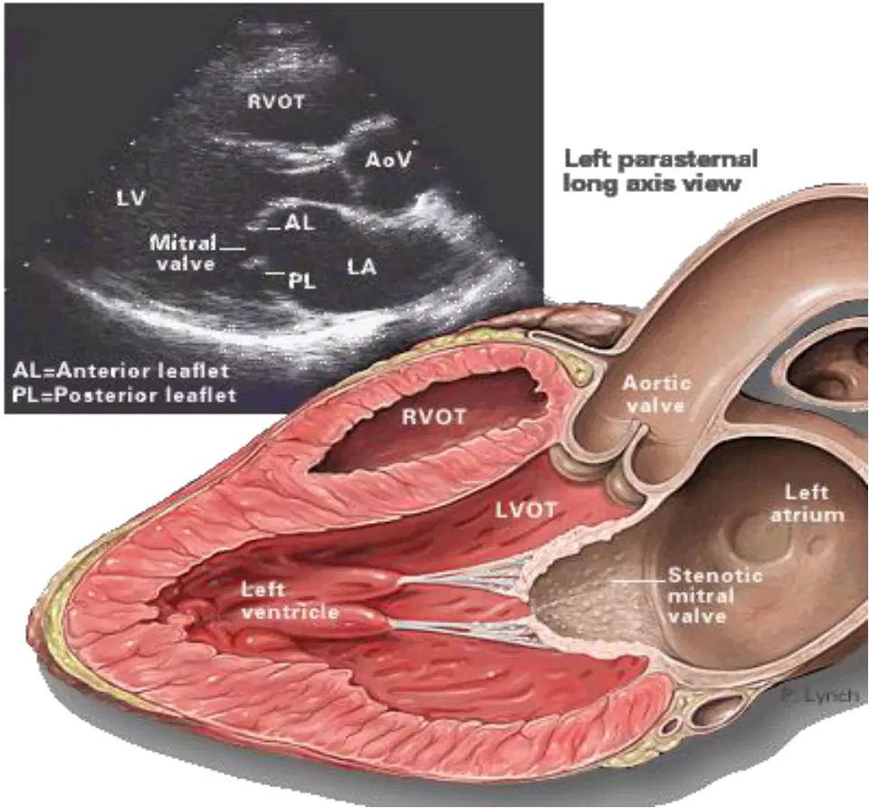
In the left parasternal long-axis view shown, which hemodynamic consequence most directly results from a stenotic mitral valve?
Enhanced left ventricular filling with lower diastolic gradient
Increased left atrial pressure causing pulmonary venous backup
Reduced right ventricular pressure preventing pulmonary hypertension
Decreased left atrial pressure with improved venous return
A patient with long-standing mitral stenosis presents with palpitations, exertional dyspnea, and intermittent hemoptysis. Which pathophysiologic sequence best explains these findings?
Pulmonary arterial vasodilation lowers right-sided pressures, improves oxygenation
Right ventricular failure increases systemic venous pressure, reduces left atrial preload
Left ventricular hypertrophy elevates systemic afterload, decreases pulmonary pressures
Atrial fibrillation leads to decreased cardiac output, pulmonary venous hypertension thickens bronchial veins
Which complication of mitral stenosis directly results from increased left atrial pressure causing blood backup into pulmonary veins?
Pulmonary hypertension due to venous backup
Left atrial thrombus from blood stasis
Embolization from intracardiac thrombus
Atrial fibrillation due to atrial stretch
A 35-year-old with symptomatic mitral stenosis has a valve area of 0.9 cm² and no severe regurgitation, calcification, or left atrial thrombus. Which surgical option is preferred?
Closed mitral valve commissurotomy
Percutaneous coronary intervention
Open mitral valve replacement
Ablation for atrial fibrillation
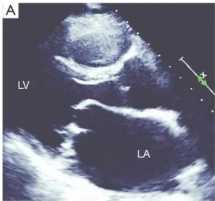
In the parasternal long-axis view shown, the anterior mitral leaflet displays a domed, ‘hockey stick’ shape during diastole. Which mechanism most directly produces this characteristic morphology?
Annular dilatation widening the valve orifice
Chordae tendineae elongation reducing leaflet tension
Papillary muscle rupture increasing leaflet mobility
Commissural fusion limiting leaflet edge opening
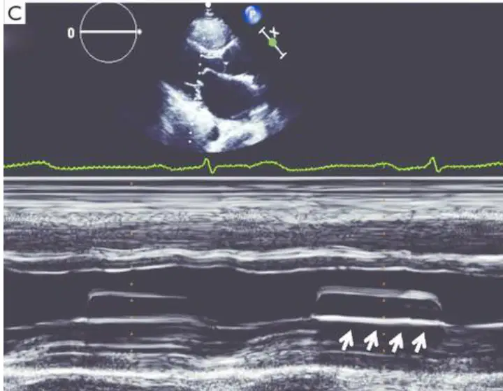
On the M-mode tracing through the mitral valve, the E–F slope is markedly decreased. What does a reduced E–F slope most strongly indicate in this context?
Normal posterior leaflet motion with preserved compliance
Impaired early diastolic leaflet closure due to stenosis
Enhanced rapid ventricular filling without obstruction
Systolic anterior motion causing LV outflow tract obstruction
Which method directly measures mitral valve orifice area using 2D echocardiography and correlates well with catheterization when performed correctly?
Proximal isovelocity surface area method for MR
Planimetry of the mitral valve orifice on PSAX
Pressure half-time formula using Doppler data
Continuity equation from LVOT velocity-time integral
During planimetry of the mitral valve, how should the imaging plane be aligned to obtain an accurate orifice area that reflects true obstruction severity?
Parallel to the mitral valve leaflet tips
Perpendicular to the mitral valve leaflet tips
Aligned with the aortic valve commissures
Oblique through the LV inflow tract

