
 Worksheets
ECG CH 9 & 10 Wayground Review
Total questions: 74
Worksheet time: 45mins
Where are the ventricular pacemaker cells found?
At the SA node
At the Purkinje fibers
At the AV node
At the bundle of His
What is the inherent rate of the Purkinje network?
100 to 150 bpm
60 to 100 bpm
20 to 40 bpm
40 to 60 bpm
In all ventricular dysrhythmias, which of the following qualities describes P-P intervals?
Biphasic
Missing
Regular
Irregular
QRS complexes that measure 0.12 seconds (120 ms) or greater and a heart rate between 20 and 40 bpm indicate that the impulses causing ventricular depolarization are coming from which location?
Purkinje fibers
Interatrial pathways
SA node
AV node
What is the term for ventricular rhythms that originate in the Purkinje network?
Junctional escape rhythms
Ventricular escape rhythms
Asystole rhythms
Agonal rhythms
Why do ventricular rhythms occur?
Because the rate impulses from the SA node are too fast for ventricular response
Because higher pacemaker sites within the heart have failed
Because the ventricular impulses are stronger than the atrial impulses
Because a first-degree AV block prevents atrial impulses from reaching the ventricles
What is the typical heart rate with accelerated idioventricular rhythm?
Less than 20 bpm
20 to 40 bpm
40 to 100 bpm
100 to 150 bpm
What percentage of patients with ventricular tachycardia become unconscious immediately?
10%
30%
50%
75%
Which of the following statements about PVCs is true?
PVCs can only originate from one site in the ventricles.
PVCs may be unifocal or multifocal in origin.
PVCs will not lead to ventricular tachycardia.
PVCs never have an effect on the conduction of normal impulses.
Which of the following rhythms does NOT necessarily require immediate emergency interventions?
Asystole
Ventricular fibrillation
Ventricular tachycardia
Premature ventricular complexes
What is the term for PVCs that occur during the normal R-R interval and do not interrupt the normal cycle?
Occasional PVCs
Interpolated PVCs
Coupling
Frequent PVCs
What is it called when more than six PVCs occur per minute?
Occasional PVCs
Ventricular tachycardia
Ventricular fibrillation
Frequent PVCs
What is the cause of agonal rhythms?
Failure of the SA node
Failure of all pacemakers of the heart
Blocked conduction at the AV node
Loose electrode wires
Which ventricular dysrhythmia has a heart rate between 20 and 40 bpm?
Asystole
Agonal rhythm
Accelerated idioventricular rhythm
Idioventricular rhythm
What is the primary difference between idioventricular rhythm and accelerated idioventricular rhythm?
Heart rate
QRS complex
P wave
R-R interval
In which ventricular dysrhythmia do three or more PVCs occur in a row with a ventricular rate greater than 100 bpm?
Ventricular tachycardia
Ventricular fibrillation
Accelerated idioventricular rhythm
SVT
Which of these terms applies specifically to PVCs that occur in varied shapes and forms?
Unifocal PVCs
Interpolated PVCs
Multifocal PVCs
Bigeminy PVCs
How is ventricular fibrillation typically described?
Wide and bizarre rhythm
Organized rhythm
Absence of rhythm
Chaotic, asynchronous rhythm
Which of the following is an ectopic impulse that occurs early in the cycle and originates from the ventricles?
PAC
PJC
SVT
PVC
Which of the following signs and symptoms are characteristic of every patient experiencing ventricular fibrillation?
Unconsciousness, apneic, and pulseless
Dizziness, chest pain, and cough
Talkativeness, hypertension, and tachycardia
Shortness of breath, hypotension, and bradycardia
What is the difference between ventricular tachycardia and ventricular fibrillation?
Ventricular tachycardia has three or more PVCs and a heart rate greater than 100 bpm; ventricular fibrillation is chaotic electrical activity with only fibrillatory waves.
Ventricular tachycardia has three or more PVCs and a heart rate greater than 100 bpm; ventricular fibrillation has no P wave and an early QRS complex that is wide and bizarre in appearance.
Ventricular fibrillation is chaotic in appearance; ventricular tachycardia has an absence of P waves, a heart rate of 40-100 bpm, and a wide and bizarre QRS complex.
Ventricular fibrillation is chaotic electrical activity with only fibrillatory waves; ventricular tachycardia has an absence of P waves, a heart rate less than 20 bpm, and wide and bizarre QRS complexes.
Which ventricular dysrhythmia is sometimes referred to as "straight-line" or "flat-line"?
Agonal
Sinus bradycardia
Asystole
Ventricular fibrillation
Which ventricular dysrhythmia has a heart rate less than 20 bpm?
Ventricular fibrillation
Idioventricular rhythm
Asystole
Agonal rhythm
Which of the following statements about agonal rhythm and asystole is FALSE?
Both rhythms demonstrate mild bradycardic arrhythmias
Both rhythms are life threatening and require basic and advanced life support
Both rhythms have an absence of P waves
In both rhythms, the patient will be unconscious
What is the term for two PVCs back to back?
Bigeminy
Coupling
Frequent PVCs
Occasional PVCs
What symptoms would a patient in asystole exhibit?
Difficulty breathing and bradycardia
Unconsciousness and apnea
Chest pains and palpitations
Alert and oriented
Which of the following is absent with apnea?
Pulse
Electrical activity
Blood pressure
Breathing
In which ventricular dysrhythmia do the ventricle walls quiver, preventing any movement of blood out of the ventricles and resulting in no cardiac output?
Ventricular tachycardia
Ventricular flutter
Supraventriculartachycardia
Ventricular fibrillation
Which of the following is a characteristic of an idioventricular rhythm?
Heart rate of 40 to 100 bpm
Wide QRS complexes
Upright P waves
Constant P–R intervals
What is the term for a PVC that occurs on the T wave or during the vulnerable period of the ventricular refractory period?
Trigeminy
T-on-R PVC
Bigeminy
R-on-T PVC
Choose the correct responses.
Ventricular Depolarization
Atrial Depolarization
Ventricular Repolarization
Which is the right atrium?
Click on the:
right ventricle

You are recording an ECG; a portion of the tracing is shown here. Your patient is pale and tells
you that she is a little dizzy. Which of the following is the least likely reason for the tracing?
Poorly attached or dried-out electrodes
Excessive patient movement
Broken lead wires
Ventricular fibrillation
Which of the following is a characteristic of the R-R interval in an idioventricular rhythm?
It depends on the pattern of PVCs.
It is regular.
It is usually irregular due to ectopic beats.
It cannot be determined.
You have been asked to perform an ECG on a hospital patient who was admitted to the cardiac
care unit this morning. When you review the ECG tracing from the emergency department
before the patient was admitted, you notice that the patient had an idioventricular rhythm. As
you prepare to enter the patient's room, you will not be surprised to find the patient in what
state?
Unconscious
Alert and oriented
Hypertensive
Combative
In accelerated idioventricular rhythm, how do the QRS complexes appear?
They have a classic wide and bizarre appearance.
They cannot be distinguished because the heart rate is too rapid.
They fall within the normal range of 0.12 to 0.20 seconds due to the heart rate.
They are irregular because of multifocal ventricular impulses.
What is the condition in which impulses generated in the atria are delayed because of damage
in the ventricular electrical pathways?
Bradycardia
First-degree AV block
Bundle branch block
Idioventricular rhythm
Which of the following rhythms might a patient be able to tolerate without signs of low cardiac
output?
Ventricular fibrillation
Accelerated idioventricular rhythm
Idioventricular rhythm
Ventricular tachycardia
Accelerated idioventricular rhythm
36
Ventricular tachycardia
Ventricular fibrillation
Idioventricular rhythm
Accelerated idioventricular rhythm
An accident victim is being treated in the emergency department. A friend who was present at
the time of the accident is in the patient's cubicle when the patient’s ECG monitor sounds an
alarm and shows a flat-line pattern. The patient's friend becomes frantic. What should you do?
Calmly escort the patient's friend out of the immediate area.
Tell the patient's friend to get out of the way so the medical team can work
Tell the patient's friend to go to the desk and ask that the physician be notified.
Ask the patient's friend to begin CPR while you get the emergency cart.
In a right bundle branch block (RBBB), how does the QRS appear?
Taller and deeper than normal
Positively deflected
Negatively deflected
Normal in length
What is the ventricular rate in asystole?
(a)
Where can right and left bundle branch blocks be differentiated?
In the bipolar leads
In the limb leads
In the precordial leads
In the augmented leads
To rule out fine ventricular fibrillation, asystole must be confirmed in at least how many leads?
(a)
The patient on which you are about to perform an ECG tells you that she sometimes feels an
occasional "thumping" in her chest. Which of the following might you expect to see on the ECG
tracing?
Ventricular tachycardia
Idioventricular rhythm
Ventricular fibrillation
Premature ventricular complexes
Why can't the atrial rate be determined in idioventricular rhythms?
The wide and bizarre QRS complex masks the P waves.
Atrial depolarization is absent.
The atrial rate is too irregular to be determined.
The P waves are buried in the QRS complexes.
What happens when one or both of the ventricular pathways are not functioning properly due
to damage or a delay from cardiac disease, drugs, or other conditions?
The patient never experiences PVCs
A bundle branch block occurs
An idioventricular rhythm occurs
A second degree AV heart block, type I occurs
What additional patient information is needed on the ECG tracing when a bundle branch block
is discovered?
Patient’s symptoms
Patient’s cardiac history
Patient’s blood pressure reading
Patient’s underlying rhythm
A bundle branch block must be considered when the QRS complex measures what length?
0.06 to 0.10 seconds
Less than 0.06 seconds
Greater than 0.12 seconds or 120 milliseconds
0.04 to 0.08 seconds
A patient’s dysrhythmia originates from above the ventricles and you notice QRS complexes on
the ECG tracing measuring 0.12 second or greater. What could be the cause of this abnormal
morphology?
Supraventriculartachycardia
Bundle branch block
Third-degree AV block
Atrial fibrillation
How is a right bundle branch block (RBBB) distinguished from a left bundle branch block
(LBBB)?
Monitor lead V1. If the majority of the ventricular depolarization is positively deflected,
it is LBBB; if the QRS is negative, it is RBBB
Monitor leads V1 to V6 and measure the PR interval
Monitor lead V1. If the majority of the ventricular depolarization is positively deflected,
it is RBBB; if the QRS is negative, it is LBBB
Monitor leads I, II, and III and observe the P wave configuration
Bundle branch block rhythm is similar to a sinus rhythm but has what distinguishing
characteristic?
QRS duration less than 0.12 seconds
QRS duration greater than 0.12 seconds
PR interval less than 0.12 seconds
PR interval greater than 0.20 seconds
You observe a wide QRS complex in lead II. Which lead should you check to evaluate the
location of the bundle branch block?
(a)
What measurement on the ECG tracing represents how long it takes for the current to travel
through the ventricular myocardial tissue?
PR interval
QRS duration
P-P interval
R-R interval
52
Multiple
A bundle branch block has which of the following characteristics?
It occurs when a bundle branch fail to conduct impulses.
It causes narrowed QRS complexes to appear on the ECG.
It causes a prolonged PR interval.
It accelerates depolarization of the ventricle it supplies.
Which of the following statements about bundle branch block is true?
Bundle branch block overrides the patient’s underlying heart rhythm.
If the patient has a bundle branch block, no P wave will be seen on the ECG tracing.
Bundle branch block is a life-threatening condition.
Any sinus, atrial, or junctional rhythm can be affected by bundle branch block.
You are about to perform an ECG on a patient known to have sinus tachycardia with right
bundle branch block. Which of the following signs and symptoms would you be likely to find in
this patient?
Congestive heart failure
Hypertension
Unconsciousness
Rapid heart rate
Which of the following is a characteristic of left bundle branch block?
The right ventricle depolarizes normally, but conduction through the left ventricle is
slowed.
Neither ventricle depolarizes normally because the impulse is not conducted through
the left bundle branch.
The left ventricle depolarizes normally, but the impulse is not spread to the right
ventricle.
Neither ventricle depolarizes normally because the impulse is not conducted through
the right bundle branch.
Neither ventricle depolarizes normally because the impulse is not conducted through
the right bundle branch.
Lead I
aVF
V1
V6
What is the term for PVCs that all have a similar shape on a tracing?
(a)
In a left bundle branch block (LBBB), how does the QRS appear?
Positively deflected
Taller and deeper than normal
Normal in length
Negatively deflected
When analyzing V1 to determine the presence of a bundle branch block, what should you look
for?
A QRS complex that measures less than 0.12 seconds with no P wave
An inverted T wave and ST segment depression
A QRS complex that measures 0.12 seconds or greater and a P wave
ST segment elevation and a pathologic Q wave
What is a QS complex?
An inverted QRS complex
A deep Q/S wave with no preceding R wave
A QRS complex with no preceding P wave
A QRS complex in which the R wave is buried in the Q wave
What is a QS complex?
A QRS complex in which the R wave is buried in the Q wave
A QRS complex with no preceding P wave
A deep Q/S wave with no preceding R wave
An inverted QRS complex
You have run a 12-lead ECG on Mrs. Simmons. In lead V1, you notice that the QRS is positive
and has a “bunny ears” appearance. What does Mrs. Simmons most likely have?
Left bundle branch block
Right bundle branch block
Complete AV block
Left ventricular hypertrophy
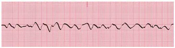
Identify the following rhythm:
(a)
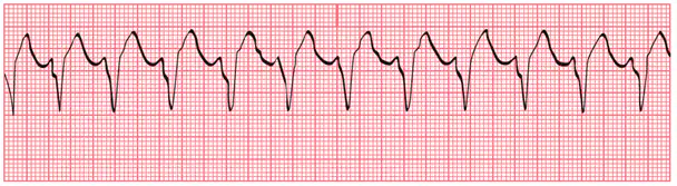
Identify the following rhythm:
(a)
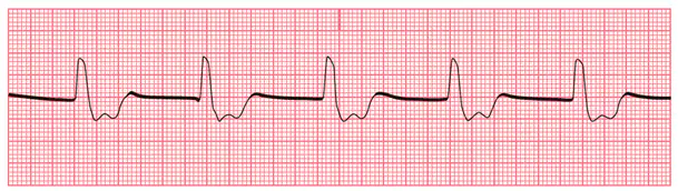
Identify the following rhythm:
(a)
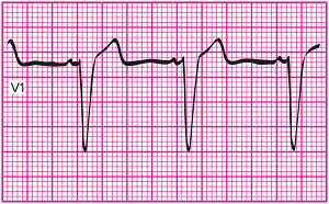
The V1 tracing shown here is from a 12-lead ECG performed on a 36-year-old male patient as
part of a yearly physical examination. The patient appears to be in good general health. What
does this tracing show?
(a)
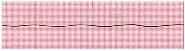
Identify the following rhythm:
(a)
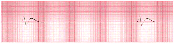
Identify the following rhythm:
(a)
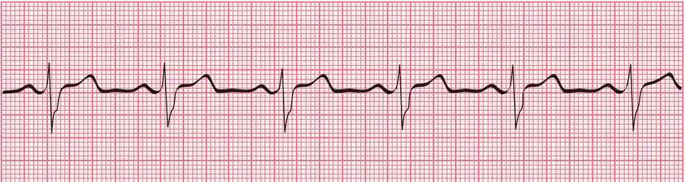
What rhythm is shown in this ECG tracing?
(a)
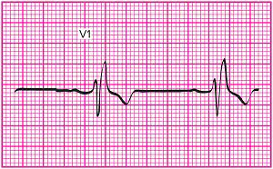
You have run a 12-lead ECG on Ms. Reilly, a patient in the cardiac care unit of the hospital
where you work. A portion of the V1 tracing is shown here. What cardiac condition would you
suspect based on this tracing?
Right bundle branch block
Complete AV block
Left ventricular hypertrophy
Left bundle branch block

