
 Worksheets
2526_Chest Anatomy and Positioning
Total questions: 93
Worksheet time: 47mins
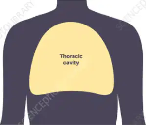
Which structure primarily protects the organs within the thoracic cavity?
Abdominal wall muscles only
Clavicle and scapulae only
Rib cage, sternum, and spine
Pelvic girdle and sacrum
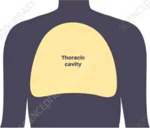
Which set lists major organs housed in the thoracic cavity?
Stomach, liver, gallbladder
Kidneys, spleen, pancreas
Lungs, heart, great vessels
Bladder, uterus, prostate
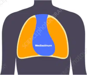
The mediastinum is best described as which of the following?
Space within abdomen behind stomach
Central compartment between lungs
Outer membrane covering lungs
Muscular sheet separating pelvis
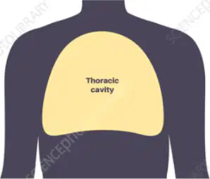
Which border forms the inferior boundary of the thoracic cavity?
Diaphragm muscle dome
Cervical fascia plane
Pelvic inlet rim
Clavicular line pair
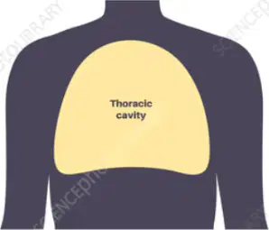
Knowing thoracic borders helps clinicians primarily with which task?
Predicting bone fracture healing
Determining blood glucose control
Evaluating lung inflation and mediastinum
Choosing antibiotics for pneumonia
Which component is part of the bony thorax?
Ilium and ischium
Tibia and fibula
Humerus and ulna
Sternum and ribs
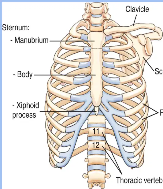
Which labeled sternum region is the small tip at the inferior end of the sternum in the diagram?
Manubrium region at superior end
Body region at central shaft
Xiphoid process at distal tip
Clavicle region near shoulders
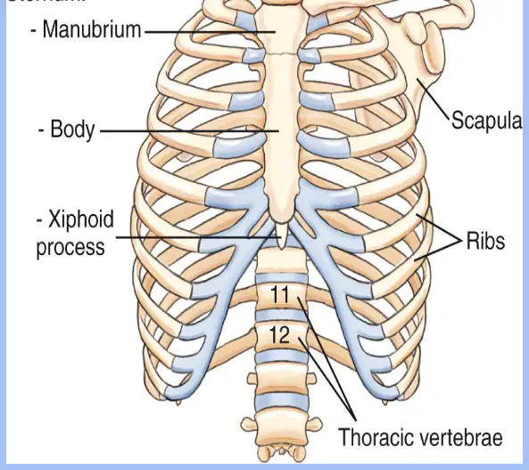
Ribs 1–7 are best classified as which type according to the diagrammed bony thorax?
True ribs directly attach to sternum
False ribs lacking any cartilage
Floating ribs with anterior fusion
Posterior ribs forming the clavicle
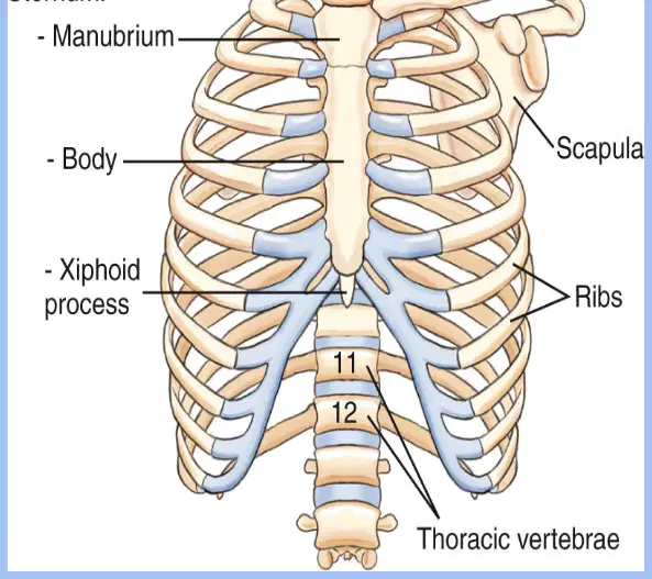
Which statement correctly matches rib numbers to their classification shown in the visual?
Ribs 11–12 are true ribs
Ribs 8–10 are false ribs
Ribs 3–9 are false ribs
Ribs 1–7 are floating ribs
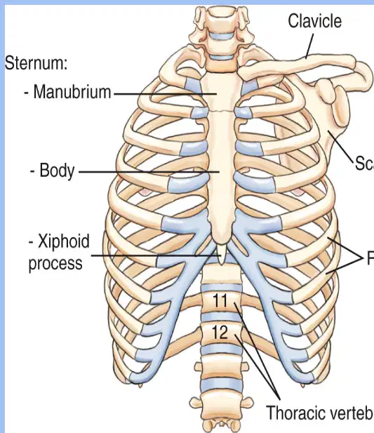
Which portion of the sternum articulates with ribs 2–7 as indicated by the image and notes?
Costal cartilage only
Manubrium superior section
Body middle section
Xiphoid inferior tip
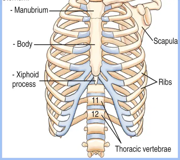
According to the illustrated thoracic spine, each thoracic vertebra typically attaches to what structure?
The scapula medially
The clavicle laterally
The humerus distally
A pair of ribs bilaterally
A patient’s radiograph shows only nine posterior ribs visible during inspiration. Using the guidance beside the diagram, what is the best interpretation?
Inspiratory effort was suboptimal
Thoracic vertebrae are fractured
Scapulae obscure the clavicles
True ribs are all missing
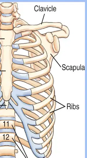
Which statement best describes the scapulae as shown?
Long curved bones of anterior chest
Small round bones of vertebral column
Flat triangular bones on posterior thorax
Short irregular bones near xiphoid
Which statement best defines the mediastinum?
Inferior abdominal cavity below the diaphragm
Central thoracic space between the lungs
Superior neck space above the clavicles
Posterior pleural cavity behind the lungs
On a normal chest X-ray, where should the trachea typically be positioned?
Obscured behind the cardiac silhouette
Midline in the upper mediastinum
Left of midline near the aortic arch
Right of midline near the vena cava
Which vessel forms the radiographic feature known as the aortic knob?
Ascending aorta inferior border
Superior vena cava contour line
Arch of the aorta on chest imaging
Pulmonary artery main segment
Which organ is typically prominent in infants and may appear as a sail sign on pediatric chest images?
Thyroid gland in adolescents
Adrenal gland in toddlers
Thymus gland in young children
Spleen shadow in neonates
Which list best represents major structures contained within the mediastinum?
Lungs, pleura, bronchioles, alveoli
Liver, stomach, pancreas, spleen
Heart, trachea, esophagus, aorta
Kidneys, ureters, bladder, urethra
Which statement best describes the lobes of the lungs?
Right has three lobes; left has two lobes
Right has two lobes; left has three lobes
Both lungs have two lobes each
Both lungs have three lobes each
The cardiac notch is a feature located on which structure?
Lateral surface of the right lung
Posterior surface of the heart
Inferior surface of the trachea
Medial surface of the left lung
Which pleural layer directly covers the lung surface?
Visceral pleura covers lung surface
Parietal pleura covers lung surface
Pleural space covers lung surface
Pericardial pleura covers lung surface
What is the main function of the pleural space during breathing?
Allows smooth lung movement with fluid
Houses cartilage to keep airway open
Stores air for emergency breathing
Provides blood flow to lung tissues
Which landmarks are typically checked on a chest X‑ray for lung assessment?
Apex beat, diaphragm dome, aorta arch
Cervical ribs, clavicular heads, atlas
Apices, bases, costophrenic angles, hilum
Thyroid gland, laryngeal inlet, esophagus
Which statement about the trachea is accurate?
Primary site of gas exchange in lungs
Main airway to carina where it divides
Muscular tube without any cartilage rings
Short segment ending above the larynx
Where does the trachea bifurcate into right and left bronchi?
At the base, a flat plate
At the hilum, a soft groove
At the apex, a smooth dome
At the carina, a sharp ridge
During intubation, where should an endotracheal tube tip be positioned relative to the carina?
Just below the left main bronchus
Exactly at the level of the carina
Approximately 2–3 cm above the carina
Firmly within the right main bronchus
Which characteristic makes the right main bronchus more likely to receive aspirated objects?
Shorter, wider, and more vertical
Longer, narrower, and more horizontal
Curved around the heart surface
Supported by fewer cartilage rings
Which lung region does the right main bronchus supply?
Only the inferior lobe of right lung
The two lobes of the left lung
The three lobes of the right lung
Both upper lobes of both lungs
Which description best fits the left main bronchus?
Longer, narrower, and more horizontal
Direct route to right upper lobe
Shorter, wider, and more vertical
Mostly straight with no curvature
A patient aspirates a peanut. Based on airway anatomy, where is it most likely to lodge first?
Pleural space due to negative pressure
Tracheal rings due to strong suction
Left main bronchus due to heart curve
Right main bronchus due to its vertical angle
Which statement best describes the diaphragm’s action during inhalation?
It relaxes and moves upward to expand lungs
It contracts and moves downward to expand lungs
It contracts and moves upward to compress lungs
It relaxes and moves downward to expel air out
On a normal chest X‑ray, what should the costophrenic angles look like?
Asymmetrical and jagged, irregular margins
Clean and crisp, sharply pointed corners
Invisible completely, flat diaphragmatic lines
Blunted and hazy, rounded lower borders
Why is the right hemidiaphragm typically higher on X‑ray than the left?
The spleen anchors it in position
The liver pushes it upward from below
The stomach bubble lifts it superiorly
The heart weight presses it inferiorly
Which description best characterizes the diaphragm’s role in body cavities?
Separates thoracic cavity from abdominal cavity
Connects pleural cavity to peritoneal cavity
Fuses heart apex with liver superior surface
Anchors ribs to vertebrae during movement
During exhalation at rest, what does the diaphragm primarily do?
Relaxes and descends, increasing lung space
Contracts and ascends, expelling tidal air
Contracts and flattens, pushing ribs inward
Relaxes and ascends, reducing lung volume
Which statement about the aorta on imaging is most accurate?
Seen as aortic arch and descending aorta
Appears only as the ascending segment
Visible solely on the right mediastinum
Forms the lower right heart border
Where does the superior vena cava primarily lie within the mediastinum?
Midline behind the descending aorta
Posterior left to the pulmonary veins
On the left side anteriorly positioned
On the right side of the mediastinum
Which vessel returns blood from the lower body and joins the right atrium just above the diaphragm?
Coronary sinus
Pulmonary trunk
Superior vena cava
Inferior vena cava
Which statement about the pulmonary arteries is correct?
Carry deoxygenated blood from right ventricle
Carry oxygenated blood to the left atrium
Run anterior to the ascending aorta mainly
Do not branch at the lung hila
Which soft tissue feature can make the lung bases appear hazy on a chest X‑ray?
Breast shadows over lower lungs
Normal rib cortical lines
Scapular borders projecting laterally
Costophrenic angles being sharp
A missing breast shadow on one side of a chest X‑ray most likely suggests which scenario?
Prior mastectomy on that side
Poor inspiratory effort only
Normal chest wall musculature
Incorrect exposure settings
Neck soft tissue evaluation on a chest X‑ray is useful primarily for detecting which finding?
Tracheal deviation or swelling
Hiatal hernia positioning
Costal cartilage calcification
Pulmonary embolism location
Thickening of neck soft tissues on a chest X‑ray can suggest which process?
Hyperinflation from asthma
Pleural effusion layering
Diaphragmatic eventration
Infection or masses present
Which body habitus typically has a high diaphragm with organs positioned more up and out?
Sthenic average build
Hypersthenic body build
Asthenic body type
Hyposthenic physique
Which body habitus is most common and shows standard organ placement?
Asthenic very thin build
Hyposthenic slender build
Hypersthenic large build
Sthenic average build
In which body habitus are the lungs characteristically long and vertical with a low diaphragm?
Sthenic average type
Asthenic very thin type
Hypersthenic broad type
Hyposthenic slender type
A patient has a lean build, slightly lower organs, and long narrow lungs. Which habitus best fits?
Hypersthenic large type
Hyposthenic slender type
Asthenic very thin type
Sthenic average type
On a chest X‑ray, shorter and wider lungs with a high diaphragm most likely indicate which habitus?
Asthenic build present
Hypersthenic build present
Sthenic build present
Hyposthenic build present
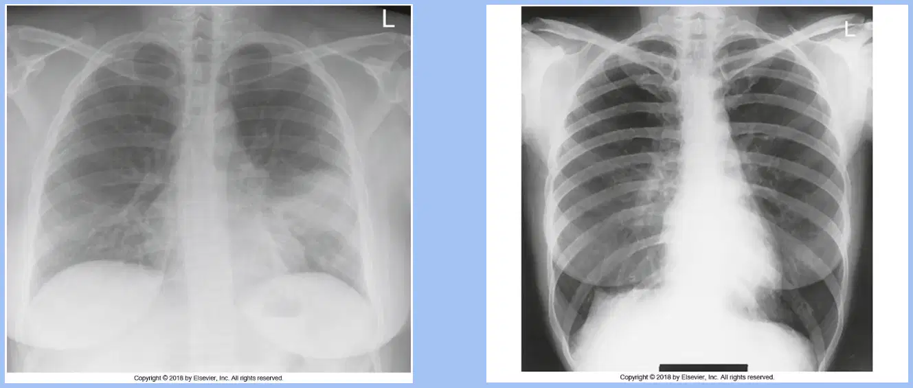
Comparing two chest X‑rays, one shows a high dome‑shaped diaphragm with wide lungs, the other a low diaphragm with long narrow lungs. Which pairing is correct?
Left sthenic, right hypersthenic
Left hypersthenic, right asthenic
Left hyposthenic, right sthenic
Left asthenic, right sthenic
For a standard PA chest projection, where should the central ray be directed?
Perpendicular to T4 slightly lateral
Perpendicular to T7 at midline
Angled 5° cephalad to T6
Angled 10° caudad to T5
What source-to-image distance is standard for routine PA and lateral chest radiographs?
84 inches distance
72 inches distance
40 inches distance
60 inches distance
During a PA chest, which maneuver best prevents scapulae from superimposing lung fields?
Roll shoulders forward fully
Keep arms relaxed at sides
Retract shoulders backward strongly
Elevate shoulders upward only
Which breathing instruction optimizes lung inflation for a PA chest?
Expose on quiet expiration
Expose on second full inspiration
Expose on first gentle inspiration
Expose during breath-hold midway
Which patient landmark level corresponds closely with T7 for chest centering?
C7 vertebra prominens level
Jugular notch level
Inferior angle of scapula level
Xiphoid process level
What field of view should be included on a properly collimated PA chest?
Lower lobes to diaphragm only
Heart only with adjacent ribs
Entire lungs from apices to costophrenic angles
Upper lobes above clavicles only
Which positioning statement is correct for a PA chest patient setup?
MSP perpendicular, MCP parallel to IR
MSP parallel, MCP perpendicular to IR
Both MSP and MCP parallel to IR
Both MSP and MCP perpendicular to IR
Which technical factor selection is most appropriate for a routine chest radiograph?
High kVp 130–140, high mAs
High kVp 110–125, low mAs
Low kVp 60–70, high mAs
Moderate kVp 80–90, high mAs
Which evaluation criterion indicates rotation on a PA chest image?
Asymmetric sternoclavicular joint distances
Superimposed posterior ribs bilaterally
Scapulae completely cleared laterally
Costophrenic angles sharply shown
What is the routine lateral chest projection typically performed?
Right lateral projection
Cross-table lateral only
Left lateral projection
Either lateral by technologist
On a true lateral chest, which structures should be seen in profile without rotation?
Posterior ribs superimposed approximately
Scapulae superimposed over lung fields
Manubrium overlapping vertebrae significantly
Sternoclavicular joints widely separated
Which positioning action helps keep the chin from obscuring apices on a PA chest?
Tilt the head downward
Rotate head toward detector
Flex the neck toward chest
Ask the patient to chin up
Why is 72-inch SID preferred for chest radiography?
Reduces magnification of heart structures
Compensates for tube angle distortion
Allows higher mAs with sharp detail
Increases magnification of lung fields
Which statement best distinguishes AP from PA chest variations listed for trauma or portable exams?
AP chest may be supine or semi-erect
AP chest excludes visualization of apices
AP chest always upright only
AP chest requires 40-inch SID routinely
Decubitus chest positions are primarily used to evaluate which condition?
Air–fluid levels or free air
Thoracic spine scoliosis
Cardiac valve stenosis only
Clavicular fractures mainly
On a properly positioned PA chest radiograph, which finding best confirms minimal rotation at the sternoclavicular (SC) joints?
Clavicles cross the upper lung apices
Left SC joint is slightly elevated superiorly
Right SC joint appears noticeably wider
SC joints appear symmetric bilaterally
Which feature indicates adequate inspiration on a PA chest image?
Posterior ribs are entirely obscured
Six anterior ribs are partly blurred
Only the first five ribs are distinct
Ten posterior ribs are clearly visible
On a PA chest exam, where should the scapulae be positioned relative to the lungs?
Outside the lung fields laterally
Overlying the upper lobes medially
Superimposed on the heart shadow
Projected across both hila centrally
Which structures should be included within the collimated field on a PA chest radiograph?
Entire lung fields and apices
Only the right lung field
Only the left lung field
Heart borders only centrally
Which anatomy should be seen in profile on a correctly positioned PA chest radiograph?
Only heart and diaphragm centrally
Lungs, heart borders, trachea, diaphragm
Spine and pelvis predominantly
Only lungs without heart borders
During lateral chest positioning, how should the patient’s midcoronal plane (MCP) be aligned to the image receptor (IR)?
Parallel to the IR surface
Perpendicular to the IR surface
Oblique at forty-five degrees
Angled posteriorly ten degrees
Which patient setup best minimizes rotation for a lateral chest projection?
Arms positioned at the patient’s sides
Left hip rotated posteriorly
Right shoulder pulled anteriorly
Shoulders and hips stacked vertically
What breathing instruction optimizes lung inflation for both PA and lateral chest images?
End expiration during exposure
Shallow breathing throughout
Full inspiration at exposure
Breath-hold after expiration
Which evaluation criterion confirms a true lateral chest radiograph?
Posterior ribs nearly superimposed
Sternum rotated anteriorly markedly
Humeri superimposed over lung fields
Hila displaced far from midline
On a lateral chest image, which region should be centered to the exposure field?
Hilar region centered mid-thorax
Right hemidiaphragm exclusively
Only the posterior costophrenic angle
Upper mediastinum without lungs
Which structures should be visualized in profile on a correct lateral chest radiograph?
Pelvis and lumbar vertebrae
Anterior ribs and clavicles
Only right lung exclusively
Left lung, side of heart, hila
Which central ray level is appropriate for an AP chest when the patient is supine?
T7, three to four inches below jugular notch
T4, at the level of the sternal angle
T10, two inches above xiphoid tip
C7, at the base of the neck
For a trauma AP chest, which SID is recommended to reduce magnification when feasible?
Eighty inches regardless of equipment
Forty inches as the standard distance
Seventy-two inches or maximum possible
Fifty-six inches for all trauma cases
Which field of view best meets AP chest positioning requirements?
Upper lobes excluding clavicles
Lower lobes with costophrenic angles
Entire lungs including apices
Mediastinum only without apices
Which patient positioning instruction is correct for an AP chest on a supine patient?
Chin up with shoulders shrugged
Chin up with shoulders lowered
Chin down with shoulders elevated
Head neutral with shoulders raised
During an AP chest exposure, when should the image be taken for optimal lung inflation?
On the first shallow inspiration
On the second full inspiration
During quiet expiration phase
During a breath-hold after exhale
Which technical factor set is most appropriate for an AP chest?
kVp 60–65 with moderate mAs
kVp 120–130 with very low mAs
kVp 70–80 with very high mAs
kVp 100–110 with low–moderate mAs
Which structure typically appears magnified on an AP chest radiograph?
Cardiac silhouette appears enlarged
Hilar vessels appear narrowed
Diaphragm appears compressed
Clavicles appear foreshortened
On a properly positioned AP chest, how should the clavicles appear?
Overlapping the lower lung bases
Rotated superiorly above manubrium
Angled downward toward sternum
Relatively horizontal across apices
How many posterior ribs should be visible above the diaphragm on a good AP chest inspiration?
Ten to eleven posterior ribs visible
Five to six posterior ribs visible
Seven posterior ribs exactly visible
Eight to nine posterior ribs visible
Which structures should be demonstrated in profile on a diagnostic AP chest radiograph?
Skull base, cervical spine, mandible
Scapulae, humerus, clavicular joints
Liver, spleen, kidneys, stomach bubble
Lungs, trachea, diaphragm, heart outline
Why does the heart appear larger on AP chest images compared with PA images?
Short SID decreases geometric blur
Increased OID from supine positioning
Higher kVp reduces subject contrast
Beam divergence toward the diaphragm
For apical lordotic imaging to project clavicles above lung apices, which positioning concept is essential?
No angulation with arms raised
Steep obliquity with caudal tilt
Caudal angulation with chin down
Cephalic angulation or patient lordosis
A semi-erect AP chest shows only seven posterior ribs above the diaphragm. What adjustment would most directly improve lung inflation visualization?
Center two inches higher toward T4
Angle beam caudally five degrees
Increase kVp and reduce mAs
Coach deeper inspiration and repeat
Apical lordotic chest is dont to visualize
The heart
Costophrenic Angles
Lower Lobes
Apices
Inspiration/Expiration views are ordered to check for what?
Pneumonia
Foreign Body
Pneumothorax
Pulmonary Embolism
Oblique Chests are completed at what angle?
30
45
60
90
The left lung is demonstrated in what two views
RAO
LAO
RPO
LPO
The right lung is demonstrated in what two views
RAO
LAO
RPO
LPO

