
Font size
 Worksheets
5 Functions of the Skeletal System
Total questions: 60
Worksheet time: 30mins
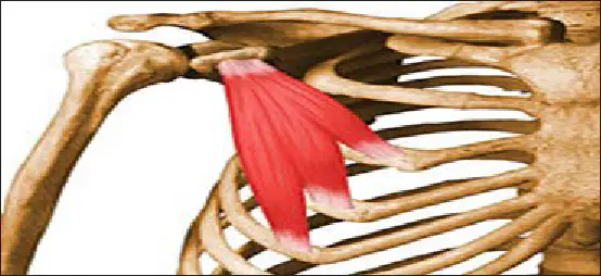
Which statement best describes how the skeletal system enables movement?
Bones provide leverage and muscle attachment points
Ligaments shorten to move bones at joints
Cartilage contracts to swing limbs through space
Bones contract to pull tendons and create force
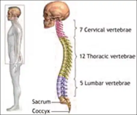
Which vertebral region is primarily responsible for supporting the upper body in humans?
The backbone including cervical through lumbar vertebrae
Only the cervical vertebrae support the rib cage
The rib cage bears the full body weight alone
The sacrum alone supports the entire upper body
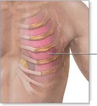
Select all functions that directly protect vital organs.
Rib cage surrounding heart and lungs
Skull encasing the brain
Bone marrow creating blood cells
Long bones storing calcium and phosphorus
Which process occurs in bone marrow tissue located in the center of many bones?
Secretion of digestive enzymes into intestines
Synthesis of red and white blood cells
Breakdown of dietary proteins and fats
Filtration of blood plasma into urine
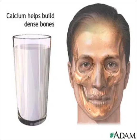
A person has a deficiency in calcium and phosphorus storage. Which skeletal function is most impaired?
Electrical signaling across nervous tissue
External defense against bacterial invasion
Primary movement through muscle contraction
Mineral reservoir for maintaining body balance

Which pair correctly matches a skeletal function with an example?
Blood creation—tendons producing platelets
Support—radius anchoring the spinal cord
Movement—cartilage contracting to bend joints
Protection—rib cage shielding the lungs
Multiple roles of the skeleton sustain homeostasis. Which are true? Select all that apply.
Vertebrae compress to pump lymph through vessels
Bones store minerals to regulate blood levels
Muscle attachment to bones enables movement
Bone marrow generates new blood cells regularly
If the rib cage were severely weakened, which outcomes would most likely occur first?
Loss of all muscle attachment in the torso
Inability to produce red blood cells anywhere
Collapse of the cervical spine under skull
Greater risk to heart and lungs from impacts
Which set of bones belongs to the axial skeleton?
Clavicle, scapula, metacarpals
Pelvis, femur, tibia, fibula
Humerus, radius, ulna, carpals
Skull, ribs, sternum, spine
Which structures are part of the appendicular skeleton?
Hips and legs
Skull and sternum
Shoulders and arms
Ribs and vertebrae
A bone with a broad, thin surface like the scapula is classified as which type?
Irregular bone with variable shape
Flat bone with broad surface
Long bone with tubular shaft
Short bone with articular surface
Which bones are typical examples of long bones?
Humerus, femur, phalanges
Vertebrae, carpals, patella
Skull, sternum, ribs
Metacarpals, tarsals, carpals
Which list correctly pairs bone types with examples?
Long bones – metatarsals, metacarpals
Irregular bones – vertebrae, patella
Flat bones – skull, sternum
Short bones – carpals, tarsals
Which layer directly covers the outer surface of a bone?
Periosteum covering bone surfaces
Compact bone forming cortex
Spongy bone in epiphyses
Bone marrow between trabeculae
Where is compact bone primarily located in a typical long bone?
Between periosteum and articular cartilage
Only at joint surfaces near cartilage
Inside medullary cavity with marrow
Beneath the periosteum forming shaft
Spongy (cancellous) bone is best described as which location?
Beneath compact bone near ends
On outer bone surface layer
Only present in short bones
Within central medullary canal
Bone marrow primarily occupies which spaces in a long bone?
Gaps among spongy bone trabeculae
Dense layers of compact bone
Articular cartilage at joint ends
Spaces beneath the periosteum
Which combination describes features of long bones listed in the material?
Thin broad surfaces throughout
Articular surfaces at the ends
Mostly spongy bone externally
Tubular shaft in the diaphysis
Which body region transition marks axial to appendicular elements in the upper limb?
Clavicle and scapula girdle area
Thoracic vertebrae and ribs area
Sacrum and coccyx midline area
Mandible and cranial vault area
Which bones are NOT categorized as flat bones in this material?
Carpals and tarsals listed
Broad thin bones listed
Skull and sternum listed
Scapula and ribs listed
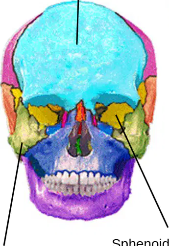
In the front-view skull diagram, which bone forms the forehead and roof of the eye sockets?
Temporal bone
Frontal bone
Parietal bone
Occipital bone
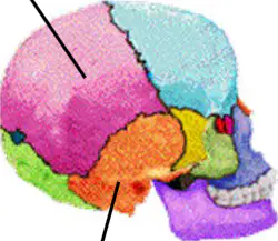
On the lateral skull diagram, which bone lies inferior to the parietal bone and houses the ear structures?
Sphenoid bone
Occipital bone
Temporal bone
Zygomatic bone
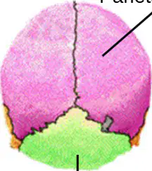
Which bones are paired and meet along the sagittal suture at the top of the skull?
Occipital bones
Frontal bones
Sphenoid bones
Parietal bones
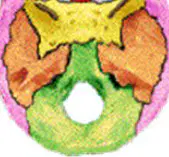
Which bone forms the posterior and inferior portion of the cranium, enclosing the foramen magnum?
Temporal bone
Frontal bone
Parietal bone
Occipital bone
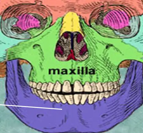
In the facial bones diagram, which bone is the upper jaw that holds upper teeth and forms part of the hard palate?
Mandible
Nasal
Maxilla
Zygomatic
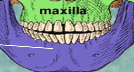
Which statement correctly contrasts maxilla and mandible in the face diagram?
Maxilla is movable; mandible is fixed
Both maxilla and mandible are movable
Maxilla forms upper jaw; mandible is movable
Maxilla forms lower jaw; mandible is upper
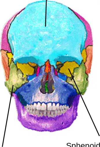
Which bone forms the cheek prominence and part of the lateral orbital rim in the front-view cranium diagram?
Occipital bone
Sphenoid bone
Zygomatic bone
Parietal bone
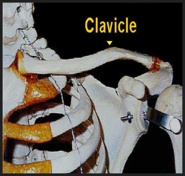
According to the clavicle slide, which function best describes the clavicle in the shoulder girdle?
Connects the scapula to the vertebral column
Protects the lungs during breathing
Holds the shoulder joint away from the torso
Anchors the humerus to the ulna

Based on the clavicle facts, which statement is accurate about its vulnerability?
It is thick like the femur and hard to break
It is thin like a little finger and breaks easily
It rarely fractures because it is flexible cartilage
It only breaks under more than 50 pounds of force
Which bones together form the shoulder girdle connecting the upper limb to the torso?
Clavicle
Scapula
Sternum
Humerus
Which region of the vertebral column contains seven vertebrae and supports the neck?
Cervical region of the neck
Thoracic region of the chest
Sacral region below lumbar
Lumbar region of the back
How many thoracic vertebrae are present, and what do they articulate with?
Four fused vertebrae forming tail
Five vertebrae bearing most weight
Seven vertebrae supporting skull
Twelve vertebrae attaching to ribs
Select all statements that are correct about the lumbar region.
It consists of five vertebrae
It forms the lower back area
It provides rib attachment points
It sits superior to the cervical region
Which sequence correctly orders the vertebral regions from superior to inferior?
Cervical, lumbar, thoracic, sacrum, coccyx
Lumbar, cervical, thoracic, sacrum, coccyx
Thoracic, cervical, lumbar, coccyx, sacrum
Cervical, thoracic, lumbar, sacrum, coccyx
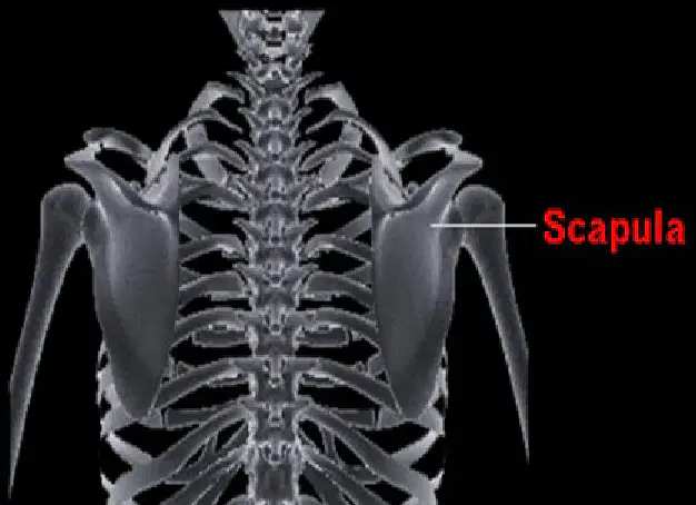
Identify the bone labeled as the shoulder blade that lies on the posterior rib cage and helps form the shoulder joint.
Scapula on the back of ribs
Sternum along the midline
Clavicle anterior to ribs
Humerus of the upper arm
Which paired statements about the sacrum and coccyx are accurate?
The sacrum consists of fused vertebrae
The sacrum lies superior to the coccyx
Both are at the base of the vertebral column
The coccyx is part of the thoracic region
A fall injures the most inferior part of the vertebral column. Which structure is most likely affected?
Coccyx at the very bottom
Thoracic vertebra T6-T8
Sacrum above the coccyx
Lumbar vertebra L3-L5
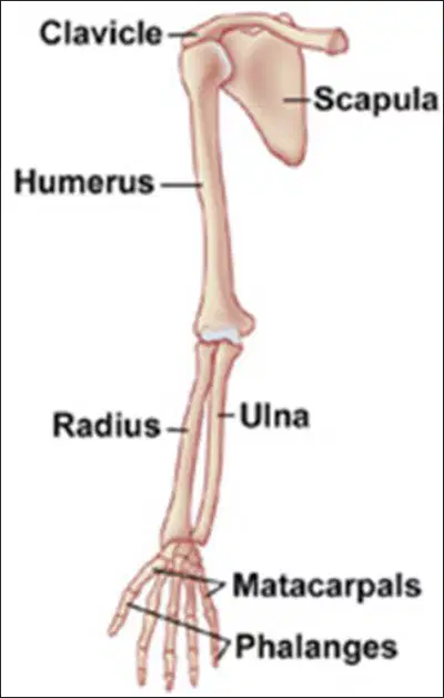
In the left diagram, which labeled bone forms the upper arm connecting the shoulder to the elbow?
Clavicle
Scapula
Humerus
Ulna
Radius
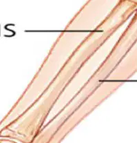
Which set lists the bones of the forearm from lateral (thumb side) to medial when the palm faces forward?
Clavicle then radius
Ulna then radius
Radius then ulna
Humerus then ulna
Scapula then humerus
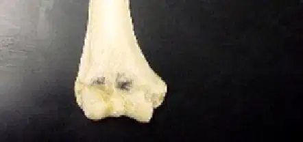
In the right photo of the single long bone, which anatomical bone is shown based on its position in the upper limb?
Metacarpal of the hand
Humerus of the arm
Radius of the forearm
Ulna of the forearm
Phalanx of a finger
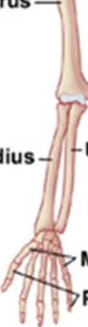
Which bones compose the palm region between the wrist and fingers?
Phalanges only
Metacarpals and carpals
Carpals and phalanges
Metacarpals only
Carpals only
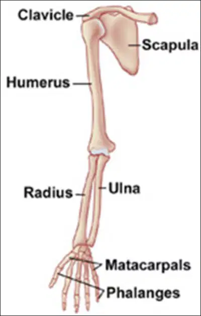
Select all bones that articulate at the wrist joint with the forearm.
Ulna
Radius
Phalanges
Metacarpals
Carpals

When giving a thumbs-up, which bone aligns with the thumb side of the forearm?
Scapula aligns with thumb
Clavicle aligns with thumb
Humerus aligns with thumb
Radius aligns with thumb
Ulna aligns with thumb
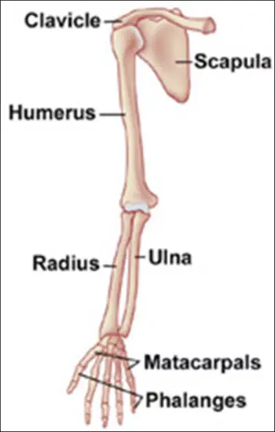
Which sequence correctly orders bones from shoulder to fingertips along one limb?
Clavicle, scapula, ulna, radius, metacarpals, phalanges
Scapula, humerus, radius, metacarpals, carpals, phalanges
Humerus, clavicle, radius, metacarpals, carpals, phalanges
Clavicle, humerus, radius, carpals, metacarpals, phalanges
Scapula, clavicle, humerus, ulna, carpals, phalanges
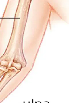
Which statement best distinguishes the radius from the ulna in standard anatomical position?
Ulna is lateral and aligns with thumb
Radius is medial and larger at elbow
Radius is medial and aligns with little finger
Ulna is lateral and larger at wrist
Radius is lateral and aligns with thumb
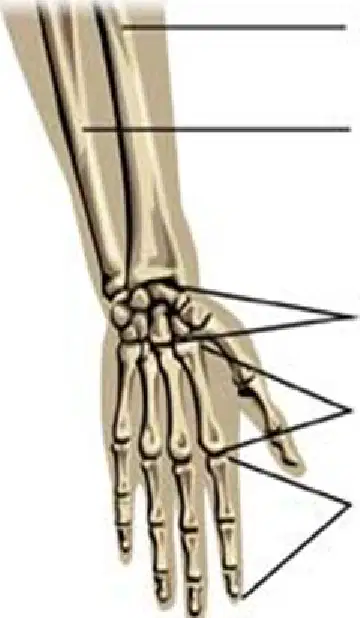
In the labeled hand diagram, which group of bones forms the palm between the carpals and the phalanges?
Phalangites
Carpioles
Metatarsals
Metacarpals
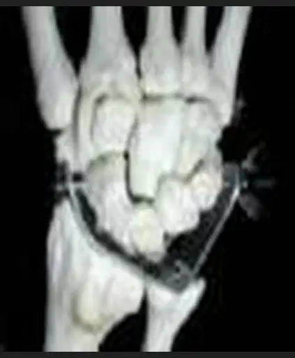
Which bones are directly proximal to the metacarpals at the wrist joint in the hand diagrams?
Ulna shafts
Radius heads
Phalanges
Carpals
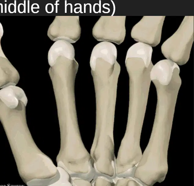
In the close-up image of the hand’s middle section, what feature best identifies metacarpals compared to phalanges?
Longer shafts with proximal bases
Shorter segments with distal tips
Flat plates with wide surfaces
Rounded sesamoid-like nodules
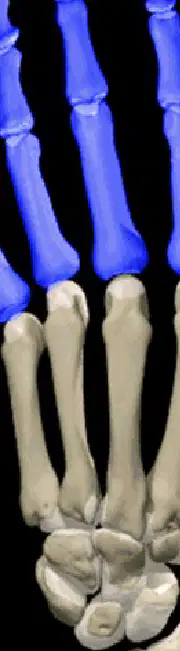
Across the highlighted bones image, which colored segments represent the phalanges of the hand?
The pale metacarpal shafts
The beige radius-ulna ends
The grey carpal cluster
The blue finger segments
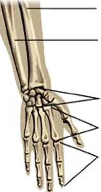
Which sequence correctly orders the bones from the forearm toward the fingertips as shown?
Carpals → Radius/Ulna → Metacarpals → Phalanges
Radius/Ulna → Metacarpals → Carpals → Phalanges
Radius/Ulna → Carpals → Metacarpals → Phalanges
Metacarpals → Carpals → Radius/Ulna → Phalanges
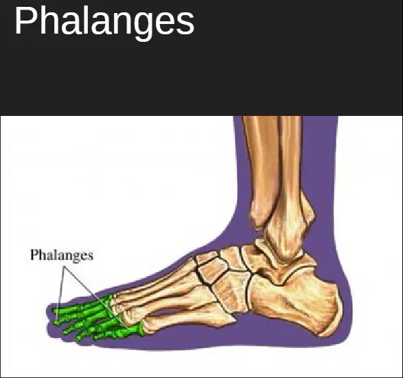
Select all statements that are accurate about phalanges in the provided images.
They form the wrist cluster beneath the palm
They are also present in toes, as labeled
They articulate directly with the radius
They lie distal to the metacarpals
They are the finger bones shown in segments
In the lower limb, which bone is the largest and bears most body weight during standing?
Femur
Patella
Tibia
Fibula
Which two long bones run parallel in the leg between the knee and the ankle?
Femur and tibia
Tibia and fibula
Fibula and patella
Femur and fibula
Which bone forms the kneecap and improves the leverage of the quadriceps tendon?
Fibula
Patella
Tarsal
Femur
Where is the tibia primarily located relative to the fibula in the lower leg?
Posterior to fibula
Lateral to fibula
Superior to fibula
Medial to fibula
Which statement best distinguishes tarsals from metatarsals in the foot?
Tarsals are long bones; metatarsals are short bones
Tarsals support body weight; metatarsals do not bear load
Tarsals are toe tips; metatarsals are heel bones
Tarsals are ankle bones; metatarsals form the foot arch
Which bone directly articulates with the acetabulum of the hip to form the hip joint?
Femur head
Fibula head
Tibia plateau
Patella surface
Which set lists only bones of the foot (not the leg)?
Metatarsals and phalanges
Tarsals and metatarsals
Fibula and metatarsals
Tibia and tarsals
During an inversion ankle sprain, which bone remains most medial in the lower leg providing main weight-bearing support?
Patella
Talus
Fibula
Tibia
Which pairing correctly matches bone with typical structural feature?
Tibia—medial malleolus at ankle
Patella—sesamoid within tendon
Fibula—primary knee weight-bearing
Femur—proximal head and neck

