Search Header Logo
The Liver

The Liver

Assessment

Presentation

•

Biology

•

University - Professional Development

•

Hard

Created by

Joseph Anderson

FREE Resource

11 Slides • 9 Questions

1

LIVER

Liver is the largest visceral structure in the abdominal cavity, and the largest gland in the human body.


Functions = synthesis of bile, glycogen storage and clotting factor production.

Slide image

2

Slide image

SUPERFICIAL LOCATION: mainly at RUQ, right hypochondriac & epigastric regions.

3

Multiple Choice

Question image

A patient is suspected with tumour of the right lobe of the liver. An early imaging diagnosis using an ultrasonography is suggested. As a sonographer, which area of the abdomen you must put the transducer to scan?

1

Right upper quadrant

2

Left upper quadrant

3

Right lower quadrant

4

Left lower quadrant

4

Liver Surface

  • Diaphragmatic surface – the antero-superior surface of the liver. Simply known as anterior view.

  • Visceral surface – the postero-inferior surface of the liver. Simply called posterior view.

Slide image

5

Slide image

6

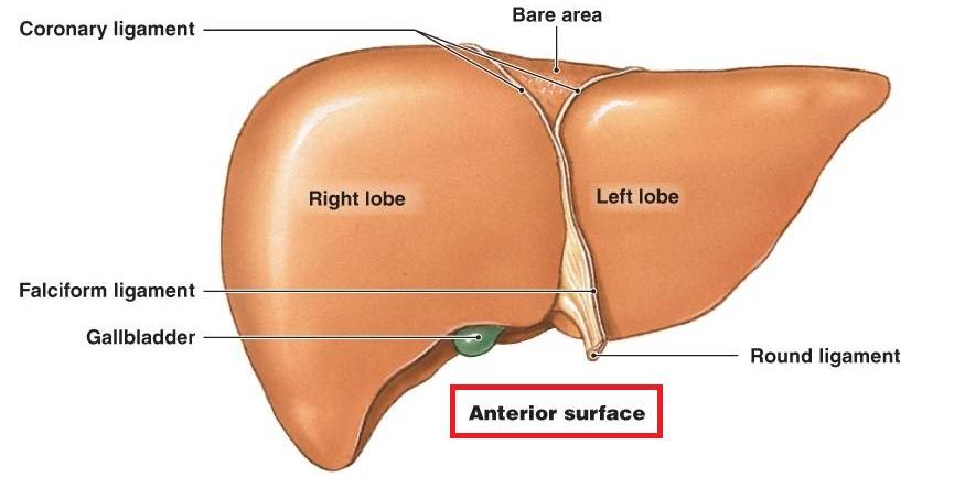
Anterior surface of liver

  • devide by right and left lobes.

  • falciform ligament, coronary ligament & round ligament between right and left lobes.

  • lower of the right lobe = lower part of gallbladder demonstrated

  • superior part between right and left lobes = bare area (diaphragmatic surface, triangle shape, not cover by peritoneum layer, attached directly to the diaphragm)

7

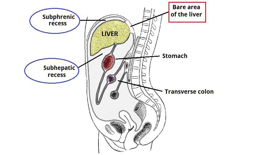
Recess

  • spaces between the liver and surrounding structures. 

  • Subphrenic recess = space between liver & diaphragm.

  • Subhepatic recess = space between liver & transverse colon

  • Hepatorenal recess = space ebtween liver and right kidney
Slide image

8

Slide image

9

Multiple Choice

Which liver surface covered by the diaphraghm?

1

Diaphragmatic surface

2

Visceral surface

10

Multiple Choice

Which ligament is NOT located between right and left lobes of the liver

1

Falciform ligament

2

Coronary ligament

3

Anterior cruciate ligament

4

Round ligament

11

Multiple Choice

The surface area on the liver that did not covered by peritoneum is called __________

1

subphrenic recess

2

subhepatic recess

3

hepatorenal recess

4

bare area

12

Multiple Choice

Hepatorenal recess or also known as Morison's pouch is a space between the liver and _________

1

duodenum

2

stomach

3

right kidney

4

left kidney

13

Slide image

14

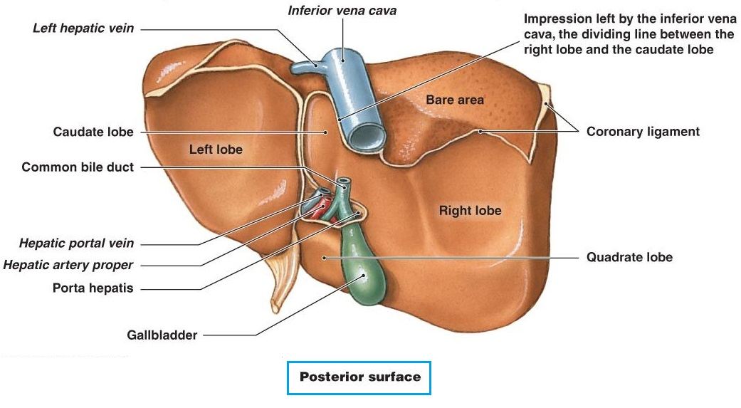
Posterior surface of liver

  • At the right lobe, can see inferior vena cava (IVC) at the upper part and gallbladder at the lower part.

  • On the right lobe, IVC separate the right lobe into another smaller lobe called = caudate lobe.

  • On the right lobe, gallbladder separate the right lobe into another smaller lobe called = quadrate lobe.

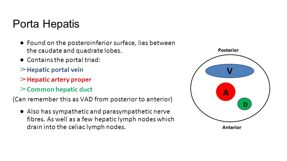
  • On the right lobe (between caudate lobe & quadrate lobe), there is a structure known as porta hepatis. This structure where 3 main vessel/duct insert into the liver which are (i) common hepatic duct, (ii) hepatic artery proper & (iii) hepatic portal vein.

15

Slide image

16

Multiple Choice

TRUE or FALSE


Based on the posterior surface of liver, abdominal aorta separate caudate lobe to right lobe of the liver

1

TRUE

2

FALSE

17

Multiple Choice

TRUE or FALSE


Based on the posterior surface of liver, gallbladder separate quadrate lobe to right lobe of the liver

1

TRUE

2

FALSE

18

Multiple Choice

TRUE or FALSE


Based on the posterior surface of liver, porta hepatis separate quadrate lobe to caudate lobe.

1

TRUE

2

FALSE

19

Multiple Choice

Which ONE structure does NOT enter the liver through porta hepatis?

1

Common hepatic duct

2

Common bile duct

3

Hepatic artery proper

4

Hepatic portal vein

20

Artery & Vein

  • dual blood supply.

  • (1) Hepatic artery proper (25%) – Derived from the coeliac trunk.

  • (2) Hepatic portal vein (75%) – supplies the liver with partially deoxygenated blood, carrying nutrients absorbed from the small intestine. 

  • Venous drainage of the liver is achieved through hepatic veins. These hepatic veins then open into the IVC

Slide image
pattern-tertiary

LIVER

Liver is the largest visceral structure in the abdominal cavity, and the largest gland in the human body.


Functions = synthesis of bile, glycogen storage and clotting factor production.

Slide image

Show answer

Auto Play

Slide 1 / 20

SLIDE