
Integumentary System
Presentation
•
Biology
•
University
•
Practice Problem
•
Medium
+1
Standards-aligned
Christine Boudreau
Used 10+ times
FREE Resource
32 Slides • 23 Questions
1
Integumentary
System
2
3
Fill in the Blanks
Type answer...
4
5
Explanation
The epidermis is avascular, meaning it lacks its own blood supply. Instead, nutrients and oxygen must travel from blood vessels in the dermis to the epidermis by diffusion. This process limits the thickness of viable cells in the epidermis and contributes to the layering pattern as keratinocytes move outward and eventually die.
6
Multiple Choice
Which combination of structures primarily helps regulate body temperature?
Sebaceous glands and stratum corneum
Sweat glands and dermal blood vessels
Nails and adipose tissue
Keratinocytes and melanocytes
7
Explanation
Thermoregulation is a critical function of the integumentary system. Eccrine sweat glands help cool the body by releasing sweat, which evaporates from the skin surface, removing heat. Meanwhile, dermal blood vessels respond to temperature changes through vasodilation (to release heat) and vasoconstriction (to conserve heat). The other choices involve structures with other roles—such as protection, cushioning, or pigmentation—but not active thermoregulation.
8
Open Ended
Explain the difference between thin skin and thick skin in terms of
structure, function, and location.
9
10
11
Multiple Choice
Which epidermal layer contains the highest number of metabolically active keratinocytes?
Stratum corneum
Stratum granulosum
Stratum basale
Stratum lucidum
12
Explanation
The stratum basale contains basal stem cells that are mitotically active and generate new keratinocytes.
13
14
Open Ended
Explain the differences between thick and thin skin.
15
Fill in the Blanks
Type answer...
16
Desmosomes are cell junctions that hold keratinocytes together and give the stratum spinosum its characteristic appearance under a microscope.
17
Multiple Select
Which specialized epidermal cell type is responsible for detecting fine touch? (Select all that apply).
Merkel cells
Meissner’s corpuscles
Pacinian corpuscles
Ruffini endings
18
Merkel cells are slow-adapting mechanoreceptors located in the stratum basale and are responsible for detecting fine touch and texture, especially in areas like fingertips.
Meissner’s corpuscles are fast-adapting receptors found in the dermal papillae that detect light touch and changes in texture.
Pacinian corpuscles are deep pressure receptors located in the dermis or hypodermis and respond to vibration and deep pressure.
Ruffini endings detect skin stretch and sustained pressure and contribute to the perception of object manipulation and grip control.
19
Fill in the Blanks
Type answer...
20
If keratinocytes do not undergo normal apoptosis, they may survive and proliferate abnormally, leading to cancerous growths such as squamous cell carcinoma.
21
Open Ended
Why do keratinocytes die as they move toward the surface of the epidermis?
22
Sample Answer
As keratinocytes migrate upward through the layers of the epidermis, they move farther away from the blood supply in the dermis, leading to reduced access to nutrients and oxygen. At the same time, they undergo terminal differentiation, during which they lose their nuclei and organelles and accumulate keratin and lipids. This process is regulated by signaling molecules and enzymes that trigger apoptosis, allowing the cells to transform into tough, flat, dead corneocytes that make up the protective outer layer of skin.
23
24
Multiple Choice
Which structures are not found in the dermis?
Sweat glands
Blood vessels
Meissner’s corpuscles
Keratinocytes
25
Keratinocytes are found in the epidermis, not the dermis. The dermis contains blood vessels, glands, and sensory receptors.
26
Open Ended
Langer’s lines are natural patterns in the skin formed by the orientation of collagen fibers in the reticular layer of the dermis. Surgeons often make incisions parallel to these lines because doing so reduces skin tension, promotes faster healing, and minimizes scarring.
Based on this information, what do you think might happen if a surgeon makes an incision perpendicular to Langer’s lines instead? Explain your reasoning.
27
Sample Answer
If a surgeon cuts perpendicular to Langer’s lines, the incision is likely to be pulled open by the natural tension in the skin. This could result in slower healing, increased tension on the wound edges, and more noticeable scarring, because the collagen fibers resist closure in that direction.
28
29
Multiple Choice
Which of the following is not a function of the hypodermis?
Insulation
Energy storage
Keratin production
Shock absorption
30
Keratin is produced by keratinocytes in the epidermis, not the hypodermis.
31
Fill in the Blanks
Type answer...
32
33
Burns are classified based on how deeply they penetrate the layers of the skin:
A first-degree burn affects only the epidermis, causing redness and mild pain (e.g., sunburn).
A second-degree burn damages the epidermis and part of the dermis, often resulting in blisters, swelling, and more intense pain.
A third-degree burn extends through the entire epidermis and dermis, and may also affect the hypodermis or deeper tissues.
Burns
34
Multiple Choice
Why is the hypodermis a preferred site for subcutaneous injections?
It is part of the epidermis and regenerates quickly.
It contains dense irregular connective tissue.
It has a rich blood supply and loose matrix for rapid absorption.
It contains melanocytes that process medication.
35
The vascularity and loose connective tissue of the hypodermis allow medications to be absorbed quickly when injected.
36
Open Ended
Which specific skin structures would likely be damaged or destroyed in a third-degree burn? Explain your reasoning.
37
Sample Answer:
In a third-degree burn, the damage includes the epidermis, dermis, and possibly the hypodermis. Structures that would be destroyed include keratinocytes, melanocytes, sensory receptors (like Merkel cells and Meissner’s corpuscles), sweat glands, sebaceous glands, blood vessels, hair follicles, and collagen fibers in the dermis. Because the nerve endings are also damaged, the area may be numb despite the severity. If the burn reaches the hypodermis, adipose tissue and larger blood vessels may also be affected.
38
39
Multiple Choice
Which structure is responsible for supplying blood to the growing hair follicle?
Arrector pili muscle
Inner root sheath
Hair papilla
Hair shaft
40
The hair papilla is located at the base of the hair bulb and contains capillaries that nourish the hair matrix.
41
Multiple Choice
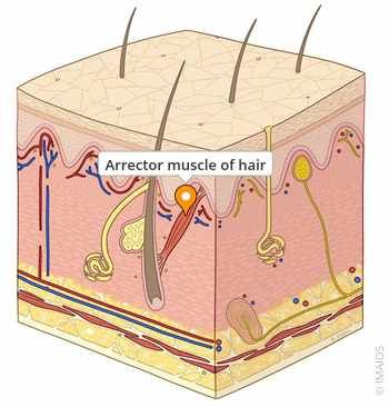
Which of the following best describes the function of the arrector pili muscle?
Protects the hair matrix from UV radiation
Detects fine touch and vibration
Anchors the hair bulb to the dermis
Contracts to raise the hair shaft during cold or emotional stimuli
42
The arrector pili is a small smooth muscle that pulls the hair upright, creating goosebumps.
43
44
Multiple Choice
Where are melanocytes located within the layers of the epidermis?
Stratum corneum
Stratum spinosum
Stratum granulosum
Stratum basale
45
Melanocytes reside in the stratum basale, where they produce melanin and transfer it to keratinocytes.
46
Multiple Choice
In albinism, melanocytes are present but unable to produce melanin due to:
Lack of keratin production
Loss of blood flow to the skin
Deficiency of the enzyme tyrosinase
Autoimmune destruction of melanocytes
47
Steps of Melanin Production (Melanogenesis)
It starts with an amino acid called tyrosine.
Melanocytes take in tyrosine from the body.An enzyme called tyrosinase turns tyrosine into dopaquinone.
This is the key step—if tyrosinase doesn’t work, melanin can’t be made (like in albinism).Dopaquinone turns into either eumelanin or pheomelanin.
Eumelanin is brown to black pigment
Pheomelanin is yellow to red pigment
Your genetics determine which type you make more of, affecting your skin and hair color.
Melanin is packed into melanosomes.
These are little vesicles (containers) that carry melanin.Melanosomes are transferred to keratinocytes.
The pigment ends up in the skin cells and sits above their nuclei to protect DNA from UV radiation.
48
49
Multiple Choice
Which type of sweat gland is primarily involved in thermoregulation?
Apocrine
Sebaceous
Eccrine
Ceruminous
50
Eccrine glands help cool the body through evaporative sweating.
51
Multiple Choice
What makes apocrine sweat thicker than eccrine sweat?
It contains high levels of glucose
It is mixed with sebum
It includes lipids and proteins
52
Apocrine sweat has proteins and lipids, making it thicker than eccrine sweat.
53
Fill in the Blanks
Type answer...
54
Sebaceous glands open into hair follicles, aiding lubrication of hair and skin.
55
Open Ended
What questions do you have?
Integumentary
System
Show answer
Auto Play
Slide 1 / 55
SLIDE
Similar Resources on Wayground
50 questions
Exam 1 Review
Presentation
•
University
48 questions
BIO111 Chp 15 Genes and Proteins
Presentation
•
University
48 questions
POPULAR MEMBER SERVICES DIAGNOSTICS, HOSPITILIZATION AND SURGERY
Presentation
•
KG
52 questions
BIO110 Biodiversity- Microorganisms
Presentation
•
University
51 questions
Sept 2024_Search strategies and library databases
Presentation
•
University
51 questions
Present Simple (have) review family
Presentation
•
University
50 questions
Ocean Lecture
Presentation
•
12th Grade
47 questions
Ser + Adjetivos
Presentation
•
12th Grade
Popular Resources on Wayground
20 questions
"What is the question asking??" Grades 3-5
Quiz
•
1st - 5th Grade
20 questions
“What is the question asking??” Grades 6-8
Quiz
•
6th - 8th Grade
10 questions
Fire Safety Quiz
Quiz
•
12th Grade
20 questions
Equivalent Fractions
Quiz
•
3rd Grade
34 questions
STAAR Review 6th - 8th grade Reading Part 1
Quiz
•
6th - 8th Grade
20 questions
“What is the question asking??” English I-II
Quiz
•
9th - 12th Grade
20 questions
Main Idea and Details
Quiz
•
5th Grade
47 questions
8th Grade Reading STAAR Ultimate Review!
Quiz
•
8th Grade