
 Worksheets
Ch 8 The Eye
Total questions: 65
Worksheet time: 43mins
Rods are light sensitive cells in the retina that
give you color vision in dim light
respond in dim light
give you color vision in bright light
respond in bright light
Cones are light sensitive cells in the retina that
give you color vision in bright light
respond in dim light
give you color vision in dim light
respond in bright light
The optic nerve transmits signals to the
temporal lobe
parietal lobe
occipital lobe
frontal lobe
True/False: The vitreous humor helps give the cornea its rounded shape.
True
False
The pupil is located at the center of the
sclera
iris
retina
nothing
The vitreous humor is
gel like and gives the eyeball its shape
watery and gives the eyeball its shape
The blind spot is created by the
retina
optic nerve leaving the eye through the retina
nothing; we don't have one
The sclera is designed for
strength
clarity
What are the functions of eyelids and eyelashes
to protect and rehydrate the eyes
keep dirt out of the eyes
keep debris out of the eys
all of the above
The human eye is surrounded by how many muscles?
4
2
6
5
What enters the eyes so you can see?
air
dust
light
blood
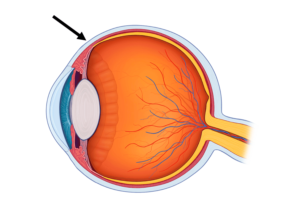
The white outer part of the eye.
Cornea
Lens
Sclera
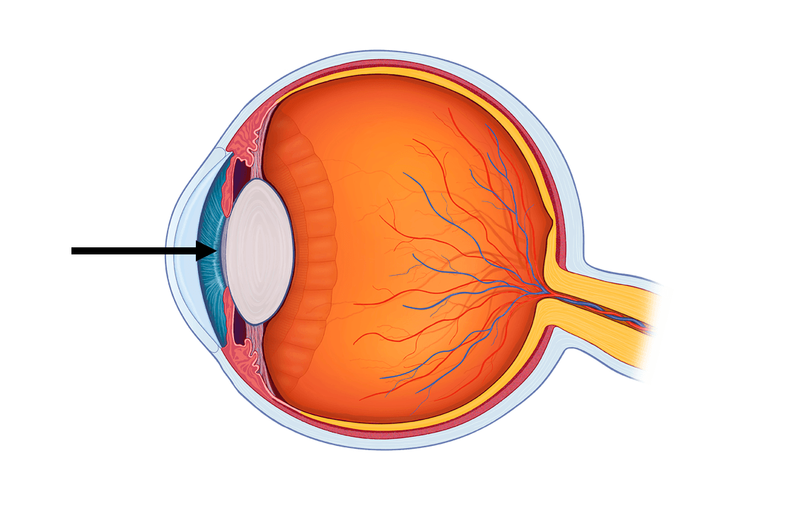
The opening of the eye where light enters
Retina
Sclera
Pupil
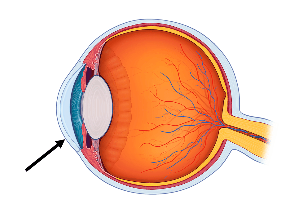
The clear part of the eye that light will first pass through.
Lens
Cornea
Sclera
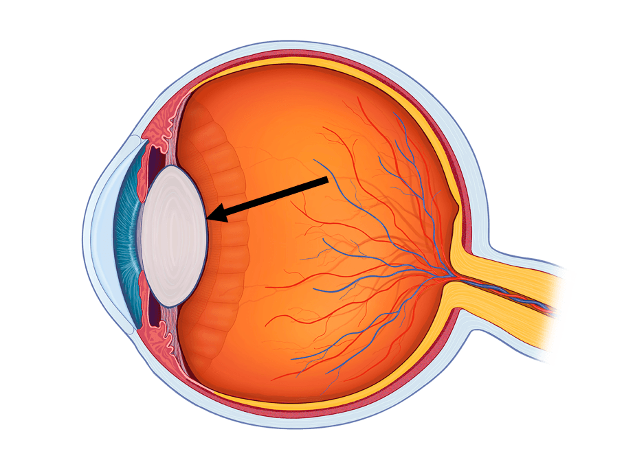
This part of the eye focuses light onto the retina.
Retina
Optic Nerve
Lens
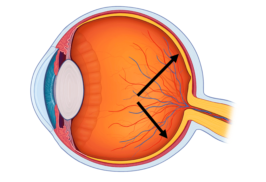
Photosensitive part of the eye that contains rods and cones.
Sclera
Retina
Optic Nerve
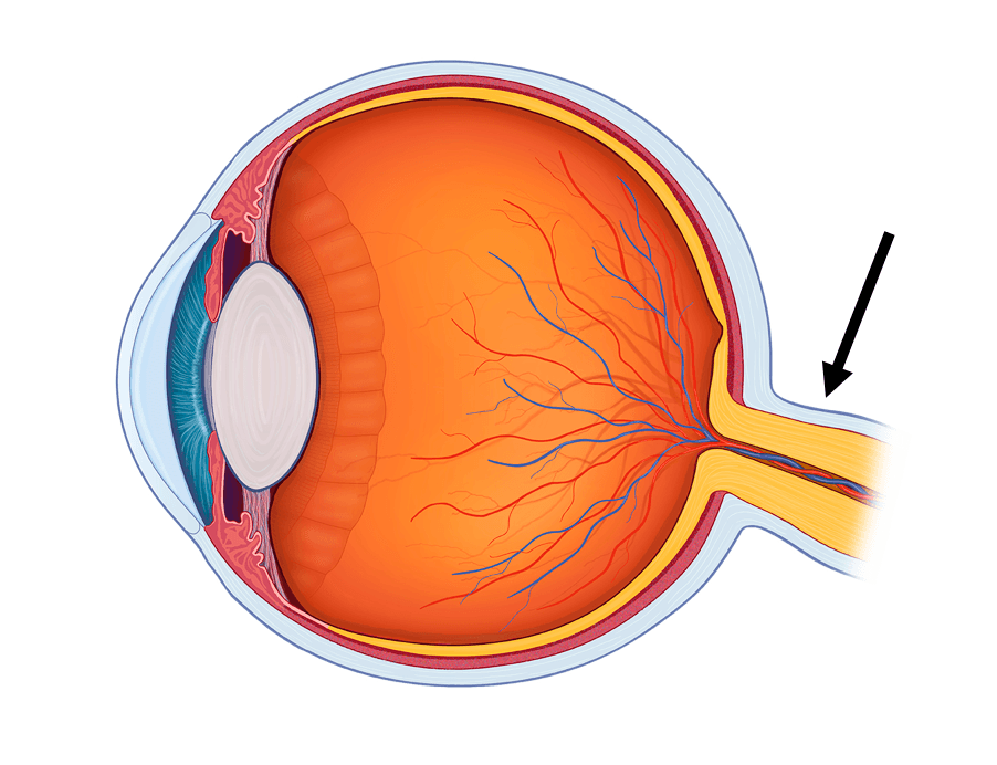
This part of the eye sends signals from the retina to the visual cortex.
Optic Nerve
Retina
Iris
Which of these is located between the cornea and lens, maintains the convex shape of the cornea with its pressure, provides nutrition for ocular tissues, and carries away the waste from said tissues?
Cornea
Aqueous Humour
Vitreous Humour
Conjuctiva

Identify the refractive error.
Presbyopia
Myopia
Hypermetropia
Astigmatism

Tears are produced by the ______________ located in the _________ part of the _________ eyelid.
lacrimal sac, medial, upper
lacrimal sac, lateral, upper
lacrimal gland, medial, upper
lacrimal gland, lateral, upper

The anterior segment includes the cornea. Which characteristic distinguishes the cornea from other tissues:
it produces tears
it has a lack of blood vessels
it drains the tears
none of the above

The small compartment between the cornea and the iris is called the __________________________.
tear lake
Canal of Schlemm
lacrimal sac
anterior chamber

The clear, jelly-like substance that fills the intraocular cavity behind the lens is _______________________.
aqueous humor
vitreous
goblet cells
none of the above

Where do the 2 optic nerves meet behind the eyes in the brain?
optic chiasm
visual cortex
frontal lobe
trabecular meshwork

Structure E is the liquid called _________.
Aqueous humor
Vitreous humor
Lens
Cornea
Structure K is the _________.
Fovea centralis
Retina
Lens
Cornea
Structure F is the _________.
Sclera
Retina
Lens
Cornea
Structure B is the _________.
Sclera
Choroid
Lens
Cornea
Ciliary body
Structure J is the _________.
Sclera
Choroid
Retina
Cornea
Ciliary body
Structure C is the _________.
Sclera
Iris
Retina
Cornea
Ciliary body
Structure H is the _________.
Sclera
Iris
Retina
Cornea
Ciliary body
Structure L is the _________.
Sclera
Iris
Retina
Optic nerve
Choroid

Structure A is the _________.
Lacrimal gland
Inferior meatus
Lacrimal sac
Lacrimal canaliculus
Structure C is the _________.
Lacrimal gland
Inferior meatus
Lacrimal sac
Lacrimal canaliculus
Structure G is the _________.
Lacrimal gland
Inferior meatus
Lacrimal sac
Lacrimal canaliculus
Structure F is the _________.
Nostril
Inferior meatus
Lacrimal sac
Lacrimal canaliculus
Structure i is the _________.
Sclera
Iris
Retina
Cornea
Choroid
Which of the following are part of the fibrous layer of the eye? Mark all that apply
Cornea
Sclera
Optic nerve
Retina
Which of the following separates the anterior chamber of the eye from the posterior chamber of the eye?
Lens
Cornea
Iris
Vitreous humor
Eye with vision in which the image focuses in front of the retina since the eye is too long.
Emmetropia
Hyperopia
Myopia
Astigmatism
Which of the following is an eye with vision in which the image focuses beyond the retina since the eye is too short?
Emmetropia
Hyperopia
Myopia
Astigmatism
This disorder is known as farsightedness.
Emmetropia
Hyperopia
Myopia
Astigmatism
This term refers to a normal eye with normal vision.
Emmetropia
Hyperopia
Myopia
Astigmatism
The fibrous layer is made up of the _________ and ________. Choose both answers.
Cornea
Retina
Sclera
Choroid coat
Iris
Structure G is the _________.
Sclera
Vitreous humor
Retina
Aqueous humor
Anterior segment
Controls the shape of the lens.
Ciliary Body
Extrinsic Eye Muscles
Ciliary Zonules
Optic Nerve
Iris
Move the entire eyeball around
Ciliary Body
Extrinsic Eye Muscles
Ciliary Zonules
Optic Nerve
Iris
Produces aqueous humor
Ciliary Body
Extrinsic Eye Muscles
Ciliary Zonules
Optic Nerve
Iris
Assist in holding the lens in an upright position so its shaped can be controlled
Ciliary Body
Extrinsic Eye Muscles
Ciliary zonules
Optic Nerve
Iris
Controls the size of the pupil
Ciliary Body
Extrinsic Eye Muscles
Ciliary Zonules
Optic Nerve
Iris
Allows light to bend into the eye from the outside environment
Optic Nerve
Fovea
Lens
Cornea
Vitreous Humor
Bends incoming light dramatically so that it can be focused on the retina
Optic Nerve
Fovea
Lens
Cornea
Vitreous Humor
Supports the posterior surface of the lens and holds the retina against the wall of the eye
Cornea
Vitreous Humor
Sclera
Aqueous Humor
Choroid
Nourishes the cornea and lens
Aqueous Humor
Ciliary Body
Vitreous Humor
Retina
Choroid
Nourishes the retina
Aqueous Humor
Ciliary Body
Vitreous Humor
Retina
Choroid
The cornea
helps protect the eye
begins bending light to make an image
both of the above
none of the above

