
 Worksheets
ECG/EKG
Total questions: 48
Worksheet time: 43mins
The PCT is monitoring the client’s electrocardiography (ECG). What does the P wave on the ECG tracing represent?
Contraction of the atria
Contraction of the ventricles
Depolarization of the atria
Depolarization of the ventricles
The PCT is reviewing a patient’s electrocardiogram. Which wave should the PCT recognize as being the first positive wave in a normal cardiac cycle?
P wave
Q wave
R wave
S wave
A patient is experiencing electrocardiogram changes. Which wave should the PCT recognize as representing ventricular repolarization?
Q wave
R wave
S wave
T wave
When disrobing the patient will be undressed from ?
Waist down with gown open to the front
Waist up and no lower leg access , gown open to the front
Waist up, lower leg access and gown open to the front
Gown open to the front, Waist up and no lower leg access
The multichannel EKG machine is a recorder that monitors
10 leads
12 leads
12 & 5 leads
5 leads
The importance of electrodes are
artifact and interfere
clean tracing.
angles and planes.
needed to transmit the impulses,
Electrocardiograph paper can be displayed
identified by darker lines
represents 0.04 seconds
graph or dot matrix format,with vertical and horizontal line or dots
five small boxes horizontal
Leads I,II and III are
travel from a negative
bipolar
negative leads
positive leads
Leads AVL,AVR and AVF are
bipolar
unipolar
augmented
tracing
P Wave:
SA Nodes contracts
Both atria contracts
Impulses reach the av node 0.10 delay allows
Repolarization or recovery of the ventricles
Impulses hit av bundle branch off into two bundles down front of heart into purkinje fibers
QRS
Repolarization or recovery of the ventricles
Impulses hit av bundle branch off into two bundles down front of heart into purkinje fibers
SA Nodes contracts
Both atria contracts
Impulses reach the av node 0.10 delay allows atria
V1
LEFT side of the sternum, directly across from V1 at the FOURTH intercostal space
LEFT side of the chest, FIFTH intercostal space, midclavicular line
RIGHT side of the sternum at the FOURTH intercostal space
LEFT side of the chest, FIFTH intercostal space, midaxillary line
V2
LEFT side of the chest, FIFTH intercostal space, midclavicular line
LEFT side of the sternum, directly across from V1 at the FOURTH intercostal space
LEFT side of the chest, FIFTH intercostal space, midclavicular line
LEFT side of the chest, FIFTH intercostal space, anterior axillary line
V3
LEFT side of the chest, FIFTH intercostal space, midaxillary line
RIGHT side of the sternum at the FOURTH intercostal space
LEFT side of the chest, FIFTH intercostal space, anterior axillary line
LEFT side of the chest, midway between V2 and V4
V4
LEFT side of the chest, FIFTH intercostal space, midclavicular line
LEFT side of the chest, midway between V2 and V4
LEFT side of the chest, FIFTH intercostal space, midaxillary line
LEFT side of the sternum, directly across from V1 at the FOURTH intercostal space
V5
LEFT side of the chest, FIFTH intercostal space, anterior axillary line
LEFT side of the chest, FIFTH intercostal space, midaxillary line
LEFT side of the chest, FIFTH intercostal space, midclavicular line
LEFT side of the sternum, directly across from V1 at the FOURTH intercostal space
V6
LEFT side of the chest, FIFTH intercostal space, midclavicular line
LEFT side of the chest, FIFTH intercostal space, anterior axillary line
LEFT side of the chest, FIFTH intercostal space, midaxillary line
LEFT side of the sternum, directly across from V1 at the FOURTH intercostal space
The paper on an ECG normally moves at:
50mm/sec
75mm/sec
25cm/sec
25mm/sec
The width of each small box represents:
0.04 seconds
0.02 seconds
0.40 seconds
0.20 seconds
The small boxes on ECG paper measure:
1 cm by 0.20 seconds
1mm by 0.20 seconds
1cm by 0.04 seconds
1mm by 0.04 seconds
The entire ECG is:
3 seconds long
6 seconds long
9 seconds long
12 seconds long
The big boxes on the ECG paper measure:
5mm by 0.20 seconds
1mm by 0.20 seconds
5mm by 0.04 seconds
1mm by 0.04 seconds
A wave that is 10 small boxes high and three small boxes wide is described as being:
1.0mm by 0.3 seconds
10mm by 0.12 seconds
12mm by 0.3 seconds
12mm by 0.10 seconds
A distance of 2 big boxes and 2 little boxes wide is described as being:
22mm wide
0.22 seconds wide
0.48 seconds wide
4.8 seconds wide
The pacemaker of the heart
AV node
SA node
Bundle of His
Bundle branches
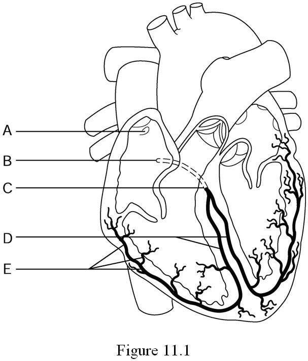
1.atria contract
2. AV node
3. Bundle of His
4.Purkinje fibers
5. SA node
6. ventricles contract

Recording of electrical changes that occur during the cardiac cycle is referred to as
blood pressure reading
ECG
QRS wave
T Wave
Atrial fibrillation is caused the failure of AV node
True
False
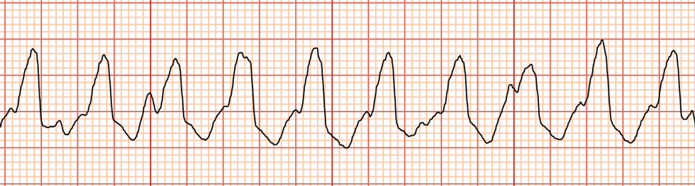
Identify the tracing
Atrial Fibrillation
Sinus Tachycardia
Ventricular tachycardia
Supraventricular tachycardia
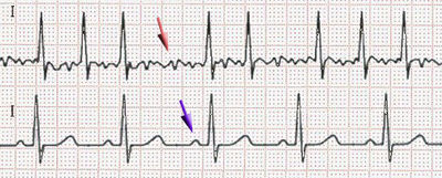
What is the ECG showing (Red Arrow)
Atrial Fibrillation
Ventricular Fibrillation
Asystole
Premature Ventricular Contraction
Heart Block
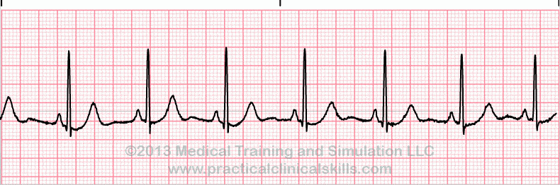
What is the ECG showing
Atrial Fibrillation
Ventricular Fibrillation
normal sinus rhythm
Premature Ventricular Contraction
Heart Block
An abnormal heart rhythm that makes the heart unable to pump blood effectively
cardiac arrest
ventricular fibrillation
asystole
none of the above
s electrical activity between two negative poles
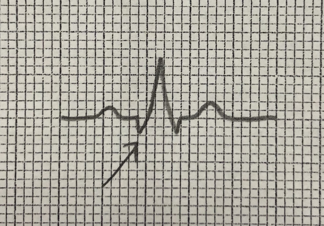
Name this part of the ekg tracing.
P
R
Q
T
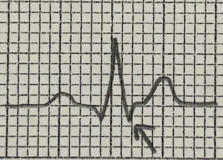
Name this part of the EKG tracing.
P
Q
R
S
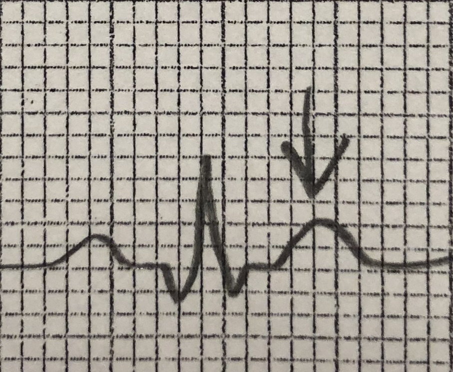
Name this part of the ekg tracing.
Q
R
S
T
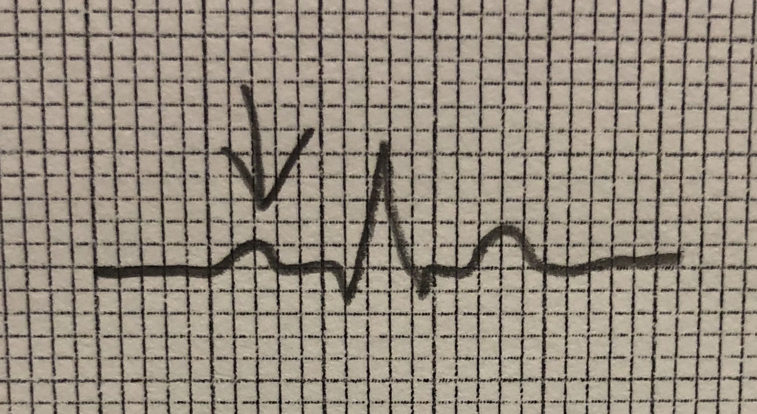
Name this part of the ekg tracing.
P
Q
R
S

