
 Worksheets
Essentials of Healthcare Semester 2 Midterm Review
Total questions: 120
Worksheet time: 5hrs 53mins

What muscle is this?


What are the 3 types of muscle tissue?
Smooth Muscle, Skeletal Muscle, Coronary Muscle
Epithelial Muscle, Skeletal Muscle, Coronary Muscle
Epithelial Muscle, Skeletal Muscle, Cardiac Muscle
Smooth Muscle, Cardiac Muscle, & Skeletal Muscle
Which muscles are involuntary?
skeletal
cardiac
smooth
Which types of muscle tissue are voluntary?
Cardiac
Skeletal
Smooth
Epithelial

Number 9 on the picture is ______.
frontalis
orbicularis oculi
sternocleidomastoid
zygomaticus

Number 4 in the picture is ____________.
zygomaticus
nasalis
masseter
orbicularis oris

Number 5 in the picture is _____.
frontalis
zygomaticus
platysma
masseter

Identify the muscle.
Vastus lateralis
Vastus medialis
rectus femorus
sartorius

Identify the muscle.
vastus lateralis
vastus medialis
sartorius
rectus femorus

Number 15 is the _____.
orbicularis oculi
orbicularis oris
zygomaticus
platysma

Identify the muscle.
masster
temporalis
zygomaticus
sternocleidomastoid

Identify the muscle.
platysma
sternocleidomastoid
tempoalis
zygomaticus

Identify the muscle.
orbicularis oculi
orbicularis oris
temporalis
platysma

Identify the muscle.
frontalis
occipitalis
temporalis
sternocleidomastoid

vastus lateralis
vastus medialis
rectus femoris
sartorius

Identify the muscle.
rectus abdominus
intercostal muscles
erector spinae
platysma

What structure carries filtrate from the renal pelvis to the bladder?
Loop of Henle
Distal convoluted tubule
Ureter
Urethra

Identify
Proximal convoluted tubule
Distal convoluted tubule
Glomerulus
Glomerular capsule

Identify
Proximal convoluted tubule
Distal convoluted tubule
Loop of Henle
Collecting duct


How is the female urethra different from the male urethra?
It is longer
It is wider
It is shorter
It is more narrow
If someone has hematuria, they are experiencing
pus in the urine
blood in the urine
glucose in the urine
urea in the urine
bladder infection
Cystitis
Urethritis
Nephritis
Glomerulonephritis
Requires dialysis & possibly kidney transplant
Renal Calculi
Urethritis
Kidney Failure
Edema
Damage to the filtration portion of the kidney
PKU
Glomerulonephritis
Cystitis
Acute Kidney failure
What disorder is a chronic, degenerative disease in which the nerve cells in the CNS that control voluntary movement degenerate resulting in weakness/muscle atrophy?
ALS
Carpal Tunnel Syndrome
Cerebrovascular Accident
Epilepsy
With this disorder, blood flow to the brain is impaired, resulting in lack of oxygen and destruction of brain tissue.
CVA
ALS
Encephalitis
Shingles
Encephalitis is ____.
inflammation of the brain caused by a pathogen
excessive accumulation of CSF in the ventricles and subarachnoid space
the loss of voluntary muscle movement and coordination in some part of the body
acute inflammation of nerve cells caused by the herpes zoster virus
Abnormal surges in electrical impulses in the neurons of the brain caused by various things (genetics, brain injury, trauma, tumors, toxins, etc) are known as ___________.
epilepsy
CVA
ALS
Carpal Tunnel Syndrome
A progressively painful hand and arm condition caused by the pinching of nerve/tendon that passes through a canal from the forearm to the hands is ___________.
Carpal Tunnel Syndrome
Cerebral Palsy
Meningitis
Parkinson's Disease

What disorder does the picture likely depict? Look at the symptoms listed and the forward leaning posture.
Parkinson's Disease
Epilepsy
Shingles
Paralysis
What happens if hydrocephalus goes untreated?
death
treatment is not necessary
excessive cerebro-spinal fluid will accumulate on the brain
there is no treatment/cure

What type of paralysis is depicted?
paraplegia
quadriplegia
hemiplegia

What type of paralysis is depicted?
paraplegia
quadriplegia
hemiplegia

What is this a picture of?
Shingles
CVA
Epilepsy
Neuralgia


Contains the medulla oblongata, pons and midbrain.
diencephalon
brain stem
cerebrum
cerebellum
protects the brain
skull
meninges
cerebrum
cerebrospinal fluid
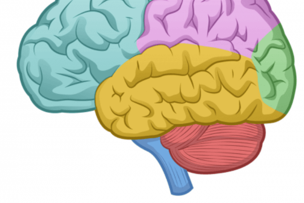
What structure is red in this picture?
Cerebrum
Cerebellum
Brain Stem
Corpus Callosum
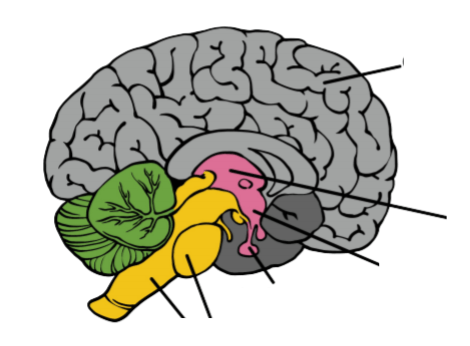
What structure is grey in this picture?
Cerebrum
Cerebellum
Brain Stem
Reticular Formation
Carries messages between the peripheral nervous system and the brain:
cerebellum
corpus callosum
spinal cord
special thalamus

Identify the human cranial nerve A.
Trigeminal
Olfactory
Abducens
Oculomotor

Identify the human cranial nerve I.
Trigeminal
Olfactory
Abducens
Oculomotor

Identify the human cranial nerve D.
Trigeminal
Olfactory
Abducens
Oculomotor
Identify the human cranial nerve B.
Vestibulocochlear
Optic
Abducens
Oculomotor
Identify the human cranial nerve H.
Vestibulocochlear
Optic
Accessory
Trochlear
Identify the human cranial nerve G.
Trigeminal
Optic
Accessory
Hypoglossal
Identify the human cranial nerve F.
Facial
Vagus
Accessory
Hypoglossal
Identify the human cranial nerve J.
Facial
Vagus
Accessory
Hypoglossal
Identify the human cranial nerve L.
Facial
Vagus
Glossopharyngeal
Hypoglossal
Identify the human cranial nerve K.
Facial
Vagus
Glossopharyngeal
Hypoglossal
Identify the human cranial nerve C.
Facial
Vagus
Glossopharyngeal
Trochlear
Identify the human cranial nerve E.
Vestibulocochlear
Vagus
Glossopharyngeal
Trochlear

This cranial nerve innervates the muscles of facial expression
trigeminal
abducens
facial
vagus
This structure is the outer coating of the front of the eye ball.
Cornea
Lens
Retina
Optic Nerve
This structure is located behind the iris and bends light onto the retina.
Retina
Lens
Optic Nerve
Pupil
This structure sends images to the brain so we see the image correctly.
Iris
Optic Nerve
Cornea
Lens
This structure is the black, center part of the eye. Light enters the eye here.
Iris
Optic Nerve
Pupil
Lens
Light travels in a __________ line until it refracts off an object.
Curved
Straight
Zig Zag
purple
This structure is the colored part of the eye.
Pupil
Iris
Retina
Optic Nerve
The path light takes through the eye....
Cornea, Retina, Optic Nerve, Iris, Pupil, Lens
Cornea, Iris, Optic Nerve, Pupil, Lens, Retina
Cornea, Iris, Pupil, Lens, Optic Nerve, Retina
Cornea, Iris, Pupil, Lens, Retina, Optic Nerve,
Rods help us see...
in color
black, white, and grey
only in yellow
only in blue
Cones help us see...
color
black, white, and grey
only purple
only with glasses on

What is letter I pointing to?
Cornea
Iris
Optic Nerve
Pupil

What is letter G pointing to?
Cornea
Iris
Optic Nerve
Lens

What is letter F pointing to?
Lens
Iris
Pupil
Optic Nerve

What is letter B pointing to?
Optic Nerve
Retina
Iris
Pupil
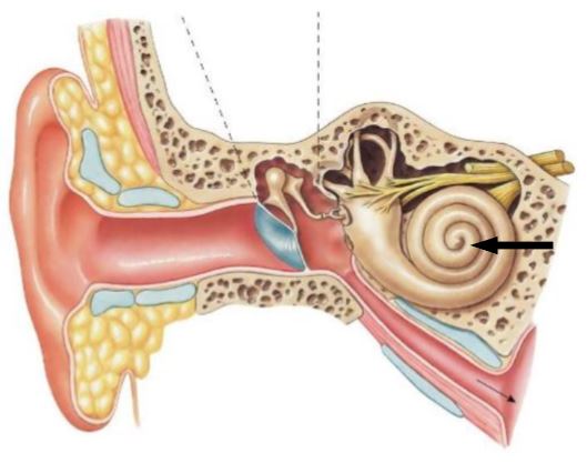
What is this?
Pinna
Hearing Nerve
Cochlea
Ear Drum
Anvil
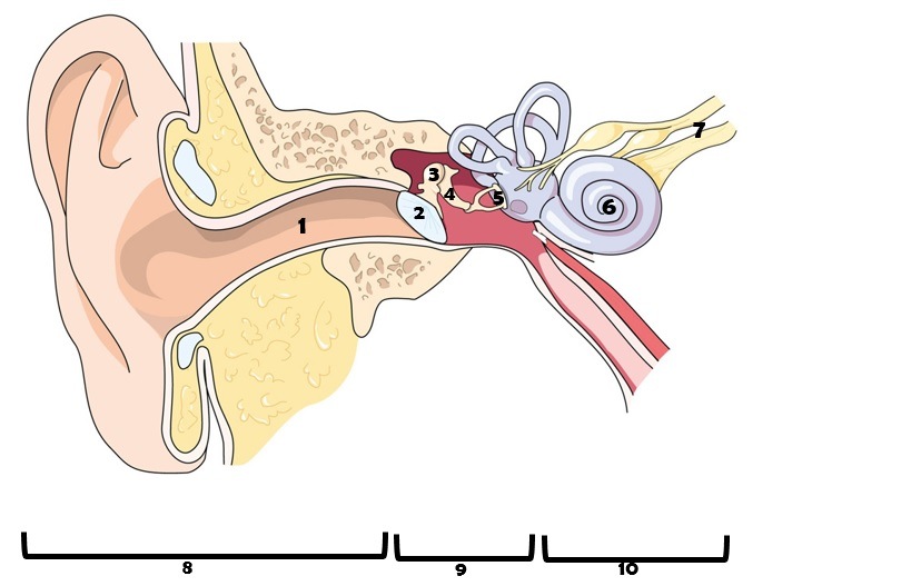
The ear drum is labeled as which number in the diagram?
1
2
3
4
5
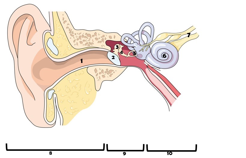
Identify the part of the ear labeled as 1 in the diagram.
auditory nerve
ear canal
eardrum
hammer

