
 Worksheets
Cardiovascular System
Total questions: 91
Worksheet time: 52mins


Arteries carry oxygen rich blood away from the heart.
True
False
Veins carry oxygen poor blood back to the heart.
False
True
How does the heart work?
It pumps blood first to the lungs, back to the heart and then around the rest of the body.
It pumps blood first to the rest of the body, back to the lungs and then to the heart.
It pumps blood around the rest of the body, back to the heart and then to the lungs,

Our heart is.....
a tissue
a body system
a group of cells
an organ

These are between the atria and ventricles that allow blood to flow through the chambers
Pulmonary Circulation
Valves
Atria
Ventricles
Septum
Small vessels that carry blood from arterioles and to small vessels called venules which then empty into veins
Arterioles
Arteries
Veins
Venules
Capillaries
The cusps (leaflets) of atrioventricular valves attach directly to
papillary muscles.
trabeculae carneae
chordae tennidae
interatrial septa
The visceral pericardium is the same as the
epicardium
mediastinum.
parietal pericardium.
The right ventricle pumps blood to the
right and left lungs
left ventricle.
left atrium.
aorta.
The left ventricle pumps blood to the
lungs.
right ventricle.
right atrium.
aorta
The bicuspid or mitral valve is located
in the opening of the aorta.
in the opening of the pulmonary trunk.
where the venae cavae join the right atrium
between the left atrium and the left ventricle

Identify the structure labeled "6."
cusp of tricuspid valce
bicuspid valve
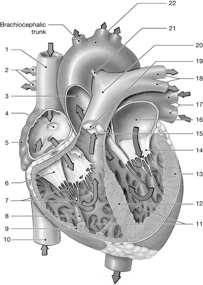
Identify the structure labeled "8."
papillary muscle
moderator band
pectinate muscles
Compared to arteries, veins
are more elastic.
have more smooth muscle in their tunica media
hold their shape better when cut.
have thinner walls
After blood leaves the capillaries, it enters the
arteries.
arterioles.
venules.
Tachy-
Slow
Rapid
Around
Vessel
Structure around the heart
Transcardia
Circumcardia
Pericoronal
Pericardium
Hematopoiesis
Structure of blood
Dilation of blood
Production of blood
Protection of blood
What would happen if a small hole developed in the septum?
The heart would stop pumping.
Blood would move between the right and left side of the heart.
The blood from the pulmonary artery would enter the aorta.
Blood would not flow into the coronary arteries
After blood leaves the right atrium, what valve prevents back flow?
tricuspid
mitral
pulmonary
aortic
What follows diastole?
phagocytosis
fibrillation
hyperbole
systole
Where does an electrical impulse go after it leaves the Bundle of His?
SA node
aorta
AV node
Purkinje fibers
What type of blood vessel is the MOST muscular and elastic?
artery
vein
venule
capillary
Why is a myocardial infarction life threatening?
A damaged heart muscle cannot pump blood efficiently.
Swollen veins lose elasticity, which decreases blood flow to vital organs.
A minor bump or fall could lead to internal bleediing.
It inhibits the exchange of oxygen and carbon dioxide in body cells.
What is the name of the circulation circuit that circulates blood to the body?
Pulmonary circuit
Hepatic circuit
Systemic circuit
Respiratory circuit
Blood pressure is the greatest in which vessel?
Artery
Vein
Capillaries
Aorta
What is known as the "pacemaker" because it initiates the heartbeat?
Bundle of His
AV node
SA node
Purkinje fibers
Blockage of a coronary artery that cuts off the supply of blood to the heart is known as?
Hypertension
Arteriosclerosis
Congestive Heart Failure
Myocardial Infarction
Valve between the left ventricle and aorta
Pulmonary valve
Aortic valve
Mitral valve
Bicuspid valve
Outer layer of the heart
Endocardium
Pericardium
Epicardium
Myocardium
Period of heart contraction, top number in BP
Diastole
Systole
Endocardium
Embolus
Smooth layer of cells lining the inside of the heart
Pericardium
Myocardium
Endocardium
Epicardium
Valve between the left atrium and left ventricle
Mitral valve
Pulmonic valve
Aortic valve
Femoral valve
Muscular middle layer of the heart
Pericardium
Myocardium
Endocardium
Epicardium
What does depolarization result in?
relaxation
contraction
repulsion
attraction
What does repolarization result in?
relaxation
contraction
attraction
repulsion
Intrinsic electrical impulses start at which of the following locations?
AV Node
Bundle of His
Purkinje fibers
SA Node
The AV node is located between which two parts of the heart?
right atria, left atria
atria, ventricle
left ventricle, left ventricle
epicardium, pericardium
Which of the following is most likely to occur if there is a blockage to the AV bundle nerve impulse?
the AV Node will not produce electrical impulses
the SA Node will not produce electrical impulses
no effect
the Purkinje fibers will not produce electrical impulses
The last place electrical impulses move through the heart is located where?
the AV Node
the Purkinje fibers
the SA Node
the AV Bundle
Where are the electrical signals for the conduction process created?
SA Node
AV Node
Purkinje Fibres
Bundles of His
What is the second structure that electrical signals pass through in the conduction process?
AV Node
SA Node
Purkinje Fibres
Bundles of His
Depolarization of a heart chamber such as the left atrium means the left atrium has
contracted
relaxed
1.atria contract
2. AV node
3. Bundle of His
4.Purkinje fibers
5. SA node
6. ventricles contract
What controls the decrease of the SA Node firing rate?
Central Nervous System
Sympathetic Nervous System
Parasympathetic Nervous System
What is another word for AV bundle?
Bundle of HIS
AV node
Pacemaker
Purkinje Fibers
Where is the SA Node located?
Anterior Right Atrium
Posterior Right Atrium
Anterior Left Atrium
Posterior Left Atrium
Which structures sends the electrical stimulus to the purkinje fibres?
SA Node
Bundles of His
AV Node
Purkinje Fibres
Which structure allows a delay in the signal between the atria and ventricles, allowing ventricles to fill with blood before contracting.
SA Node
AV Node
Purkinje Fibres
Bundles of His
What is the correct sequence of electrical conduction of the heart?
AV node, internodal pathways, SA node, Bundle of his, right and left bundle branch, Purkinje fibers.
SA node, internodal pathways, AV node, Purkinje fibers, right and left bundle branches, Bundle of his.
SA node, internodal pathways, AV node, Bundle of his, right and left bundle branches, Purkinje fibers.
None of the options are correct.
Which is located within the walls of the ventricles?
Purkinje fibers
SA node
AV node
AV bundle and branches

