
 Worksheets
Human Heart and Heart Conduction System
Total questions: 32
Worksheet time: 2hrs 49mins
Name
Class
Date
1.

The pointed, inferior portion of the heart, known as the ________ rests on the diaphragm and is oriented towards the left hip.
a)
base
b)
mediastinum
c)
apex
d)
pericardium
2.
The two superior receiving chambers of the heart are known as the _________
a)
atria
b)
ventricles
3.
The valves between the atria and ventricles are known as the ________ valves.
a)
atrioventricular
b)
aortic
c)
semilunar
d)
pulmonary
4.
Blood leaves the right ventricle through the ___________.
a)
pulmonary trunk
b)
aorta
c)
superior vena cava
d)
coronary sinus
5.
The term that means heart contraction is ________.
a)
diastole
b)
systole
c)
tachycardia
d)
fibrillation
6.
Large blood vessels that carry blood away from the heart are called:
a)
arteries
b)
capillaries
c)
veins
d)
venae cavae
7.
Arteries are normally depicted as red while veins are colored blue. The exceptions to this rule are the _______ arteries and veins.
a)
systemic
b)
hepatic
c)
coronary
d)
pulmonary
8.
Pulmonary circulation involves blood flow to and from the heart and the _______.
a)
body
b)
digestive organs
c)
brain
d)
lungs
9.
When the ventricles contract, the bicuspid (mitral) valve prevents blood from flowing from the ________.
a)
right ventricle to right atrium
b)
left ventricle to left atrium
c)
left atrium to the right atrium
d)
right atrium to the left atrium
10.
The sinoatrial node is located in the ________.
a)
aorta
b)
right atrium
c)
left atrium
d)
right ventricle
11.
Which one of the following represents the correct path for the transmission of an impulse in the intrinsic conduction system of the heart?
a)
AV node, Bundle of His, SA node, Purkinje fibers, bundle branches
b)
SA node, AV node, Bundle of His, bundle branches, Purkinje fibers
c)
SA node, Bundle of His, AV node, Purkinje fibers, bundle branches
d)
SA node, Bundle of His, bundle branches, Purkinje fibers, AV node
12.
Exchanges between blood and tissue cells occur in capillary beds.
a)
True
b)
False
13.
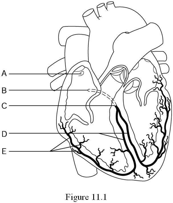
Which label is representing the Purkinje fibers?
a)
A
b)
C
c)
D
d)
E
14.

What does letter D represent?
a)
Aorta
b)
Left Atrium
c)
Left ventricle
d)
Right Atrium
15.

Separates the chambers of the heart.
a)
mitral valve
b)
septum
c)
aortic valve
16.

Unoxygenated blood enters the right atrium from the top of the heart.
a)
inferior vena cava
b)
superior vena cava
c)
aorta
17.

name of the valve that separates right atrium from right ventricle.
a)
tricuspid
b)
aortic
c)
mitral
18.

Unoxygenated blood enters the right atrium from the bottom of the heart.
a)
inferior vena cava
b)
superior vena cava
c)
pulmonary veins
19.

Name of the valve that separates left atrium from left ventricle.
a)
mitral valve
b)
tricuspid
c)
pulmonary valve
20.

Major artery at the top of the heart sends oxygenated blood from the left ventricle to the rest of the body.
a)
aorta
b)
pulmonary artery
c)
inferior vena cava
21.

A flap of tissue that prevents blood from flowing backward
a)
Aorta
b)
Atrium
c)
Septum
d)
Valve
22.

Where does blood go after the right atrium?
a)
left atrium
b)
left ventricle
c)
right ventricle
d)
aorta
23.
name of the valve that separates right atrium from right ventricle.
a)
tricuspid
b)
aortic
c)
mitral
24.

Which chamber in the heart is the strongest?
a)
Left Atrium
b)
Left Ventricle
c)
Right Atrium
d)
Left Descending Coronary Artery
25.
TYPE of blood flow that cycles from the right ventricle to the lungs and back to the left atrium
a)
systemic circulation
b)
pulmonary circulation
26.
TYPE of blood flow that cycles to the body tissues; allows for oxygen to be distributed to all the cells of the body.
a)
systemic circulation
b)
pulmonary circulation
27.
First part of Cardiac Conduction System: This *PACEMAKER* sends action potentials to both atria causing them to contract.
a)
Sinoatrial (SA) Node
b)
AV node
28.
Second part of Cardiac Conduction System: This structure receives a signal and passes it on to the Bundle of His
a)
Purkinje fibers
b)
Atrioventricular (AV) node
c)
Sinoatrial (SA) Node
29.
Third part of Cardiac Conduction System: This structures branches down from septum and out towards each ventricle. They *relay* the signal to the Purkinje fibers.
a)
Atrioventricular (AV) node
b)
Sinoatrial (SA) Node
c)
Bundle of His
d)
Purkinje fibers
30.
A echocardiogram is a tool that is used to:
a)
Measure the electrical events in a heart beat
b)
Measure the movement of blood through the heart using sound waves
c)
Measure the movement of blood through the heart using X-rays and specific dyes
d)
Measure the oxygenation of your blood
31.
Which is a measure of the electrical events that occur in a single heart beat?
a)
Electrocardiogram
b)
Echocardiogram
c)
Angiogram
d)
Magnetic Resonance Imagery (MRI)
32.
These blood vessels are the first branches of the aorta and feed the heart with freshly oxygenated blood:
a)
Pulmonary Arteries
b)
Plumonary Veins
c)
Vena Cava
d)
Coronary Arteries
100 %

