
 Worksheets
Lab Exam 1 - A&P2
Total questions: 70
Worksheet time: 47mins
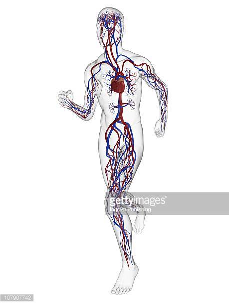
What is the main organ of the circulatory system?
small intestine
bone
lungs
heart
Arteries carry oxygen rich blood away from the heart.
True
False
Veins carry oxygen poor blood back to the heart.
False
True
The heart is about the size and shape of your . . .
head
fist
hand
foot


What is the correct path through the circulatory system which describes the passage of a blood clot originating in the left leg?
Vena cava → left atrium → left ventricle → lungs → right atrium → right ventricle → aorta
Vena cava → right atrium → right ventricle → lungs → left atrium → left ventricle → aorta
Vena cava → left atrium → right atrium → lungs → left ventricle → right ventricle → aorta
Vena cava → right atrium → left atrium → lungs → right ventricle → left ventricle → aorta
What divides the left side of the heart from the left side?
Atrium
Ventricles
Wall
Septum
Rank the following blood vessels in order of their average pressure, from highest to lowest: artery, vein, arteriole, venule, aorta, capillary.
Capillary > arteriole > venule > artery > vein > aorta
Capillary > vein > venule > arteriole > artery > aorta
Aorta > arteriole > venule > artery > vein > capillary
Aorta > artery > arteriole > capillary > venule > vein
How can the circulatory system promote heat retention/conservation, such as on a cold day?
Vasoconstriction
Vasodilation
Decreasing tunica media contraction
Increasing capillary surface area

Identify:
Left Ventricle
Right Atrium
Right Ventricle
Left Atrium

Identify:
Vena Cava
Aorta
Pulmonary veins
Pulmonary artery
What do red blood cells do?
Carry oxygen from the lungs to the body cells and transport carbon dioxide from the cells back to the lungs
Collects blood from the capillaries and drain it into the veins
Keeps blood flowing in one direction to the heart and prevent blood from flowing backwards
Bring nutrients to the cells and carries away waste materials
What valve is found in between the left atrium and left ventricle?
Aortic Semilunar Valve
Pulmonary Semilunar Valve
Tricuspid
Mitral
When blood exits the left ventricle, it must go through which valve?
Aortic
Pulmonic
Mitral
Tricuspid
The part of the cardiac conduction system that goes down the interventricular septum is the:
AV node
SA node
Bundle of His
Purkinje Fibers
What portion of the conduction system is known as the pacemaker?
AV node
Purkinje fibers
SA node
Bundle of His
Contraction of the heart is also know as:
Diastole
Systole
The semilunar valves are the:
Aortic and pulmonary
Pulmonary and Tricuspid
Aortic and mitral
Mitral and Tricuspid
The string like feature that attaches to and helps close the AV valves is the:
Chordae tendinae
Papillary muscles
Interventricular septum
Foramen ovale

Structure C is the _______.
Left atrium
Right atrium
Right ventricle
Left ventricle
Chordae tendinae
Structure S is the _______.
Tricuspid valve
Right ventricle
Myocardium
Left ventricle
Pericardium
Structure F is the _______.
Tricuspid valve
Bicuspid valve
Aortic valve
Pulmonary valve
Aorta
Structure K is the _______.
Left pulmonary artery
Right pulmonary artery
Aorta
Left ventricle
Left pulmonary vein
Structure Q is the _______.
Right atrium
Right ventricle
Aorta
Left ventricle
Left pulmonary vein
Structure J is the
Right atrium
Right ventricle
Aorta
Left ventricle
Superior vena cava
Structure I is the
Inferior vena cava
Superior vena cava
Aorta
Left ventricle
Right ventricle
Structure G is the
Inferior vena cava
Superior vena cava
Aorta
Left ventricle
Right ventricle
Structure D is the ______.
Right pulmonary vein
Superior vena cava
Aorta
Left ventricle
Right pulmonary artery
Structure O is the ______.
Right pulmonary vein
Superior vena cava
Aorta
Left ventricle
Mitral valve
Structure N is the ______.
Right pulmonary vein
Tricuspid valve
Aorta
Aortic valve
Mitral valve
Structure A is the ______.
Right pulmonary vein
Inferior vena cava
Aorta
Aortic valve
Superior vena cava

Structure BCE is the _______
Left atrium
Right atrium
Left ventricle
Right ventricle
Structure C is the _______
Right pulmonary artery
Right atrium
Aorta
Left pulmonary vein
Structure BE is the _______
Superior vena cava
Right atrium
Aortic arch
Left pulmonary vein
Structure AC is the _______
Right ventricle
Right atrium
Aortic arch
Left atrium
Structure BC is the _______
Left common carotid artery
Aorta
Superior vena cava
Left pulmonary artery
Structure CDE is the _______
Left common carotid artery
Apex
Right ventricle
Inferior vena cava
The five different types of leukocytes are neutrophils, basophils, eosinophils, lymphocytes, and __________.
triglycerides
monocytes
thrombocytes
erythrocytes
Mrs. Smith has been diagnosed with hypertension. This means that the upper number in her blood pressure reading is over 140mmHg. The upper number in a blood pressure reading is called __________.
pulse pressure
systolic
pulse
diastolic
The main function of leukocytes are to ____________.
become sticky and create a clot
transport oxygen from the lungs to the tissues
protect the body against infection
create clotting proteins
Lower than normal number of red blood cells.
anemia
phlebotomist
medical lab tech
sickle cell anemia
pulse oximeter
Genetic disorder that causes abnormally shaped hemoglobin
anemia
phlebotomist
medical lab tech
sickle cell anemia
pulse oximeter
The formed elements in blood are red blood cells, white blood cells, and platelets.
true
false
It is important to know both the blood type and the Rh factor before giving blood to a patient.
true
false
When a blood vessel is damaged, platelets become sticky and clump together to form a clot.
true
false
Neutrophils make up the majority of white blood cells in the body.
true
false
This blood type has “a” antigens on the surface of the RBC and “b” antibodies floating in the plasma:
Type O
Type B
Type AB
Type A
This blood type is considered the “universal recipient”:
Type O
Type B
Type AB
Type A
The sac that surrounds the outside of the heart is called the endocardium.
true
false
There are three chambers in the heart.
true
false
The chambers that receive blood into the heart are called ventricles.
true
false
On an EKG the contractions of the atria is represented by the ______________.
T wave
PR interval
QRS complex
P wave
On an EKG the contractions of the ventricles is represented by the ______________.
T wave
PR interval
QRS complex
P wave
On an EKG the relaxation of the ventricles is represented by the ______________.
T wave
PR interval
QRS complex
P wave
The largest blood vessel in the body is the _________.
aorta
superior vena cava
inferior vena cava
femoral artery
Which of the following is/are a major risk factor(s) for a heart attack?
stress
lack of physical activity
age
high cholesterol diet

