
 Worksheets
The Elbow Joint
Total questions: 50
Worksheet time: 5mins

What is the name of the motion in the picture?
Pronation
Supination
Elbow flexion
Elbow extension

What is the name of the motion in the picture?
Pronation
Supination
Elbow flexion
Elbow extension

What is the name of the motion in the picture?
Pronation
Supination
Elbow flexion
Elbow extension

What is the name of the motion in the picture?
Pronation
Supination
Elbow flexion
Elbow extension

The angle formed by the longitudinal axes of the humerus and forearm.
Pronation
Supination
Elbow flexion
Elbow extension
Carrying angle
The normal carrying angle in females is:
Between 10 and 15 degrees
Between 5 and 10 degrees
5 degrees
Between 15 and 20 degrees
The normal carrying angle in males is:
Between 10 and 15 degrees
Between 5 and 10 degrees
5 degrees
Between 15 and 20 degrees

The raised portion on the inferior lip of the glenoid fossa that provides attachment for the long head of the tricep muscles.
Infraglenoid Tubercle
Supraglenoid Tubercle
Supraspinatus fossa
Infraspinatus fossa
The raised portion on the superior lip of the glenoid fossa that provides attachment for the long head of the bicep muscle.
Infraglenoid Tubercle
Supraglenoid Tubercle
Supraspinatus fossa
Infraspinatus fossa

What is labeled #1 in the picture?
Trochlea
Capitulum
Medial Epicondyle
Lateral Epicondyle
Lateral Supracondylar Ridge
What is labeled #2 in the picture?
Trochlea
Capitulum
Medial Epicondyle
Lateral Epicondyle
Lateral Supracondylar Ridge
What is labeled #3 in the picture?
Trochlea
Capitulum
Medial Epicondyle
Lateral Epicondyle
Olecranon Fossa
What is labeled #4 in the picture?
Trochlea
Capitulum
Medial Epicondyle
Lateral Epicondyle
Olecranon Fossa
What is labeled #5 in the picture?
Trochlea
Capitulum
Medial Epicondyle
Lateral Epicondyle
Olecranon Fossa
What is labeled #6 in the picture?
Trochlea
Capitulum
Medial Epicondyle
Lateral Epicondyle
Olecranon Fossa

Which bony land mark is located at the proximal end of the ulna, on the posterior surface; forms the prominent point of the elbow and provides attachment for the tricep muscles? (Hint: The top part the the "U")
Olecranon Procession
Coronoid Process
Radial Notch
Ulnar Tuberosity
Styloid Process
Which bony land mark is located just below the trochlear notch; with the ulnar tuberosity, provides attachment for the brachialis muscle? (Hint: "the bottom part of the "U")
Olecranon Procession
Coronoid Process
Radial Notch
Ulnar Tuberosity
Styloid Process
Which bony land mark is labeled #3 in the picture?
Olecranon Procession
Coronoid Process
Radial Notch
Ulnar Tuberosity
Styloid Process
Which bony land mark is labeled #5 in the picture?
Olecranon Procession
Coronoid Process
Radial Notch
Ulnar Tuberosity
Styloid Process
Which bony land mark is labeled #6 in the picture?
Olecranon Procession
Coronoid Process
Radial Notch
Ulnar Tuberosity
Styloid Process
Which bony land mark is at the distal end on the lateral surface; the ulnar notch of the radius pivots around it during pronation and supination?
Olecranon Procession
Coronoid Process
Radial Notch
Head of the ulna
Styloid Process

Which bony landmark is labeled #1 in the picture?
Radial head
Ulnar head
Radial tuberosity
Radial styloid process
Ulnar styloid process
Which bony landmark is labeled #2 in the picture?
Radial head
Ulnar head
Radial tuberosity
Radial styloid process
Ulnar styloid process
Which bony landmark is labeled #3 in the picture?
Radial head
Ulnar head
Radial tuberosity
Radial styloid process
Ulnar styloid process

Which ligament attaches on the medial epicondyle of the humerus and runs obliquely to the medial sides of the coronoid process and olecranon process of the ulna?
Medial collateral ligament
Lateral collateral ligament
Annular ligament
Joint capsule

Which ligament attaches proximally on the lateral epicondyle of the humerus and distally on the annular ligament and the lateral side of the ulna.
Medial collateral ligament
Lateral collateral ligament
Annular ligament
Joint capsule
Which ligament attaches anteriorly and posteriorly to the radial notch of the ulna, encompassing the head of the radius and holding it against the ulna?
Medial collateral ligament
Lateral collateral ligament
Annular ligament
Joint capsule

What attaches around the distal end of the humerus and encompasses the trochlea and capitulum, and the fossas located above them.
Medial collateral ligament
Lateral collateral ligament
Annular ligament
Joint capsule

Origin: Distal half of humerus, anterior surface Insertion: Coronoid process and ulnar tuberosity of the ulna Action: Elbow flexion Nerve: Musculocutaneous
Brachialis
Biceps brachii
Brachioradialis
Triceps brachii
Anconeus

Origin: Long head: supraglenoid tubercle of scapula Short head: coracoid process of scapula Insertion: Radial tuberosity of radius Action: Elbow flexion, forearm supination Nerve: Musculocutaneous
Brachialis
Biceps brachii
Brachioradialis
Triceps brachii
Anconeus

Origin: Long head: supraglenoid tubercle of scapula Short head: coracoid process of scapula Insertion: Radial tuberosity of radius Action: Elbow flexion, forearm supination Nerve: Musculocutaneous
Brachialis
Biceps brachii
Brachioradialis
Triceps brachii
Anconeus

Origin: Long head: infraglenoid tubercle of scapula Lateral head: inferior to greater tubercle on posterior humerus Medial head: posterior surface of humerus Insertion: Olecranon process of ulna Action: Elbow extension Nerve: Radial
Brachialis
Biceps brachii
Brachioradialis
Triceps brachii
Anconeus

Origin: lateral epicondyle of humerus Insertion: Lateral and inferior to olecranon process of ulna Action: Assists in elbow extension Nerve: Radial
Supinator
Pronator quadratus
Brachioradialis
Pronator teres
Anconeus

Origin: medial epicondyle of humerus and coronoid process of ulna Insertion: lateral aspect of radius at its midpoint Action: forearm pronation, assistive in elbow flexion Nerve: Median
Supinator
Pronator quadratus
Brachioradialis
Pronator teres
Anconeus
Origin: Distal one-fourth of ulna Insertion: Distal one-fourth of radius Action: Forearm pronation Nerve: Median
Supinator
Pronator quadratus
Brachioradialis
Pronator teres
Anconeus

Origin: Lateral epicondyle of humerus and adjacent ulna Insertion: Anterior surface of the proximal radius Action: Forearm supination Nerve: Radial
Supinator
Pronator quadratus
Brachioradialis
Pronator teres
Anconeus
This injury is when the radius and ulna move out of place and separate posteriorly. This is usually caused by falling on an outstretched hand.
Elbow dislocation
Ulnar Collateral ligament tear
Medial epicondylitis
Lateraal epicondylitis
Little league elbow
This injury is when there is pain and inflammation on the inside of the elbow and caused by repetitive flexion of the wrist. Also known as "golfer's elbow".
Elbow dislocation
Ulnar Collateral ligament tear
Medial epicondylitis
Lateraal epicondylitis
Little league elbow
This injury is when there is pain and inflammation on the outside of the elbow and caused by repetitive extension of the wrist. Also known as "tennis elbow".
Elbow dislocation
Ulnar Collateral ligament tear
Medial epicondylitis
Lateraal epicondylitis
Little league elbow
This injury primarily affects the growth plate on the medial side of the elbow and is caused by overuse of the elbow.
Elbow dislocation
Ulnar Collateral ligament tear
Medial epicondylitis
Lateraal epicondylitis
Little league elbow
This injury is a tear to the ligament that connects the inside of your upper arm (humerus) to the inside of your forearm (ulna) and helps support and stabilize your arm.
Elbow dislocation
Ulnar Collateral ligament tear
Medial epicondylitis
Lateraal epicondylitis
Little league elbow
This injury is a break in the bone at the smallest part of the humerus bone. This is also the most common fracture in children. These fractures can be caused when you fall on your elbow or getting directly hit on it.
Supracondylar fracture
Ulnar Collateral ligament tear
Medial epicondylitis
Radial fracture
Ulnar fracture
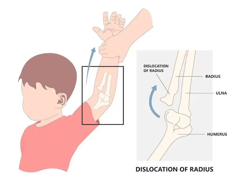
When the radial head is dislocated from the elbow
Nursemaid's elbow
Olecranon bursitis
Ulnar collateral ligament tear
Little league elbow
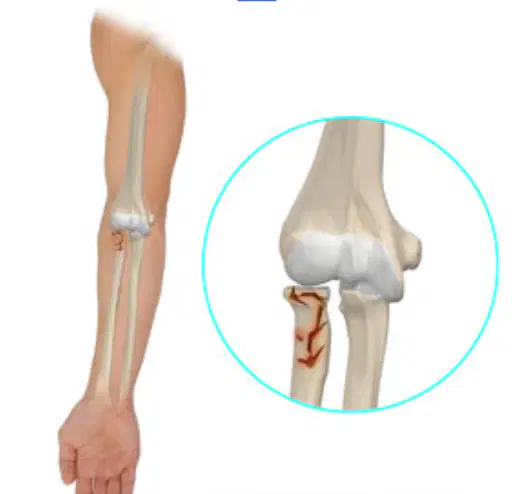
Fracture in the proximal (top) end of the radius, near the circular area of the bone
Nursemaid's elbow
Olecranon bursitis
Radial head fracture
Little league elbow
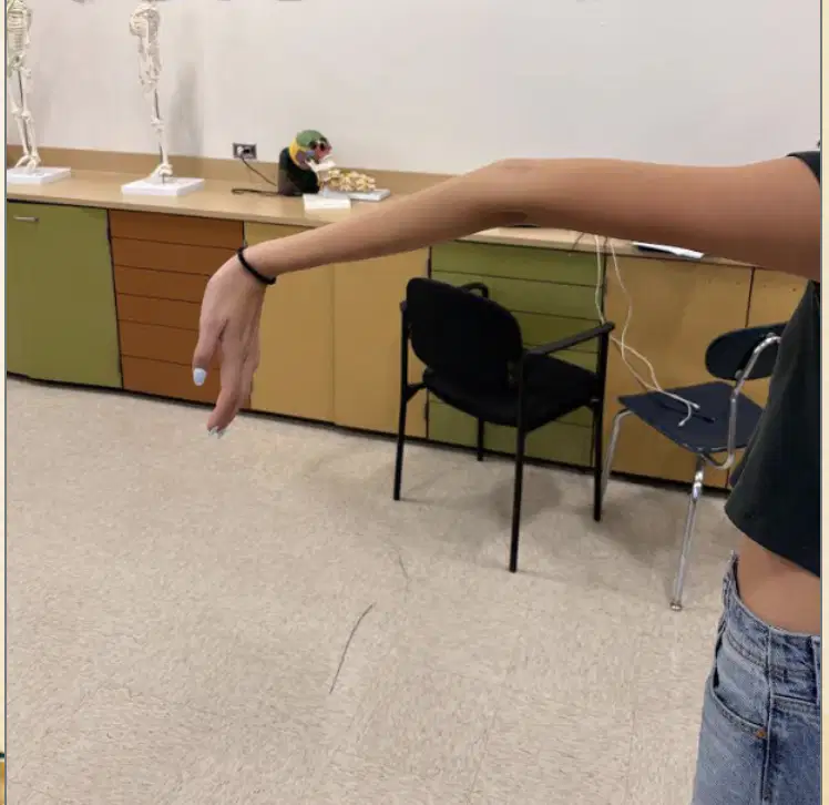
When the elbow moves beyond its normal range of motion (180 degrees)
Lateral epicondylitis
Elbow hyperextension
Radial head fracture
Meidal epicondylitis
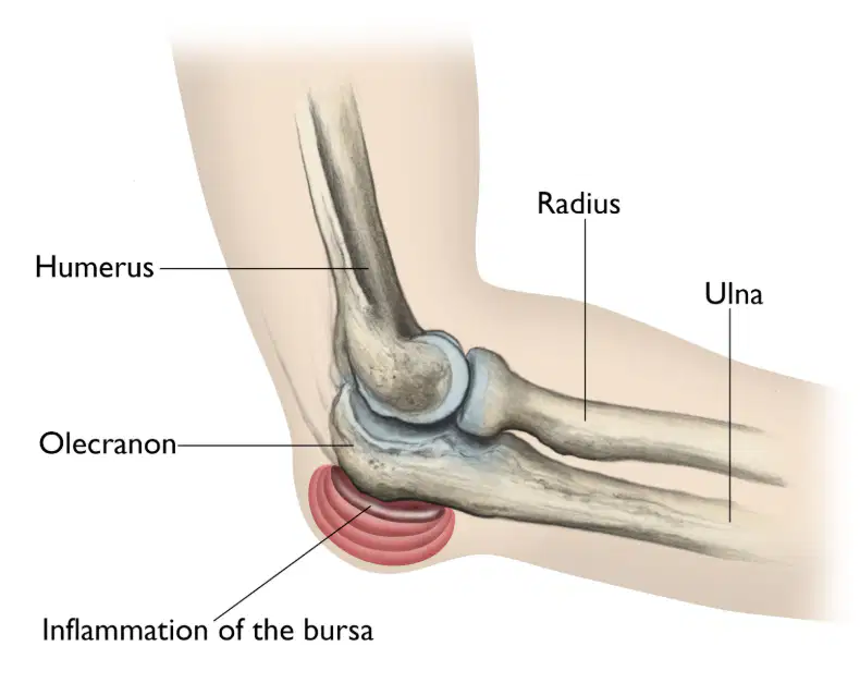
Inflammation of the bursa on the tip of the elbow (olecranon process)
Olecranon bursitis
Elbow hyperextension
Radial head fracture
Meidal epicondylitis
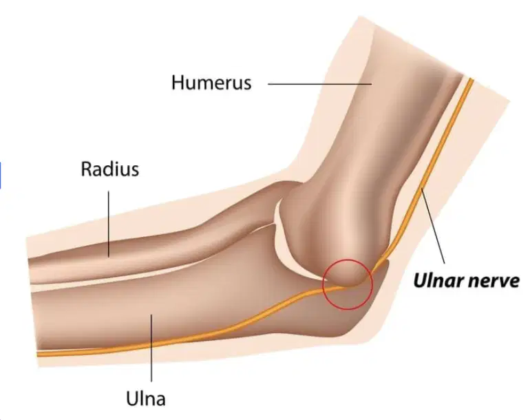
When the ulnar nerve gets irritated or compressed in the cubital tunnel (between the humerus and ulna).
Medial Collateral ligament tear
Elbow hyperextension
Cubital tunnel syndrome
Meidal epicondylitis
What ligament is torn and reconstructed in Tommy John surgery?
Ulnar collateral ligament
Radial collateral ligament
Annular ligament
Joint capsule
The Tommy John surgery is called "Tommy John" because...
Tommy John was the first baseball player to get the surgery
Tommy John was the first surgeon to perform the surgery
Tommy John was the most famous baseball player in history
Tommy John won the record for the most wins in baseball history
What tendon is typically used to reconstruct the UCL in Tommy John surgery?
Palmaris longus
Radial collateral ligament
Annular ligament
Extensor longus tendon

