
 Worksheets
Basic Cardiac Rhythm
Total questions: 50
Worksheet time: 52mins
The nurse is reviewing a patient’s electrocardiogram. Which wave should the nurse recognize as being the first positive wave in a normal cardiac cycle?
P wave
Q wave
R wave
S wave
A patient is experiencing electrocardiogram changes. Which wave should the nurse recognize as representing ventricular repolarization?
Q wave
R wave
S wave
T wave
After reviewing an electrocardiogram the nurse determines that an electrical impulse originated in a patient’s the sinoatrial node. What did the nurse see on this tracing?
An upright T wave
An inverted T wave
A positive P wave before a QRS complex
A negative P wave before a QRS complex
The nurse is determining the functioning of a patient’s sinoatrial node. How many times per minute should the nurse expect the node to normally fire?
20 to 40
25 to 55
40 to 85
60 to 100
The nurse determines that a patient’s electrocardiogram is demonstrating normal sinus rhythm. What did the nurse observe to come to this conclusion? (Select all that apply.)
Regular rhythm
P wave before each QRS complex
Wide QRS intervals
Heart rate 88 beats per minute

Lead II is the standard lead in viewing the EKG.
True
False
A common cardiac dysrhythmia characterized by premature heartbeats originating in the atria.
A-Fib
A-Flutter
PAC's
SA Block
Arrhythmia resulting from impulses coming from a locus of tissue in the area of the AV node, the "junction" between atria and ventricles.
1st Degree AV Block
Junctional Rhythm
2nd Degree AV Block Type I
2nd Degree AV Block Type II
Occurs when the natural pacemaker site shifts between SA node, the atria, and/or the AV node.
1st Degree AV Block
SA Block
Sinus Pause
Wandering Atrial Pacemaker
Rate: 150 – 250 bpm
Rhythm: regular
Accelerated Junctional Rhythm
ST
Accelerated IVR
SVT
The paper on an ECG normally moves at:
50mm/sec
75mm/sec
25cm/sec
25mm/sec
The width of each small box represents:
0.04 seconds
0.02 seconds
0.40 seconds
0.20 seconds
The small boxes on ECG paper measure:
1 cm by 0.20 seconds
1mm by 0.20 seconds
1cm by 0.04 seconds
1mm by 0.04 seconds
The big boxes on the ECG paper measure:
5mm by 0.20 seconds
1mm by 0.20 seconds
5mm by 0.04 seconds
1mm by 0.04 seconds
A distance of 2 big boxes and 2 little boxes wide is described as being:
22mm wide
0.22 seconds wide
0.48 seconds wide
4.8 seconds wide
In reference to the electrical activity of the heart, what signals the end of the cardiac cycle?
Atrial depolarization
Atrial repolarization
Ventricular depolarization
Ventricular repolarization
The period of time from the onset of the P wave to the beginning of the QRS complex.
Diastole
Contraction
Relaxation
PR Interval
Greater than 0.12 seconds is considered abnormal.
1st Degree AV Block
2nd Degree AV Block
3rd Degree AV Block
Bundle Branch Block
Used to calculate ventricular rate
6 Second Strip
P Wave
PR Interval
R-R Interval
Seen as a result of the repolarization of the ventricles.
P Wave
T Wave
QRS Complex
ST Segment
Used for irregular rhythms
1500
300
6 Second Strip
R-R Interval

Using the 6 second strip method, what is the HR of this EKG?
50
52
55
58
What does the P wave on the ECG tracing represent?
Contraction of the atria
Contraction of the ventricles
Depolarization of the atria
Depolarization of the ventricles
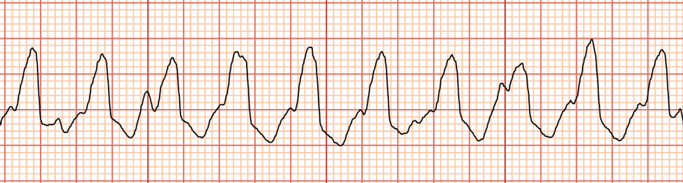
Identify the tracing
Atrial Fibrillation
Sinus Tachycardia
Ventricular tachycardia
Supraventricular tachycardia
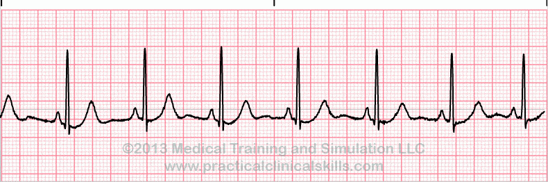
What is the ECG showing
Atrial Fibrillation
Ventricular Fibrillation
normal
Premature Ventricular Contraction
Heart Block

Interpret this Rhythm:
1st Degree AV Block
Idioventricular Rhythm
STEMI
Sinus Bradycardia

Interpret this Event:
PVC's
Multifocal PVC's
PAC's
Couplets

Interpret this Rhythm:
Atrial Fibrillation
Wenckebach
Mobitz
Muscle Artifact
Atria are irritable and quivering, causing fibrillatory waves between QRS complex
asystole
atrial flutter
A fib
V fib
Atria firing at very fast rate, causing flutter waves between normal QRS complexes
asystole
atrial flutter
atrial fib
V tach
Every other beat is a PVC
A fib
Atrial Flutter
Asystole
bigeminy
Everything is normal except the PR interval is greater then .20 seconds
Second Degree Heart Block
Third Degree Heart Block
First Degree Heart Block
Complete Heart Block
Only wide and bizarre complexes at a very slow rate
First Degree Heart Block
Sinus Bradycardia
PVC's
Idioventricular
Every beat on the strip has an absent or inverted P wave
Idioventricular
Junctional Rhythm
PJC
PAC
Premature or early beat that has an absent or inverted P wave
PAC
PJC
PVC
Premature pacemaker spike
Premature early beat that is wide and bizarre
PAC
PJC
PVC
Premature heart block
PR interval gets progressively longer and longer until there is a P wave and no QRS
First Degree Heart Block
Second degree Block Type I (Wenckebach/Mobitz I)
Second degree Block Type II (Classical/Mobitz II)
Third degree Block
PR interval is constantly the same, but some P waves have no QRS
First Degree Block
Second Degree Block Type I (Wenckebach/Mobitz I)
Third Degree Block
Second Degree Block Type II (Classical/MobitzII)
R to R interval will change (be irregular) in a cycle, not just one beat
Sinus bradycardia
Sinus arrhythmia
Sinus tachycardia
Sinus pacemaker
Everything is normal except rate is less than 60
Sinus bradycardia
Sinus tachycardia
Sinus arrhythmia
Sinus block
Everything is normal except the rate is greater than 100
Sinus arrhythmia
Sinus bradycardia
Sinus tachycardia
Sinus block
Extra P waves visible, PR constantly changes, block between atria and ventricles
First Degree Block
Second Degree Block
Third Degree Block
Fourth Degree Block
Every third beat is a PVC
Bigeminy
Trigeminy
Couplet
Triplet
Three PVC's in a row
Triplet
Trigeminy
Couplet
Bigeminy
Ventricles are irritable and no longer contract but quiver, there are no definite complexes
A fib
V fib
V tach
Atrial flutter
Ventricles are contracting but at a very fast rate, only see wide and bizarre complexes
Atrial tachycardia
Ventricular Fib
Atrial Fib
V tach
PVC's originate from different sites, they look different
multifocal
unifocal
couplet
triplet
PVC originates from the same site and they are identical to each other
Bigeminy
Trigeminy
Multifocal
Unifocal

