
 Worksheets
Comprehensive ECG Interpretation
Total questions: 62
Worksheet time: 31mins

Name the EKG
Normal Sinus Rhythm
Sinus Tachycardia
Sinus Bradycardia
Atrial fibrillation

Name the EKG
Normal Sinus Rhythm
Sinus Tachycardia
Sinus Bradycardia
Atrial fibrillation

Name the EKG
Atrial fibrillation
Atrial flutter
Super ventricular tachycardia
Asystole
Everything is normal, rate is 60 -100
Sinus tachycardia
Sinus bradycardia
Sinus arrhythmia
Normal sinus rhythm
Atria are irritable and quivering, causing fibrillatory waves between QRS complex
asystole
atrial flutter
A fib
V fib
Every other beat is a PVC
A fib
Atrial Flutter
Asystole
bigeminy
Two PVC's in a row
Couplet
Trigeminy
Bigeminy
Triplet
Everything is normal except the PR interval is greater then .20 seconds
Second Degree Heart Block
Third Degree Heart Block
First Degree Heart Block
Complete Heart Block
Premature beat, starts in the atria, but not SA node, normal P wave but different from others
PJC
PVC
Premature pacemaker
PAC
Premature early beat that is wide and bizarre
PAC
PJC
PVC
Premature heart block
PR interval gets progressively longer and longer until there is a P wave and no QRS
First Degree Heart Block
Second degree Block Type I (Wenckebach/Mobitz I)
Second degree Block Type II (Classical/Mobitz II)
Third degree Block
PR interval is constantly the same, but some P waves have no QRS
First Degree Block
Second Degree Block Type I (Wenckebach/Mobitz I)
Third Degree Block
Second Degree Block Type II (Classical/MobitzII)
R to R interval will change (be irregular) in a cycle, not just one beat
Sinus bradycardia
Sinus arrhythmia
Sinus tachycardia
Sinus pacemaker
Everything is normal except rate is less than 60
Sinus bradycardia
Sinus tachycardia
Sinus arrhythmia
Sinus block
Extra P waves visible, PR constantly changes, block between atria and ventricles
First Degree Block
Second Degree Block
Third Degree Block
Fourth Degree Block
Every third beat is a PVC
Bigeminy
Trigeminy
Couplet
Triplet
Ventricles are irritable and no longer contract but quiver, there are no definite complexes
A fib
V fib
V tach
Atrial flutter
Ventricles are contracting but at a very fast rate, only see wide and bizarre complexes
Atrial tachycardia
Ventricular Fib
Atrial Fib
V tach

EKG Interpretation:
Course V-Fib
Fine V-Fib
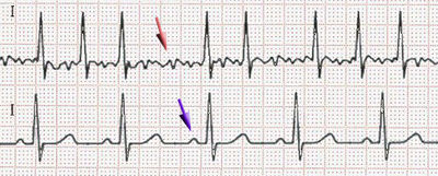
What is the ECG showing (Red Arrow)
Atrial Fibrillation
Ventricular Fibrillation
Asystole
Premature Ventricular Contraction
Heart Block

What is the ECG showing
Atrial Fibrillation
Ventricular Fibrillation
Asystole
Premature Ventricular Contraction
Heart Block

What is the ECG showing
Atrial Fibrillation
Ventricular Fibrillation
normal
Premature Ventricular Contraction
Heart Block

What is the ECG showing
Atrial Fibrillation
Ventricular Fibrillation
normal
Premature Ventricular Contraction
Heart Block

What is the ECG showing (arrow)
Atrial Fibrillation
Ventricular Fibrillation
normal
Premature Ventricular Contraction
Heart Block
Heart rate over 100 beats per minutes is
tachycardia
bradycardia
depolarization
repolarization
Which of these is not considered when making a diagnosis?
P-R interval
rate
rhythm
T wave
When making a diagnosis in the classroom, we are looking at which view?
Lead I
Lead II
Lead III
V1
With sinus dysrhythmia
the rate is abnormal
the rhythm is abnormal
the p wave is abnormal
the t wave is abnormal
In atrial flutter the atria will
not beat at all
beat as often as the ventricles beat
beat 3 or 4 more times than the ventricles
be normal
MI stands for
myocardial infarction
muscle infarction
mycocordial infarction
muscle infraction
If our patient's heart rate is 75bpm, rhythm is regular, P-R interval is normal, QRS is normal, then our diagnosis is
Sinus tachycardia
Sinus dysrhythmia
normal sinus rhythm
Sinus arrest

Interpret this EKG strip
Sinus Rhythm, Hr.70
Sinus Rhythm, Hr.90
Sinus Bradycardia, Hr.50
Sinus Tachycardia, Hr. 110

Interpret this Rhythm:
Normal sinus rhythm
Ventricular fibrillation
Sinus Brady with PAC's
Sinus Bradycardia
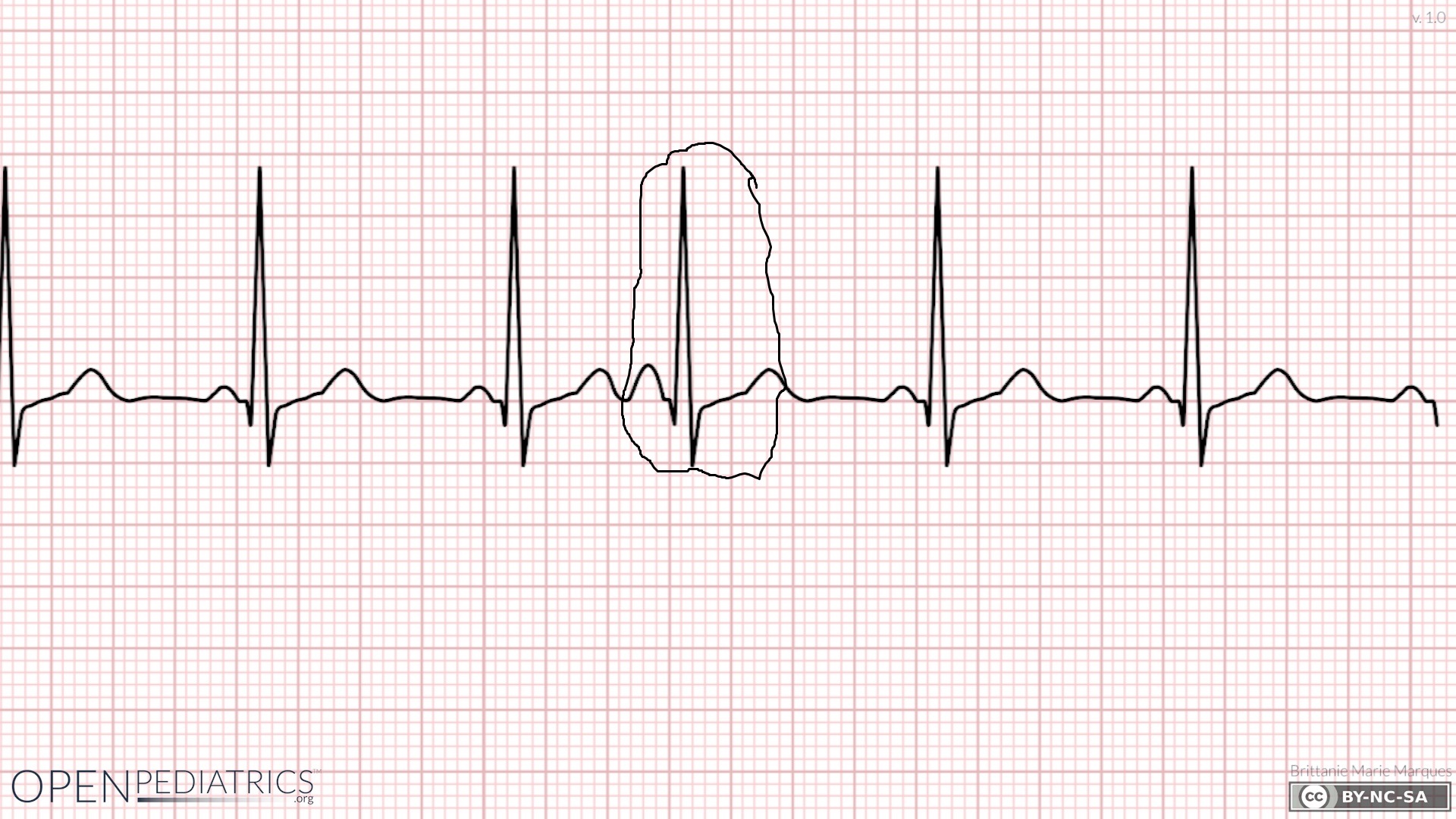
Identify what the rhythm?
Normal sinus rhythm with PAC
Sinus tachycardia with PAC
Normal sinus rhythm with PVC
Sinus tachycardia with PVC

Identify the cardiac rhythm strip pictured above
Ventricular Tachycardia
Normal Sinus Rhythm
Atrial Fibrillation
Sinus Tachycardia
This rhythm is best described as "irregularly irregular".
atrial fibrillation
atrial flutter
premature ventricular tachycardia
supraventricular tachycardia
This is not an actual rhythm, but an ectopic beat that causes the atrial to beat before it should.
atrial fibrillation
atrial flutter
premature atrial contraction
supraventricular tachycardia

Identify the cardiac rhythm strip pictured above
Atrial Flutter
Normal Sinus Rhythm
Atrial Fibrillation
Sinus Tachycardia

Identify the cardiac rhythm strip pictured above. This is a six second strip.
Atrial Flutter
Sinus Tachycardia
Atrial Fibrillation
Supraventricular Tachycardia

Identify the cardiac rhythm strip pictured above
Atrial Flutter
Normal Sinus Rhythm
Atrial Fibrillation
Sinus Tachycardia

Which of the following best describes this strip?
Tachycardia
Normal Sinus Rhythm
Bradycardia
Asystole

The nurse sees the above rhythm on the monitor. What should the nurse do first?
Defibrillate the patient
Initiate chest compressions
Check for the presence of a pulse
Advise the patient not to get out of bed
Name the EKG
Normal Sinus Rhythm
Sinus Tachycardia
Sinus Bradycardia
Atrial fibrillation
The QRS complex is normally __________ seconds in duration
0.06 to 0.10
0.04 to 0.10
0.08 to 0.12
0.10 to 0.12

Name this Rhythm
Torsades
V fib
a fib
agonal

What rhythm is here?
idioventricular
agonal
asystole
sinus bradycardia

What rhythm is here?
idioventricular
agonal
asystole
sinus bradycardia

Which best describes this rhythm?
First-degree AV Block
Second-degree AV Block Type 1
Second-degree AV Block Type 2
Third-degree AV Block

Which best describes this rhythm?
First-degree AV Block
Second-degree AV Block Type 1
Second-degree AV Block Type 2
Third-degree AV Block

Which best describes this rhythm?
Atrial Fibrillation
Atrial Flutter
Asystole
PEA

Which best describes this rhythm?
Atrial Fibrillation
Atrial Flutter
Asystole
PEA

Which best describes this rhythm?
Asystole
PEA
Pulseless Ventricular Tachycardia
Ventricular Fibrillation

Which best describes this rhythm?
Asystole
PEA
Pulseless Ventricular Tachycardia
Ventricular fibrillation

Which best describes this rhythm?
Normal Sinus Rhythm
Sinus Bradycardia
Ventricular Tachycardia
Supraventricular Tachycardia

Identify the waveform after the second heartbeat?
Premature atrial contraction
Sinus Tachycardia
Premature Ventricular Contraction
Asystole

Identify the cardiac rhythm seen above
Ventricular tachycardia
Sinus bradycardia
Atrial Fibrillation
Atrial Flutter
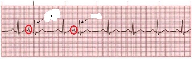
Identify what is occurring at the red circled areas?
Asystole
Normal Sinus Rhythm
Premature Ventricular Contraction
Premature Atrial Contraction
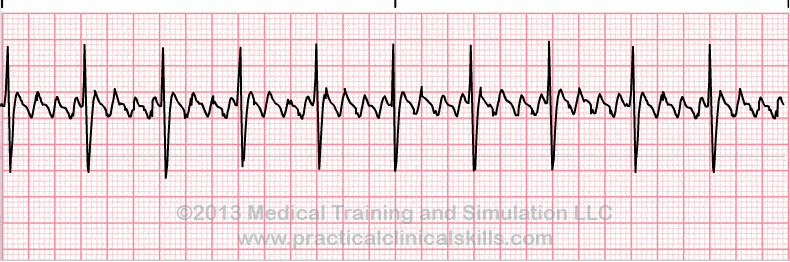
Identify the cardiac rhythm strip seen above:
Ventricular Fibrillation
Atrial Flutter
Ventricular Tachycardia
Normal Sinus Rhythm
The ECG tech notices varying R to R intervals on a patient's ECG. The ECG is otherwise within normal limits.
Call the doctor
This can be normal with breathing
Tell the patient to bear down
Call a code
Which of the following are NOT unipolar?
aVR, aVL, aVF
I, II, III
V1, V2,V3
V4, V5, V6
You have a flat line in leads II and III but normal ECG in other leads. Which of the following is probably loose?
right arm
left arm
left lower
right lower
The ECG is hooked up but all leads seem flat line and patient is awake and talking. Which lead should the tech troubleshoot?
V1
right leg
left leg
right arm

