
 Worksheets
Fall Final Exam Anatomy and Physiology 21-22
Total questions: 212
Worksheet time: 4hrs 32mins
All of the following are characteristics of all living things EXCEPT
Respond to stimulus
Grow and develop
Made of cells
Movement
What is the meaning of Gratitude?
Being kind to people
Letting others know you see how they've helped you
Being truthful and honest in what you say
Loving other people
What is the job of the mitochondria?
Makes Energy
Uses Sunlight in Plants
Creates Protein
Protects the cell
This organelle digests dead cell parts and contains enzymes
lysosome
golgi body
smooth ER
Rough ER

What organelle is represented by this image?
rough endoplasmic reticulum
smooth endoplasmic reticulum
ribosome
nucleus
Which organelle contains DNA and controls the cell?
Cell membrane
nucleus
nucleolus
lysosme

What function does this organelle carry out in the cell?
detoxification
packaging materials for release from the cell
breaking down old cellular components
storing genetic material
Proteins are produced at this organelle.
golgi body
ribosome
cytoskeleton
cilia
This organelle makes useable energy for the cell
chloroplast
mitochondria
ribosome
nucleolus

Which organelle is this?
Golgi Apparatus
Mitochondria
Nucleus
Chloroplast
This organelle makes lipids for the cell.
Rough ER
Smooth ER
Golgi body
nucleus
What organelle controls and regulates cellular activity?
Ribosome
Nucleus
Mitochondria
Lysosome
The ____________ packages and processes material before it exports out of the cell.
Vacuoles
mitochondria
Golgi apparatus
nucleus
What am I? I'm a real "powerhouse". I break down glucose to make ATP.
Cell membrane
ribosome
mitochondria
lysosome
which of the following transports material within the cell?
Golgi apparatus
Endoplasmic reticulum
cell wall
ribosomes
Rough endoplasmic reticulum has ________________ attached to it.
ribosomes
chromosomes
Golgi apparatus
nucleolus

packages and distributes proteins
proteins may be modified here
golgi body
chloroplast
ribosomes
lysosome
What is cell differentiation?
Process by which cells produce more cells
Process by which cells become specialized
Process by which cells become unspecialized
Process by which cells die
What is one goal of cellular differentiation/specialization?
To create specialized cells to help organism maintain homeostasis.
To many cells.
To make stem cells
to make single celled organisms
Stem cells are
cells that grow uncontrollably
undifferentiated and can be any type of cell
never go through cell division
only found in embryos

what is the role of stem cells in the body
fight against infection
provide specialized roles in the body
used for fertility treatment
they produce new specialized cells in the body to replace old cells that are used up
Name the cellular process responsible for repair, growth, and development in living organisms.
meiosis
mitosis
transcription
translation

This type of cell sends messages throughout the body in the form of electrical impulse.
nerve cell
sperm cell
muscle cell
skin cell

This type of cell carries oxygen throughout the body.
gamete
platelet
white blood cell
red blood cell
This type of cell carries oxygen throughout the body.
gamete
platelet
white blood cell
red blood cell

This type of cell fights infection.
red blood cell
platelet
white blood cell
antibacterial cell

This type of cell is found in males and is responsible for reproduction.
sperm cell
egg cell
muscle cell
nerve cell
In what way is the DNA of a nerve cell different from the DNA of a muscle cell?
The DNA is the same but each cell contains different genes
The DNA is the same but different genes are turned on and turned off in each cell
The DNA is different, but the same genes are turned on and off in each cell
The DNA is different, but each cell contains the same genes
All our cells, except sex cells like eggs or sperm, have ___________ DNA
different
similar
exactly the same
half the amount of
A group of similar cells that come together to perform a specific function is a/an
organ
organism
organ system
tissue
Which of the following is the correct order of organization in living things from most basic (simple) to most complex?
a. Organism, cell, tissue, organ, organ system
a. Organ system, organ, tissue, cell, organism
a. Cell, tissue, organ, organ system, organism
a. Cell, organ, tissue, organ, organism, organ system
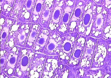
This is a example of ........
cell
tissue
organ
organ system
Most of the cell membrane is made of...
Lipids
Carbs
Proteins
Nucleic acids

Hydrophobic region of the cell membrane?
E
B
C
A
The cell membrane is made up of ????
proteins
atp
adp
proteins and lipids

During what phase of Mitosis do the chromosomes line up in the Middle
Metaphase
Prophase
Telophase
Anaphase
PMAT helps us remember the correct order of the phases of Mitosis. Which of the following has the phases of Mitosis in the correct Order
Interphase, Mitosis, Cytokinesis
Prophase, Metaphase, Anaphase, Prophase
Anaphase, Telophase, Prophase, Telophase
Prophase, Metaphase, Anaphase, Telophase
In which phase of Mitosis do the Chromosomes pull Apart
Metaphase
Prophase
Anaphase
Telophase
_____________ deals with the FORM or STRUCTURE (morphology) of the body and its ORGANIZATION.
Physiology
Anatomy
Body composition
BMI
__________________ studies the FUNCTION of these the parts/structures of the body.
Physiology
Anatomy
Body composition
BMI
_________________ is a condition of a stable internal environment
body composition
gross anatomy
homeostasis
radiographic anatomy
physiology
Axillary is also know as ________
back of the head
between the hips
armpit
belly button
Lumbar is also known as ________.
back of the knee
lower back
cheek
chest
Carpal is also known as ___________.
upper arm
around the eye
chest
wrist
Orbital is also known as _______.
around the mouth
around the eye
cheek
back of the head
Femoral is also known as _______.
the foot
thigh
back of the knee
ankle
The abdominal region is ______ to the pelvic region.
superior
inferior
The skin is _______ to the muscle.
deep
superficial
The ______ plane divides the body into left and right halves.
transverse
frontal
sagittal
The fingers are _____ to the wrist.
proximal
distal
What is the proper order for a negative feedback loop?
Sensor, control, effector, stimulus
control, effector, stimulus, sensor
stimulus, sensor, control, effector
effector, sensor, control, stimulus
Which of the following best describes the anatomical position?
sitting
standing erect with palms facing backward
standing erect with palms facing forward
curled up like a fetus
Which picture below best represents the anatomical position?


Which term best completes the sentence:
The thigh is ___________ to the pelvis (hip)?
deep
superficial
inferior
medial
Which term best completes the sentence:
The heel is ________ to the toes.
lateral
posterior
inferior
anterior
Which term best completes the sentence:
The ears are ___________ to the nose.
distal
superior
lateral
superficial
Which term best completes the sentence:
The ankle is __________ to the knee.
distal
deep
superior
medial
Which term best completes the sentence:
The muscles are ______ to the skin.
anterior
proximal
lateral
deep

What plane is this?
sagittal
frontal
transverse
coronal

What plane is this?
midsagittal
frontal
transverse
parasagittal

What plane is this?
parasagittal
oblique
transverse
frontal
Finish this sentence:
Homeostasis is the ability to maintain a __________ internal environment.
stable
fluctuating
an elevated
decreased

What type of feedback loop is this if it is showing blood sugar levels rising and then decreasing over time?
negative feedback
positive feedback
medial feedback
homeostasis

If this graph is showing homeostasis at approx 100 W/n, then what type of feedback is this showing?
negative feedback
positive feedback
neutral feedback
binary feedback
The key to negative feedback is that the correction is __________ the deviation. Example: the way a thermostat works
amplified
eliminated
opposite
there is no correction
Ventral cavity that houses the lungs.
Thoracic
Abdominal
Pelvic
Cranial

Which cardinal plane is highlighted in green?
Transverse
Sagital
Midsagital
Coronal

What type of view is this?
Midsagital
Transverse
Coronal

Cranial Cavity
Pleural Cavity
Pericardial Cavity
Vertebral Canal

The cavity surrounded by a box is?
Abdominal Cavity
Pelvic Cavity
Pleural Cavity
Cranial Cavity

The cavity surrounded by a box is?
Cranial Cavity
Abdominal Cavity
Pelvic Cavity
Abdominopelvic Cavity

The cavity surrounded by a box is?
Abdominal Cavity
Pelvic Cavity
Thoracic Cavity
Pericardial Cavity

The cavity surrounded by a box is?
Pericardial Cavity
Pleural Cavity
Abdominal Cavity
Pelvic Cavity

This type of tissue lines the internal organs and covers the outside of the body as skin
epithelial
connective
muscular
nervous

This type of tissue includes ligaments, tendons, and cartilage
epithelial
connective
muscular
nervous
Epithelial tissue is
Bone, cartilage,dense connective tissue, loose connective tissue, and blood
Highly specialized tissue that contracts or shortens to produce movement
Lining and covering tissue of the body such as skin
Which of the following is a type of muscle tissue
Skeletal
Cardiac
Smooth
All of the above
Blood is an example of what type of tissue?
muscle
epithelial
nervous
connective
Adipose tissue, cartilage, and ligaments are what type of tissue?
muscle
connective
nervous
epithelial
This type of tissue covers the internal organs and also makes up the skin
epithelial
connective
muscular
nervous
This type of tissue includes ligaments, tendons, and cartilage
epithelial
connective
muscular
nervous

The shaft of the bone is also called the
epiphysis
diaphysis
endosteum
periosteum
The hardest, most dense part of the bone tissue is the
periosteum
spongy bone
osteoblast
compact bone

What are functions of the skeleton?
Protection
Blood cell production
Movement
All of these
This type of bone is solid throughout except for the central canals and lacunae.
Compact Bone
Spongy or Cancellous Bone
Which of the following is filled with yellow marrrow?
medulary cavity
spongy bone
periosteum
compact bone
If cartilage remains in this "specific" area, then growth is still occurring; it is located at the end of long bone
Epipyseal plate
Medulary Cavity
Diaphysis
Endosteum
The ENDS of the long bone are anatomically referred to as the:
Epiphyseal plate
Medulary Cavity
Periosteum
Epiphysis

Identify 1:
Proximal Epiphysis
Diaphysis
Distal Epiphysis
Epiphyseal Line

Identify 2
Proximal Epiphysis
Diaphysis
Distal Epiphysis
Epiphyseal Line

Identify 4
Articular Cartilage
Proximal Epiphysis
Distal Epiphysis
Epiphyseal Line
Identify 5, the type of bone tissue with trabeculae is called
Spongy Bone
Compact Bone
Articular Cartilage
Epiphyseal Line
Identify the thin plate labeled #6.
Spongy Bone
Compact Bone
Articular Cartilage
Epiphyseal Line
Structure 7 is the outer covering of the bone made up of fibrous connect tissue called
Medullary Cavity
Compact Bone
Yellow Marrow
Periosteum
Identify 9, the hollow cavity inside the shaft of the bone.
Medullary Cavity
Compact Bone
Yellow Marrow
Periosteum

Structure #4 is a single unit of bone, called a/an
osteon
osteocyte
lacuna
osteoblast

Mature bone cells that live in tiny cavities within the bone are called?
osteocytes
osteoblasts
osteoclasts
osteons
Which of these is a long bone?
Femur
Patella
Scapula
Carpal

Structure #3 is a/n
osteon
perforating (Volkmann's) canals
Central (Haversian) canals
canaliculi

Structures #5, which are situated within structures #8, are
osteons
lamellae
lacunae
osteocytes

Structures #6 are
osteons
lamellae
lacunae
canaliculi

Structures #8, the cavities surrounding structures #5, are the
osteons
lamellae
lacunae
osteocytes
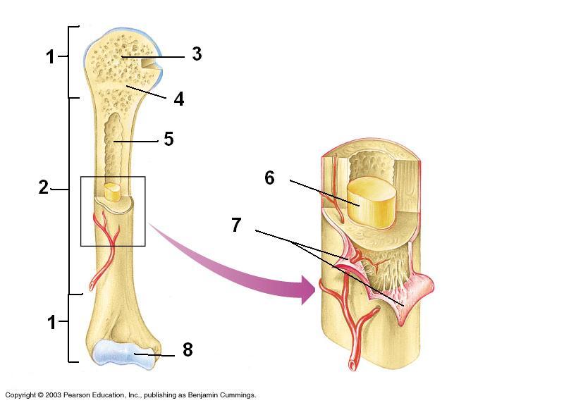
Structure #7 is the
medullary cavity
periosteum
epiphysis
diaphysis
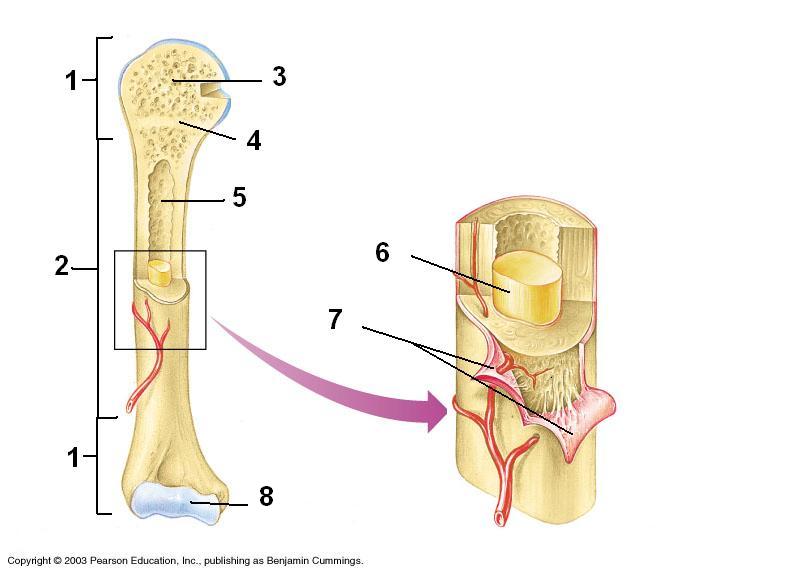
In adults, the substance at #6 would be
red marrow
yellow marrow
spongy bone
clotted blood

Number 8 is the
maxilla
mandible
frontal bone
parietal bone

Number 1 is the
parietal bone
occipital bone
frontal bone
temporal bone

Number 19 is the
fibula
tibia
patella
phalanges

Number 21 is the
fibula
tibia
femur
phalanges

Number 18 is the
fibula
tibia
femur
phalanges

Number 11 is the
radius
ulna
humerus
femur

Number 10 is the
scapula
sternum
pelvis
frontal bone

Number 25 is the
scapula
sternum
clavical
mandible

Bones 4, 5, and 6 are all
phalanges
metacarpals
carpals
tarsals

Bones numbered 3 are
carpals
metatarsals
metacarpals
phalanges

A, B, C & D are
tarsals
metatarsals
phalanges
carpals

E represents
Phalanges
tarsals
metatarsals
fibula

The number of bones in the human skeleton is
260
206
266
602
mandible
cheek bone
jaw bone
ankle bone
shoulder bone
clavicle
collar bone
cheek bone
shoulder bone
tail bone
patella
cheek bone
tail bone
hip bone
kneecap
scapula
collar bone
shoulder bone
hip bone
tail bone
carpal
breast bone
ankle bone
wrist bone
tail bone
frontal bone
forehead
cheek bone
back of the head
neck bone

bone highlighted in green
sphenoid
occipital
parietal
ethmoid

suture highlighted in red
coronal
squamosal
lamboidal
metopic

suture highlighted in red
frontal
sagittal
coronal
squamosal

bone highlighted in green
ethmoid
occipital
coronal
temporal

suture highlighted in red
coronal
lamboidal
sagittal
metopic

suture highlighted in red
lamboidal
frontal
sagittal
squamosal
The following is NOT one of the 3 Joint Classifications..
Cartilaginous
Cartilage
Fibrous
Synovial
The joints that allow the most movement are called
Fixed
Fibrous
Cartilaginous
Synovial
Examples of a Ball & Socket Joint are found in the
Shoulder
Knee
Hip
Ankle
A hinge joint allows
A wide range of movements
Circumduction
Rotation
Movement in only one direction (ie. backwards & forwards)
An example of a Saddle joint is in the
Knee
Hip
Thumb
Vertebral column
When kicking a football the knee
Flexes
Extends
Abducts
Adducts

Identify the joint action being performed.
Extension
Flexion
Abduction
Adduction


Identify the joint action being performed.
Inversion
Eversion
Plantarflexion
Dorsiflexion
Hip and shoulder have examples of this type of joint
ball and socket
condyloid
hinge
gliding
The elbows, knees and phalanges are examples of this type of joint
ball and socket
condyloid
gliding
hinge
Carpals and tarsals (flat bones) are examples of this type of joint
condyloid
gliding
hinge
saddle

The knee joint is
Hinge
Gliding
Ball and socket
Pivot
Fibrous joints are?
freely moveable
partially moveable
immovable

What are joints
A place where one bone meets
A place where two bones meet
Bones don't meet
A place where three bones meet

Turning the hand so that the palm is upward or facing anteriorly (in anatomical position).
supination
pronation
flexion
dorsiflexion
Joints between skull bones
diarthrotic/synovial
fibrous/suture
amphiarthrotic/epiphyseal disk
joint between atlas and axis (hint: they move more than the rest of the vertebral column)
synovial
cartilaginous
fibrous
all examples are diarthrosis
fibrous
cartilaginous
synovial
Pubic symphysis
fibrous/epiphyseal disk
cartilaginous/suture
cartilaginous/symphysis
synovial/symphysis
Often contains a fluid-filled cushion
fibrous
cartilaginous
synovial
Which type of joint gives you the largest range of motion?
Pivot joint
Ball-and-socket joint
Hinge joint
Gliding joint

What joint is displayed in picture
Hinge Joint
Saddle joint
Ball and socket joint

What type of joint is displayed in the picture?
Pivot joint
Ellipsoid joint
Hinge joint

What joint is displayed in the picture?
Synovial Joint
Pivot joint
Hip joint

Which is one of the functional ways a joint is classified
Fibrous—tend to be immovable
Amphiarthroses—slightly movable joints
Synovial –tend to be freely movable

Which is one of the structural ways a joint is classified?
Cartilaginous—both immovable and slightly
movable
Synarthroses—immovable joints
Diarthroses—freely movable joints

Where is the lamboidal suture located
between right and left parietal bones
next to the temporal bones
between the parietal and occipital bones
___are thin filaments.
troponin
tropomyosin
actin
myosin
Calcium binds to ___of the troponin-tropomyosin complex.
troponin
tropomyosin
actin
myosin
___are thick filaments.
actin
myosin
troponin
tropomyosin
Calcium is stored in the ___.
sarcoplasm
sarcolemma
sarcoplasmic reticulum
transverse tubules
A ___extends from one Z disc to another Z disc.
Sarcolemma
Neuromuscular junction
Sarcomere
Motor end plate
___forms a crossbridge with actin.
actin
myosin
calcium
ATP
___gets shorter during contraction.
actin
myosin
troponin-tropomyosin
none of these
___provides energy for muscle contraction and relaxation.
cAMP
ADP
ATP
M line
___slides inward during muscle contraction.
actin
myosin
Thick filaments are in a myofibril are
ATPase
Sarcoplasmic reticulum
Actin
Myosin

The plasma membrane of a skeletal muscle cell is called the:
sarcomere
myofilament
sarcolemma
sarcoplasm
The functional unit of the muscle cell is the:
Sarcomere
Osteon
Myofilament
Muscle fiber
What two chemicals are necessary for muscle contraction to occur?
Niacin and Potassium
Calcium and ATP
Sodium and Potassium
Ach and Potassium
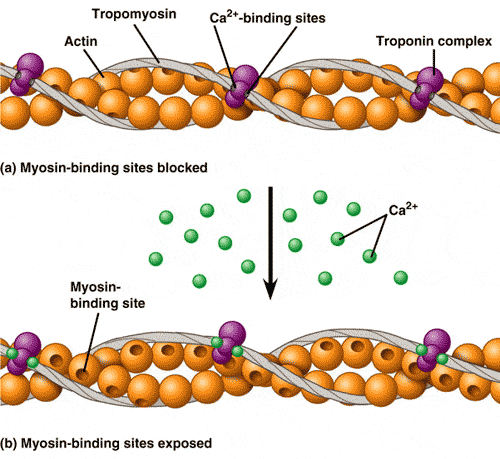
Calcium:
causes ATP binding to actin
increases the action potential along the sarcolemma
binds to proteins on myosin
triggers the binding of myosin to actin
What is a crossbridge?
Where myosin binds to actin
Where the calcium crosses into the cytoplasm
Where calcium binds with troponin
None of the answers are correct.
The energy needed to perform a muscle contraction comes directly from
ATP
Oxygen
Glucose
Calcium
The way in which the structures of the sarcomere work together in order to contract is known as the _______________.
The Sliding Filament Model
Action Potential
Sarcolemme
Muscle Fiber
Rigor Mortis
Skeletal Muscle contraction is initiated when the ________ sends a message to the muscle cell.
Muscle cell
Neuron
Gland
None of the above
The sliding filament theory of muscle contraction says
Actin and myosin filaments condense, thus causing the filament to shorten which in turn shortens the muscle
Myosin rotates and wraps actin around it making the muscle shorter
Friction is reduced between thin and thick filaments causing the thick filaments to move inside the thin
Actin filaments slide over myosin filaments, making the muscle shorter

Which number represents the actin filament?
1
2
3
4

Which number represents the myosin filament?
1
2
3
4

The neurotransmitter stored in the axon terminal identified as #3
Calcium Ion (Ca2+)
Protein Channel
Acetylcholine
Synaptic Cleft
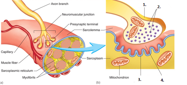
The gap between a neuron and a muscle cell: (#4 in the diagram)
synaptic cleft
sarcolemma
vesicle
Ca2+ ion
Skeletal Muscle contraction is initiated when the ________ sends a message to the muscle cell.
Muscle cell
Neuron
Gland
None of the above
Please select the correct order
1. Muscle cell releases calcium
2. Neurotransmitter releases acetylcholine
3. Sodium & Potassium pump opens
4. Acteylcholine signals cell membrane gates to open
1. Neurotransmitter releases acetylcholine
2. Acteylcholine signals cell membrane gates to open
3. Sodium & Potassium pump opens
4. Muscle cell releases calcium
1. Sodium & Potassium pump opens
2. Acteylcholine signals cell membrane gates to open
3. Muscle cell releases calcium
4. Neurotransmitter releases acetylcholine
1. Neurotransmitter releases acetylcholine
2. Acteylcholine signals cell membrane gates to open
3. Muscle cell releases calcium
4.Sodium & Potassium pump opens

Identify the muscle.
Rectus femoralis
Biceps brachii
Pectoralis major
Gastrocnemius

Abdominal
External Oblique
Erectors
Satorius

Buccinator
Occuli
Pectorals
Soulius

Hamstring
Quadriceps
Bicep
Satorius

Biceps Brachii
Triceps Brachii
Occuli
Trapezius

What muscle is highlighted in the picture?
Triceps Brachii
Biceps Brachii
Quads
Deltoids
Involuntary
skeletal
cardiac
smooth
smooth and cardiac

Trapezius
2
1
18
8
Latissimus dorsi
2
1
14
3

What is the name of this muscle?
Pectoralis major
Deltoid
Pectoralis minor
Triceps brachii

What is the name of this muscle?
Gluteus maximus
Gluteus medius
Gluteus minimus
Piriformis

What is the name of this muscle?
Infraspinatus
Supraspinatus
Subscapularis
Teres minor

What is the name of this muscle?
Semitendinosus
Biceps femoris
Semimembranosus
Rectus femoris

Identify the muscle
Sartorius
Biceps femoris
semitendinousus
Gracilis

Identify the muscle
Gluteus Maximus
Gracilis
Vastus Lateralis
Rectus Femoris

occipitalis
temporalis
frontalis
orbicularis oris

Mastoid
Sternocleidomastoid
Trapezius
Sternohyoid

semimembranosus
semitendinosus
biceps femoris
gluteus medius

Orbicularis Oris
Orbicularis Oculi
Corrugator
Frontalis

Orbicularis Oris
Orbicularis Oculi
Corrugator
Occipitals

Buccinator
Zygomaticus Major
Zygomaticus Minor
Masseter

Bright Red
Occipitalis
Frontalis
Corrugator
Zygomaticus Minor

#10?
orbicularis oculi
zygomaticus
buccinator
orbicularis oris

biceps brachii
triceps brachii
pronator
extensor digitorum

vastus medialis
vastus lateralis
rectus femoris
gracilis

Identify the Muscle.
semitendinosus
vastus medialis
biceps femoris
rectus femoris
vastus lateralis

Identify the Muscle.
semitendinosus
vastus medialis
biceps femoris
rectus femoris
vastus lateralis

rhomboid minor
trapezius
rhomboid major
serratus posterior

What muscle is this?
Deltoid
Masseter
Brachialis
Brachioradialis

What muscle is this?
palmaris longus
flexor carpi ulnaris
pronator teres
extensor carpi ulnaris
extensor digitorum

