
 Worksheets
AP Mod10 Ch16 Quiz
Total questions: 168
Worksheet time: 3hrs 48mins
blood vessels have 3 layers
tunica interna (intima)
tunica media
tunica externa
blood vessel layer that is the innermost layer, adjacent to lumen. made of simple squamous epithelium and is like the endothelium
tunica interna (intima)
tunica media
tunica externa
blood vessel layer that is the middle layer, smooth muscle and elastic fibers with CT
tunica interna (intima)
tunica media
tunica externa
blood vessel layer that is the outermost CT layer, adjacent to surrounding tissue and protects blood vessels
tunica interna (intima)
tunica media
tunica externa

A is
tunica interna
tunica media
tunica externa
B is
tunica interna
tunica media
tunica externa
C is
tunica interna
tunica media
tunica externa
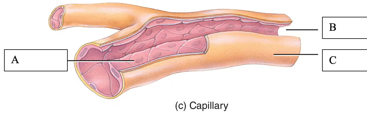
A is
endothelium
lumen
basement membrane
B is
endothelium
lumen
basement membrane
C is
endothelium
lumen
basement membrane
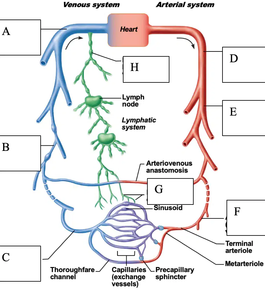
A is
large veins (capacitance vessels)
small veins (capacitance vessels)
postcapillary venule
elastic arteries (conducting arteries)
B is
large veins (capacitance vessels)
small veins (capacitance vessels)
postcapillary venule
elastic arteries (conducting arteries)
C is
large veins (capacitance vessels)
small veins (capacitance vessels)
postcapillary venule
elastic arteries (conducting arteries)
D is
large veins (capacitance vessels)
small veins (capacitance vessels)
postcapillary venule
elastic arteries (conducting arteries)
E is
muscular arteries (distributing arteries)
arterioles (resistance vessels)
lymphatic capillaries
large lymphatic vessels
F is
muscular arteries (distributing arteries)
arterioles (resistance vessels)
lymphatic capillaries
large lymphatic vessels
G is
muscular arteries (distributing arteries)
arterioles (resistance vessels)
lymphatic capillaries
large lymphatic vessels
H is
muscular arteries (distributing arteries)
arterioles (resistance vessels)
lymphatic capillaries
large lymphatic vessels
the large artery that comes out of the heart
aorta
lymphatic capillaries
pull out extra fluid and bring back to vena cava before it gets to the heart
aorta
lymphatic capillaries
carry blood away from the heart to the tissues: the walls of the ___ are elastic which allows them to absorb the pressure created by ventricles of the heart as they pump blood into the ___; because of the smooth muscle in the tunica media, the diameter of ___ can be regulated
arteries
veins
large thick-walled arteries with elastin in all 3 tunics. aorta and its major branches. large lumen = low resistance. inactive in vasoconstriction. act as pressure reservoirs-expand and recoil as blood ejected from heart (smoothes the pressure downstream)
elastic arteries
muscular arteries
arterioles
capillaries
distal to elastic arteries - deliver blood to body organs. thick tunica media with more smooth muscle. active in vasoconstriction
elastic arteries
muscular arteries
arterioles
capillaries
smallest arteries. lead to capillary beds (gas/fluid exchange). control flow into capillary beds via vasodilation and vasoconstriction
elastic arteries
muscular arteries
arterioles
capillaries
microscopic vessels that usually connect arterioles and venules. capillary walls are composed of a single layer of cells and a basement membrane. because their walls are so thin, capillaries permit the exchange of nutrients and wastes between blood and tissue cells
elastic arteries
muscular arteries
arterioles
capillaries
anastomoses = a fusion of 2 arteries that supply the same region of the body (arterial and venous). helps provide an alternate route for blood - like a failsafe. arteries that don't form an anastomosis are called "end arteries" - necrosis (death of a tissue) can occur
true
false
capillaries branch to form an extensive capillary network throughout the tissues and are found near almost every cell in the body
true
false
a muscle that opens and closes an opening in a tube or duct. can contract/relax them to control bloodflow through capillary beds
sphincter
anastomoses
types of capillaries
continuous capillaries
fenestrated capillaries
sinusoid capillaries
veins
types of capillaries that are the most common, least leaky, found throughout body, least porous, tight junctions
continuous capillaries
fenestrated capillaries
sinusoid capillaries
types of capillaries that are more porous/leaky, found in areas that require a lot of exchange between blood and tissues (small intestines, kidneys). fluid can pass through. towards middle of blood vessel
continuous capillaries
fenestrated capillaries
sinusoid capillaries
types of capillaries that are most porous/leaky. when want to move LARGE substances - cells, not just fluid. only found in areas where large particles, including cells, can pass through vessel walls. spleen, liver, bone marrow (think blood cells moving in and out)
continuous capillaries
fenestrated capillaries
sinusoid capillaries
veins and venules are under very low pressure. have to go against gravity to bring blood back to the heart. walls are thinner than arteries. have less muscle, elastic tissue (usually have none), thinner tunica externa. have uni-directional valves inside
true
false
small vessels that are formed by the union of several capillaries. drain blood from capillaries into veins. small veins
veins
venules
formed from the union of several venules. compared to arteries, they have a thinner tunica interna and media and a thicker tunica externa (have less elastic tissue and less smooth tissue than arteries). contain valves. go from small to large
veins
venules
at rest, the largest portion of blood is in systemic veins and venules (blood reservoirs). blood spends much longer time in veins than capillaries because of low pressure
true
false
substances cross capillary walls by:
diffusion
transcytosis
bulk flow
substances such as oxygen, carbon dioxide, glucose (facilitated diffusion through channels), amino acids, and some hormones cross capillary walls via simple ___ (gases. follow high-->low concentration gradient). small particles
diffusion
transcytosis
bulk flow
large, lipid-insoluble molecules (like insulin) cross capillary walls in vesicles via ___. absorbed from inside bloodstream. exocytosis
diffusion
transcytosis
bulk flow
is a passive process in which large numbers of ions, molecules, or particles in a fluid move together in the same direction. occurs from an area of higher pressure to an area of lower pressure, and continues as long as a pressure difference exists. use pressure-fluid. *more important for regulation of the relative volumes of blood and interstitial fluid. largest volume of fluid moved by this
diffusion
transcytosis
bulk flow
pressure-driven movement of fluid and solutes from blood capillaries into interstitial fluid. blood hydrostatic pressure (BHP) - (blood pressure) and interstitial fluid osmotic pressure (IFOP) promote filtration
filtration
reabsorption
pressure-driven movement of fluid and solutes from interstitial fluid into blood capillaries. interstitial fluid hydrostatic pressure (IFHP) and blood colloid osmotic pressure (BCOP) promote reabsorption
filtration
reabsorption
force that drives a fluid through a semi-permeable membrane like the wall of a capillary. "pushing" pressure
BHP
IFOP
IFHP
BCOP
sucking. more concentrated solution is going to draw fluid towards it through osmosis. "pulling" pressure
BHP
IFOP
IFHP
BCOP
osmotic pressure or concentration of a solution is a strong determinant for developing osmotic pressure (pulling pressure)
true
false
osmotic pressure is much higher in blood than in tissue fluid. it draws fluid back in
true
false
at the arterial end, blood pressure is much higher here than at the end of the capillary. net filtration
true
false
at the venous end, blood pressure drops significantly. net reabsorption
true
false
NFP = BP > OP
arterial end
venous end
NFP = HP < OP
arterial end
venous end
under normal conditions, the volume of fluid and solutes reabsorbed is almost as large as the volume filtered. NFP = (BHP + IFOP) - (BCOP + IFHP)
if positive, have outflow-arterial end. if negative, have reabsorption going back into blood at venous end
starling's law of capillaries
factors affecting blood flow
blood flow is the volume of blood that flows through any tissue in a given time period (mL/min). total blood flow is cardiac output (CO), the volume of blood that circulates through systemic (or pulmonary) blood vessels each minue
CO = heart rate (HR) x stroke volume (SV)
CO = mean arterial pressure (MAP) / resistance (R)
contraction of ventricles generates BP. BP is determined by CO, blood volume, and vascular resistance. higher the BP, the greater the blood flow
true
false
arterial blood pressure:
systolic pressure
diastolic pressure
pulse pressure
highest. pressure exerted in aorta during ventricular contraction (systole). avg. 120mm Hg in normal adult
systolic pressure
diastolic pressure
pulse pressure
lowest level of aortic pressure. ventricular diastole. ~80
systolic pressure
diastolic pressure
pulse pressure
difference between systolic and diastolic pressure: S-D. throbbing of arteries (pulse). highest pressure to lowest pressure
systolic pressure
diastolic pressure
pulse pressure
resistance (R): opposition to blood flow due to friction between blood and the walls of blood vessels. the higher it is, the smaller the blood flow. basically anything that counteracts blood flow. R depends on:
size of the blood vessel lumen (constrict muscle). most control over this part
blood viscosity - thicker blood = more blood cells, slower blood will flow. pressure/R will increase. counteract upflow
total blood vessel length
further from heart, the more friction built up, pressure and velocity decrease
true
false
R factors that remain relatively constant: blood viscosity (stickiness of blood due to formed elements and plasma proteins; increased viscosity = increased resistance); blood vessel length (longer vessel = greater resistance encountered); blood vessel diameter (greatest influence on resistance)
true
false
if constrict blood vessel, more R. if dilate BV (double), R drops by 1/16. efficient way of slowing down or speeding up bloodflow in different areas depending on needs
true
false
venous return (preload), the volume of blood flowing back to the heart through the systemic veins, occurs due to the pressure generated by contractions of the heart's left ventricle. it's assisted by:
valves
respiratory pump
skeletal muscle pump
blood flow is the volume of blood that flows through a tissue in a given period of time. blood flow is inversely related to the cross-sectional area of blood vessels. the larger the BV, have low R and velocity will be higher
true
false
more R = increased BP
true
false
the medulla oblongata contains a cardiovascular center, which is a group of neurons that regulate heart rate, contractility, and blood vessel diameter
true
false
barorecptors are important pressure-sensitive sensory neurons that monitor stretching of the walls of blood vessels and the atria
true
false
cardio-accelatory - excitatory sympathetic. cardio-inhibitory - vagus nerve, parasympathetic
true
false
short-term neural and hormonal controls
counteract fluctuations in blood pressure by altering peripheral resistance (vasoconstriction/dilation) and CO (rise = symp.; decrease = parasymp.)
counteracts fluctuations in blood pressure by altering blood volume by retaining or getting rid of fluid
long-term renal regulation
counteract fluctuations in blood pressure by altering peripheral resistance (vasoconstriction/dilation) and CO (rise = symp.; decrease = parasymp.)
counteracts fluctuations in blood pressure by altering blood volume by retaining or getting rid of fluid
increase in fluid =
increase in BP
decrease in BP
decrease in fluid =
increase in BP
decrease in BP
direct renal mechanism alters blood volume independently of hormones
increased BP or blood volume causes elimination of more urine, thus reducing BP
decreased BP or blood volume causes kidneys to conserve water, and BP rises
indirect mechanism: triggered by low BP. renin-angiotensin-aldosterone mechanism (RAA system) will increase BP
lower arterial pressure --> release of renin
renin catalyzes conversion of angiotensinogen from liver to angiotensin I (prohormone - hormone precursor)
angiotensin converting enzyme, especially from lungs, converts angiotensin I to angiotensin II (ACE). mediator. causes vasoconstriction- increase BP
functions of angiotensin II (tense up blood vessels). causes vasoconstriction directly increasing BP. increases blood volume:
stimulates aldosterone secretion
causes ADH release - stop getting rid of water and increase BP
triggers hypothalamic thirst center - increase fluid volume drinking water = increase in retention of water and increase BP
aldosterone tells tubules in kidneys and blood vessels - moves sodium into blood. water follows sodium - "salt sucks"
true
false
autoregulation is the ability of a tissue to automatically adjust its own blood flow to match its metabolic demand for delivery of oxygen and nutrients and removal of wastes. physical and chemical stimuli can lead to autoregulation (alarm chemicals- inflammation: increase bloodflow - vasodilation)
true
false
measurement of BP
systolic blood pressure (SBP)
diastolic blood pressure (DBP)
circulatory routes
systemic circulation
pulmonary circulation
hepatic portal circulation
vena cava are the
smallest veins
largest veins

A is
aorta
superior vena cava
inferior vena cava
internal and external iliac veins
B is
aorta
superior vena cava
inferior vena cava
internal and external iliac veins
C is
aorta
superior vena cava
inferior vena cava
internal and external iliac veins
D is
aorta
superior vena cava
inferior vena cava
internal and external iliac veins
E is
superior mesenteric artery
inferior mesenteric artery
common iliac artery
internal and external iliac arteries
F is
superior mesenteric artery
inferior mesenteric artery
common iliac artery
internal and external iliac arteries
G is
superior mesenteric artery
inferior mesenteric artery
common iliac artery
internal and external iliac arteries
H is
superior mesenteric artery
inferior mesenteric artery
common iliac artery
internal and external iliac arteries
brachiocephalic trunk is only on the __ side (arteries)
right
left
common hepatic artery brings oxygenated blood to the liver
true
false
mesenteric = intestines arteries
true
false

A is
right common carotid
right subclavian
brachiocephalic trunk
ascending aorta
B is
right common carotid
right subclavian
brachiocephalic trunk
ascending aorta
C is
right common carotid
right subclavian
brachiocephalic trunk
ascending aorta
D is
right common carotid
right subclavian
brachiocephalic trunk
ascending aorta
E is
common hepatic
right renal
left common carotid
left subclavian
F is
common hepatic
right renal
left common carotid
left subclavian
G is
common hepatic
right renal
left common carotid
left subclavian
H is
common hepatic
right renal
left common carotid
left subclavian
I is
axillary
left brachial
left gastric
splenic
J is
axillary
left brachial
left gastric
splenic
K is
axillary
left brachial
left gastric
splenic
L is
axillary
left brachial
left gastric
splenic
M is
abdominal aorta
left deep artery of the thigh (deep femoral) and left femoral
left gastric
splenic
N is
abdominal aorta
left deep artery of the thigh (deep femoral) and left femoral
left gastric
splenic
left common carotid and left subclavian come directly off the aorta
true
false

the three major branches off the aorta
brachiocephalic trunk
left common carotid
left subclavian

A is
right common carotid
right subclavian
brachiocephalic trunk
arch of aorta
B is
right common carotid
right subclavian
brachiocephalic trunk
arch of aorta
C is
right common carotid
right subclavian
brachiocephalic trunk
arch of aorta
D is
right common carotid
right subclavian
brachiocephalic trunk
arch of aorta
E is
left subclavian
left common carotid
pulmonary trunk (deoxygenated blood to lungs)
arch of aorta
F is
left subclavian
left common carotid
pulmonary trunk (deoxygenated blood to lungs)
arch of aorta
G is
left subclavian
left common carotid
pulmonary trunk (deoxygenated blood to lungs)
arch of aorta
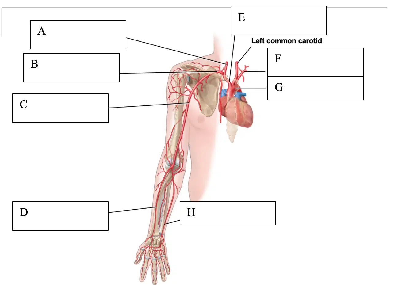
A is
right common carotid
right subclavian
right brachial
right radial
B is
right common carotid
right subclavian
right brachial
right radial
C is
right common carotid
right subclavian
right brachial
right radial
D is
right common carotid
right subclavian
right brachial
right radial
E is
brachiocephalic trunk
left subclavian
arch of aorta
right ulnar
F is
brachiocephalic trunk
left subclavian
arch of aorta
right ulnar
G is
brachiocephalic trunk
left subclavian
arch of aorta
right ulnar
H is
brachiocephalic trunk
left subclavian
arch of aorta
right ulnar
right internal carotid
brain
face/neck
right external carotid
brain
face/neck

A is
right internal carotid
right external carotid
right common carotid
brachiocephalic trunk
B is
right internal carotid
right external carotid
right common carotid
brachiocephalic trunk
C is
right internal carotid
right external carotid
right common carotid
brachiocephalic trunk
D is
right internal carotid
right external carotid
right common carotid
brachiocephalic trunk
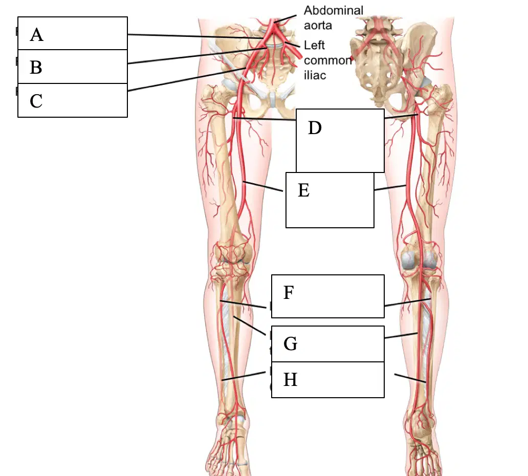
A is
right common iliac
right internal iliac
right external iliac
right deep artery of thigh (deep femoral)
B is
right common iliac
right internal iliac
right external iliac
right deep artery of thigh (deep femoral)
C is
right common iliac
right internal iliac
right external iliac
right deep artery of thigh (deep femoral)
D is
right common iliac
right internal iliac
right external iliac
right deep artery of thigh (deep femoral)
E is
right femoral
right anterior tibia
right posterior tibial
right fibular (peroneal)
F is
right femoral
right anterior tibia
right posterior tibial
right fibular (peroneal)
G is
right femoral
right anterior tibia
right posterior tibial
right fibular (peroneal)
H is
right femoral
right anterior tibia
right posterior tibial
right fibular (peroneal)

A is
right internal jugular
right external jugular
right brachiocephalic
superior vena cava
B is
right internal jugular
right external jugular
right brachiocephalic
superior vena cava
C is
right internal jugular
right external jugular
right brachiocephalic
superior vena cava
D is
right internal jugular
right external jugular
right brachiocephalic
superior vena cava
E is
hepatics
left brachiocephalic
inferior vena cava
left common iliac
F is
hepatics
left brachiocephalic
inferior vena cava
left common iliac
G is
hepatics
left brachiocephalic
inferior vena cava
left common iliac
H is
hepatics
left brachiocephalic
inferior vena cava
left common iliac
I is
left internal iliac
left external iliac
left femoral
left common iliac
J is
left internal iliac
left external iliac
left femoral
left common iliac
K is
left internal iliac
left external iliac
left femoral
left common iliac
veins are
away from the heart
up towards the heart
internal jugular is much __ than the external jugular
smaller
larger
internal jugular is the main vein bringing blood down from the brain
true
false
right and left pulmonary arteries are the only arteries in the body with deoxygenated blood
true
false

A is
right axillary (brachial)
right brachiocephalic
superior vena cava
B is
right axillary (brachial)
right brachiocephalic
superior vena cava (right atria)
C is
right axillary (brachial)
right brachiocephalic
superior vena cava (right atria)

A is
right subclavian
right brachiocephalic
right axillary
right brachial
B is
right subclavian
right brachiocephalic
right axillary
right brachial
C is
right subclavian
right brachiocephalic
right axillary
right brachial
D is
right subclavian
right brachiocephalic
right axillary
right brachial
E is
right radials
superior vena cava
right ulnars
right brachial
F is
right radials
superior vena cava
right ulnars
right brachial
G is
right radials
superior vena cava
right ulnars
right brachial

A is
inferior vena cava
right common iliac
right internal iliac
right external iliac
B is
inferior vena cava
right common iliac
right internal iliac
right external iliac
C is
inferior vena cava
right common iliac
right internal iliac
right external iliac
E is
right femoral
right anterior tibial
right posterior tibial
right great saphenous (not fibular!! - medial side!!)
F is
right femoral
right anterior tibial
right posterior tibial
right great saphenous (not fibular!! - medial side!!)
D is
inferior vena cava
right common iliac
right internal iliac
right external iliac
G is
right femoral
right anterior tibial
right posterior tibial
right great saphenous (not fibular!! - medial side!!)
H is
right femoral
right anterior tibial
right posterior tibial
right great saphenous (not fibular!! - medial side!!)

