
 Worksheets
Chapter 4 Part 4 Images
Total questions: 61
Worksheet time: 31mins
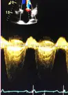
This is a way to find out if the patient has MS or MR, and is also used to calculate the MVA, mean PG and pressure half-time if MS is present.
CWD
PWD
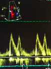
This gives us a sample of inflow at the tips of the MV leaflets and is used for the diastolic function assessment.
CWD
PWD
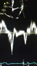
This is a mode of echo that measures the velocity of the heart muscle through the phases of one or more heartbeats by the Doppler effect of the reflected ultrasound
CWD
PWD
Tissue Doppler (TDI)
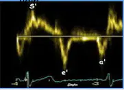
The chambers will affect the velocity and shape of the Doppler inflow signal. This is a relationship between early and late filing, how rapid flow velocity declines in early diastole, and how long it takes for filling of the ventricle to start after the ventricle relaxes.
CWD
PWD
Tissue Doppler to assess diastolic function and dysfunction
TDI
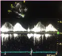
This is to evaluate pulmonary vein flow reversal and the hemodynamics due to MR.
CWD
PWD- PV4
TDI
Tissue Doppler
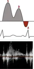
What is going on in this image?
Normal
Systolic Blunting
Flow Reversal
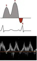
What is going on in this image?
Normal
Systolic Blunting
Flow Reversal
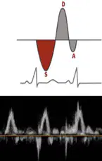
What is going on in this image?
Normal
Systolic Blunting
Flow Reversal
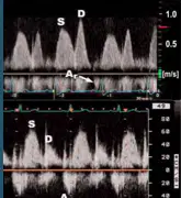
What is S?
Systolic
Diastolic
Time of peak reversal flow (atrial contraction
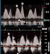
What is D?
Systolic
Diastolic
Time of peak reversal flow (atrial contraction
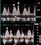
What is Ar?
Systolic
Diastolic
Time of peak reversal flow (atrial contraction
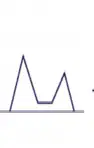
What is the name and stage of this?
Normal, 0
Impaired Relaxation, I
Pseudonormal, II
Restrictive (Reversible), III
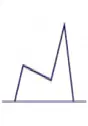
What is the name and stage of this?
Impaired Relaxation, I
Pseudonormal, II
Restrictive (Reversible), III
Restrictive (Non-reversible), IV
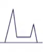
What is the name and stage of this?
Impaired Relaxation, I
Pseudonormal, II
Restrictive (Reversible), III
Restrictive (Non-reversible), IV
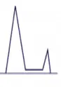
What is the name and stage of this?
Impaired Relaxation, I
Pseudonormal, II
Restrictive (Reversible), III
Restrictive (Non-reversible), IV
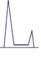
What is the name and stage of this?
Impaired Relaxation, I
Pseudonormal, II
Restrictive (Reversible), III
Restrictive (Non-reversible), IV
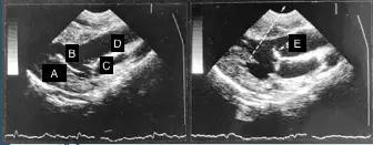
What is A?
PM
LV
LA
AO
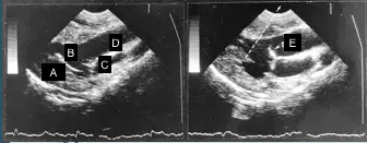
What is B?
PM
LV
LA
AO
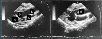
What is C?
LV
LA
AO
SAM
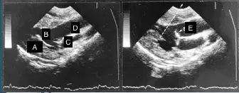
What is D?
LV
LA
AO
SAM
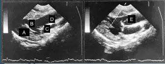
What is E?
LV
LA
AO
SAM
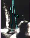
What is 1?
Focus of AOV
Focus of IVRT
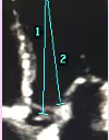
What is 2?
Focus of AOV
Focus of IVRT
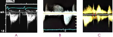
What is A?
CWD-AOV
CWD- AR/AS
CWD-IVRT
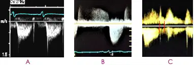
What is B?
CWD-AOV
CWD- AR/AS
CWD-IVRT
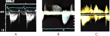
What is C?
CWD-AOV
CWD- AR/AS
CWD-IVRT
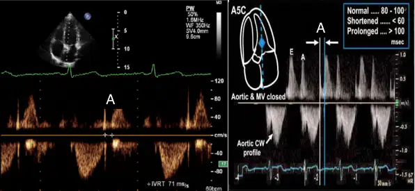
What is A?
IVCT
Systole
Diastole
IVRT
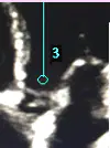
What is 3?
Focus-AOV
Focus- IVRT
PWD LVOT gate
PWD LVIT gate
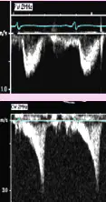
What is the top picture?
PWD- normal LVOT
CWD- LVOTO
PWD LVOT gate
PWD LVIT gate
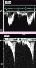
What is the bottom image?
PWD- normal LVOT
CWD- LVOTO
PWD LVOT gate
PWD LVIT gate
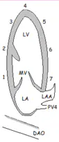
What is 1?
Basal Inferior Wall
Mid Inferior Wall
Apical Inferior Wall
Apical Cap
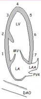
What is 2?
Basal Inferior Wall
Mid Inferior Wall
Apical Inferior Wall
Apical Cap
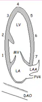
What is 3?
Basal Inferior Wall
Mid Inferior Wall
Apical Inferior Wall
Apical Cap
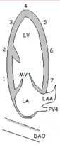
What is 4?
Basal Inferior Wall
Mid Inferior Wall
Apical Inferior Wall
Apical Cap
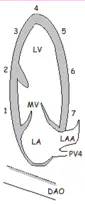
What is 5?
Apical Cap
Apical Anterior Wall
Mid Anterior Wall
Basal Anterior Wall
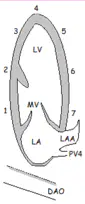
What is 6?
Apical Cap
Apical Anterior Wall
Mid Anterior Wall
Basal Anterior Wall
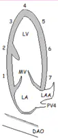
What is 7?
Apical Cap
Apical Anterior Wall
Mid Anterior Wall
Basal Anterior Wall
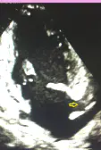
What is the arrow is pointing to?
LAA
LA area
Dilated CS
Length
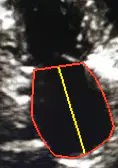
What is the red and yellow?
Red: LA Area
Yellow: Length
Red: Length
Yellow: LA Area
Red: Circumference
Yellow: Length
Red: Length
Yellow: Circumference
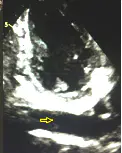
What is the arrow pointing to?
LAX SUPRA
LAX SUB
LAX DAO
LAX AAO
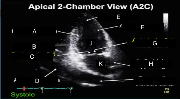
What is A?
Inferior Wall (LV)
Diaphragm
Posterior leaflet mitral valve
Abdominal aorta
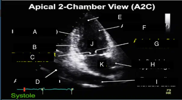
What is B?
Inferior Wall (LV)
Diaphragm
Posterior leaflet mitral valve
Abdominal aorta
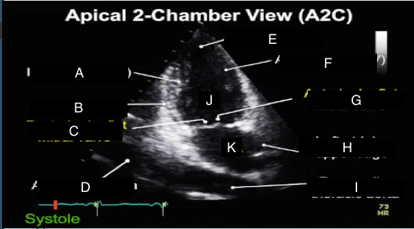
What is C?
Inferior wall (LV)
Diaphragm
Posterior leaflet mitral valve
Abdominal aorta
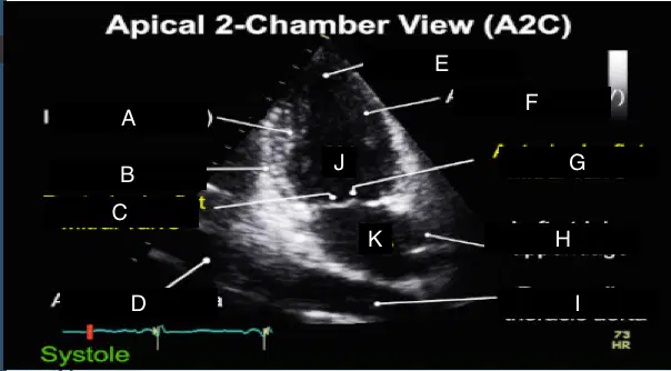
What is D?
Inferior wall (LV)
Diaphragm
Posterior leaflet mitral valve
Abdominal aorta
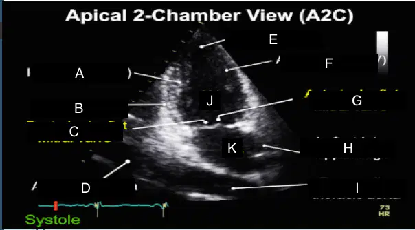
What is E?
LV apex
Anterior wall (LV)
Anterior leaflet mitral valve
Left atrial appendage
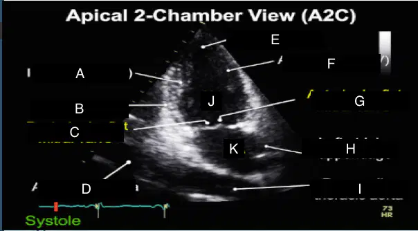
What is F?
LV apex
Anterior wall (LV)
Anterior leaflet mitral valve
Left atrial appendage
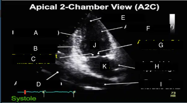
What is G?
Anterior wall (LV)
Anterior leaflet mitral valve
Left atrial appendage
Descending thoracic aorta
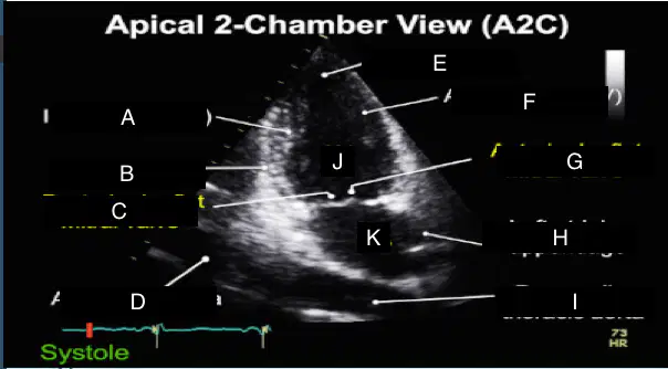
What is H?
Anterior wall (LV)
Anterior leaflet mitral valve
Left atrial appendage
Descending thoracic aorta
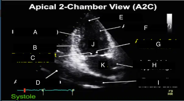
What is I?
Anterior wall (LV)
Anterior leaflet mitral valve
Left atrial appendage
Descending thoracic aorta
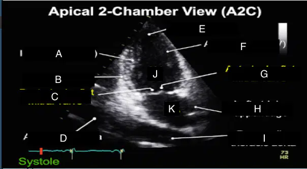
What is J?
RV
LV
LA
RA
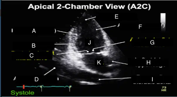
What is K?
LV
LA
RA
RV
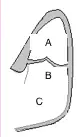
What is A?
RV
TV
MV
RA
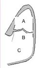
What is B?
RV
TV
MV
RA
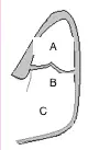
What is C?
RV
TV
MV
RA
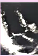
What is going on in this image?
Off-axis 3C
3C RVIT
TR
CWD- TV & Peak TRV
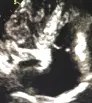
What is going on in this image?
Off-axis 3C
3C RVIT
TR
CWD- TV & Peak TRV
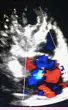
What is going on in this image?
Off-axis 3C
3C RVIT
TR
CWD- TV & Peak TRV
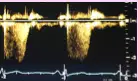
What is going on in this image?
Off-axis 3C
3C RVIT
TR
CWD- TV & Peak TRV
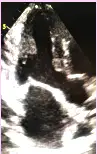
What is the name of this view?
4C
3C
5C
2C
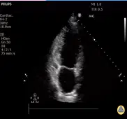
What is the view of this image?
4C
3C
5C
2C
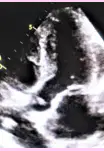
What is the name of this view?
3C
2C
5C
4C

